Introduction
Compact MeV X-ray sources are useful for several applications such as radiography and active interrogation of nuclear materials (Courtois et al., Reference Courtois, Edwards, La Fontaine, Aedy, Barbotin, Bazzoli, Biddle, Brebion, Bourgade, Drew, Fox, Gardner, Gazave, Lagrange, Landoas, Le Dain, Lefebvre, Mastrosimone, Pichoff, Pien, Ramsay, Simons, Sircombe, Stoeckl and Thorp2011). Intense lasers can generate multi-MeV hot electrons when interacting with a ~mm-thick high-Z foils such as tungsten or tantalum. In these targets, the laser couples its energy to the hot electrons mainly on the target surface via several physical processes (Malka and Miquel, Reference Malka and Miquel1996; Wilks and Kruer, Reference Wilks and Kruer1997; Santala et al., Reference Santala, Zepf, Watts, Beg, Clark, Tatarakis, Krushelnick, Dangor, McCanny, Spencer, Singhal, Ledingham, Wilks, Machacek, Wark, Allott, Clarke and Norreys2000). Subsequently, the hot electrons generate MeV bremsstrahlung X rays as they traverse through the rest of the high-Z foil. Several experiments have characterized such intense laser-driven bremsstrahlung X-ray sources (Perry et al., Reference Perry, Sefcik, Cowan, Hatchett, Hunt, Moran, Pennington, Snavely and Wilks1999; Edwards et al., Reference Edwards, Sinclair, Goldsack, Krushelnick, Beg, Clark, Dangor, Najmudin, Tatarakis, Walton, Zepf, Ledingham, Spencer, Norreys, Clarke, Kodama, Toyama and Tampo2002; Clarke et al., Reference Clarke, Neely, Edwards, Wright, Ledingham, Heathcote, McKenna, Danson, Brummitt, Collier, Hatton, Hawkes, Hernandez-Gomez, Holligan, Hutchinson, Kidd, Lester, Neville, Norreys, Pepler, Winstone, Wyatt and Wyborn2006; Hayashi et al., Reference Hayashi, Fukumi, Matsukado, Mori, Kotaki, Kando, Chen, Daito, Kondo, Kanazawa, Yamazaki, Ogura, Nishiuchi, Kado, Sagisaka, Nakamura, Li, Orimo, Homma and Daido2006; Galy et al., Reference Galy, Maucec, Hamilton, Edwards and Magill2007; Courtois et al., Reference Courtois, Edwards, La Fontaine, Aedy, Barbotin, Bazzoli, Biddle, Brebion, Bourgade, Drew, Fox, Gardner, Gazave, Lagrange, Landoas, Le Dain, Lefebvre, Mastrosimone, Pichoff, Pien, Ramsay, Simons, Sircombe, Stoeckl and Thorp2011; Courtois et al., Reference Courtois, Edwards, La Fontaine, Aedy, Bazzoli, Bourgade, Gazave, Lagrange, Landoas, Le Dain, Mastrosimone, Pichoff, Pien and Stoeckl2013; La Fontaine, Reference La Fontaine2014; Liang et al., Reference Liang, Bauer, Cimeno, Ferrari, Galtier, Granados, Lee, Liu, Nagler, Prinz, Rokni, Tran and Woods2016; Chen et al., Reference Chen, Hermann, Kalantar, Martinez, Di Nicola, Tommasini, Landen, Alessi, Bowers, Browning, Brunton, Budge, Crane, Di Nicola, Doppner, Dixit, Erbert, Fishler, Halpin, Hamamoto, Heebner, Hernandez, Hohenberger, Homoelle, Honig, Hsing, Izumi, Khan, LaFortune, Lawson, Nagel, Negres, Novikova, Orth, Pelz, Prantil, Rushford, Shaw, Sherlock, Sigurdsson, Wegner, Widmayer, Williams, Williams, Whitman and Yang2017; Yang et al., Reference Yang, Qiu, Li, Lu, Wu and Li2017).
Here, we have characterized such a MeV bremsstrahlung X-ray source from 120 TW (80 J, 650 fs) Trident laser at the Los Alamos National Laboratory interacting with a 1 mm-thick tantalum foil. Our measurements show X-ray temperature of 2.5 MeV, flux of 3 × 10¹² photons/sr/shot, beam divergence of ~0.1 sr, conversion efficiency of ~1%, that is, ~1 J of MeV X rays out of 80 J incident laser, and source size of 80 µm. Our measurement also shows that MeV X-ray yield and temperature is largely insensitive to nanosecond laser contrast up to 10−5.
In contrast, numerical simulations have shown that a double-foil scheme, where laser-driven hot electrons from a thin foil that undergoes relativistic transparency (Kaw and Dawson, Reference Kaw and Dawson1970; Palaniyappan et al., Reference Palaniyappan, Hegelich, Wu, Jung, Gautier, Yin, Albright, Johnson, Shimada, Letzring, Offermann, Ren, Huang, Horlein, Dromey, Fernandez and Shah2012, Reference Palaniyappan, Huang, Gautier, Hamilton, Santiago, Kreuzer, Sefkow, Shah and Fernandez2015) impinging onto a separate high-Z converter foil, could generate more efficient K α X rays than the single-foil scheme (Sefkow et al., Reference Sefkow, Bennett, Geissel, Schollmeier, Franke and Atherton2011). The same reasoning can also be extended to MeV X-ray generation. Our preliminary measurements of MeV X-ray source using the double-foil scheme, where the laser-driven hot electrons from a thin foil (110 nm aluminum foil) undergoing relativistic transparency (Kaw and Dawson, Reference Kaw and Dawson1970; Palaniyappan et al., Reference Palaniyappan, Hegelich, Wu, Jung, Gautier, Yin, Albright, Johnson, Shimada, Letzring, Offermann, Ren, Huang, Horlein, Dromey, Fernandez and Shah2012, Reference Palaniyappan, Huang, Gautier, Hamilton, Santiago, Kreuzer, Sefkow, Shah and Fernandez2015; Cobble et al., Reference Cobble, Palaniyappan, Johnson, Shimada, Huang, Gautier, Clark, Falk and Jung2016) impinging onto a second high-Z converter foil separated by 50–400 µm, show MeV X-ray yield more than an order of magnitude lower compared with the single-foil results discussed above. We believe that a better understanding of the hot electron transport in vacuum from the thin foil to the converter foil could help optimize the double-foil scheme. Also, reducing the gap between the two foils down to 20 µm or lower as indicated in Sefkow et al. (Reference Sefkow, Bennett, Geissel, Schollmeier, Franke and Atherton2011) could also help mitigate the hot electron transport issues. Although understanding and optimizing the double-foil scheme could potentially yield a better MeV bremsstrahlung X-ray source, it is beyond the scope of the present work.
Experimental setup
The experiments reported here were conducted at the Trident laser facility at the Los Alamos National Laboratory, USA (Batha et al., Reference Batha, Aragonez, Archuleta, Archuleta, Benage, Cobble, Cowan, Fatherley, Flippo, Gautier, Gonzales, Greenfield, Hegelich, Hurry, Johnson, Kline, Letzring, Loomis, Lopez, Luo, Montgomery, Oertel, Paisley, Reid, Sanchez, Seifter, Shimada and Workman2008). The Trident laser (80 J, 650 fs FWHM, 1053 nm wavelength, s-polarization) is focused onto the target using an f/3 off-axis parabola to a spot size of 10 µm diameter (first Airy minimum containing 65% laser energy) with a peak laser intensity of 2 × 10²⁰ W/cm2 (a 0 ≈ 13). Plasma mirrors were not used in the experiment.
The laser is incident on a 1 mm-thick tantalum foil that is rotated 10° off the laser axis (see Fig. 1a). A 0.2 T magnet, 5.5 cm long, located right behind the tantalum foil deflected the charged particles away from reaching the X-ray detector. The X-ray transmission through a tungsten wedge, thickness ranging from 0 to 5.08 cm over a length of 17.8 cm, was measured using a calibrated image plate detector (Fernandez et al., Reference Fernandez, Gautier, Huang, Palaniyappan, Albright, Bang, Dyer, Favalli, Hunter, Mendez, Roth, Swinhoe, Bradley, Deppert, Espy, Falk, Guler, Hamilton, Hegelich, Henzlova, Ianakiev, Iliev, Johnson, Kleinschmidt, Losko, McCary, Mocko, Nelson, Roycroft, Cordoba, Schanz, Schaumann, Schmidt, Sefkow, Shimada, Taddeucci, Tebartz, Vogel, Vold, Wurden and Yin2017). An additional 0.5 mm-thick tantalum foil covered the entire image plate to block X rays below 92 keV (<1% transmission at 92 keV).
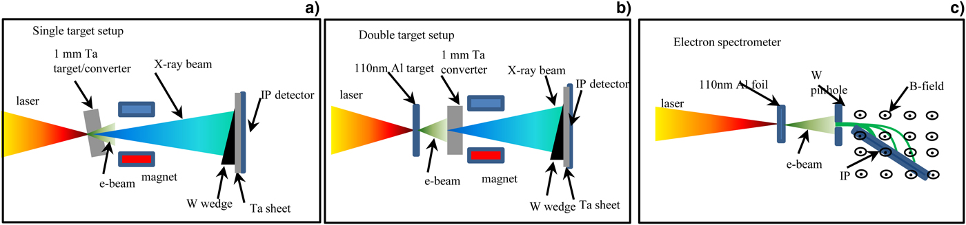
Fig. 1. Schematic representation of experimental setup (f/3 laser focus onto 110 nm Al foil). (a) The 0.12 PW Trident laser is focused with f/3 off-axis-parabola (peak intensity – 2 × 10²⁰ W/cm2) onto a 1 mm-thick tantalum foil (foil normal rotated 10° off to laser propagation axis). The tantalum foil acts both as electron source and X-ray converter. A 0.2 T magnet deflects the charged particles away. The X-ray transmission through the tungsten wedge (5.08 cm × 5.08 cm × 17.8 cm) is measured using a calibrated BAS-SR Fuji image plate (20 cm × 40 cm). The image plate was covered with a 0.5 mm-thick tantalum sheet (20 cm × 40 cm) to block X rays below 92 keV (<1% transmission at 92 keV) reaching the image plate detector. The X-ray spectrum is retrieved from the transmission data via Expectation-Maximization algorithm. (b) Double-foil scheme: same setup as before except the laser impinges onto a 110 nm-thick aluminum foil. The electron beam from the aluminum foil subsequently impinges onto a 1 mm-thick tantalum X-ray converter foil. The spacing between the foils was varied from 50 to 400 µm. (b) Schematic representation of the magnetic electron spectrometer used to measure the electron spectrum from 110 nm Al foil. A 1 mm diameter tungsten pinhole was placed 15 cm away from the converter foil to sample the electron beam.
For the double-foil scheme, shown in Figure 1(b), the intense laser, incident normally on a 110 nm-thick aluminum foil that undergoes relativistic transparency, drives a multi-MeV electron beam. These hot electrons impinge onto a 1 mm-thick tantalum foil that is 50–200 µm away from the aluminum foil. The electron beam generates bremsstrahlung MeV X rays as they traverse through the high-Z converter foil. These X rays are measured the same way as discussed above.
We also used a separate magnetic spectrometer, 0.8 T magnetic field over 10 cm long, to measure the electron spectrum from the aluminum foil (Fig. 1c). The tungsten pinhole (1 mm diameter) was placed 15 cm away from the converter foil. The image plate detector (BAS-TR) was placed inside the magnet opening at 33.7° angle with respect to the incident electron trajectory to capture the deflected electrons. The lower (higher) energy cut-off of the magnetic spectrometer was 0.2 MeV (28.2 MeV).
Results and Discussion
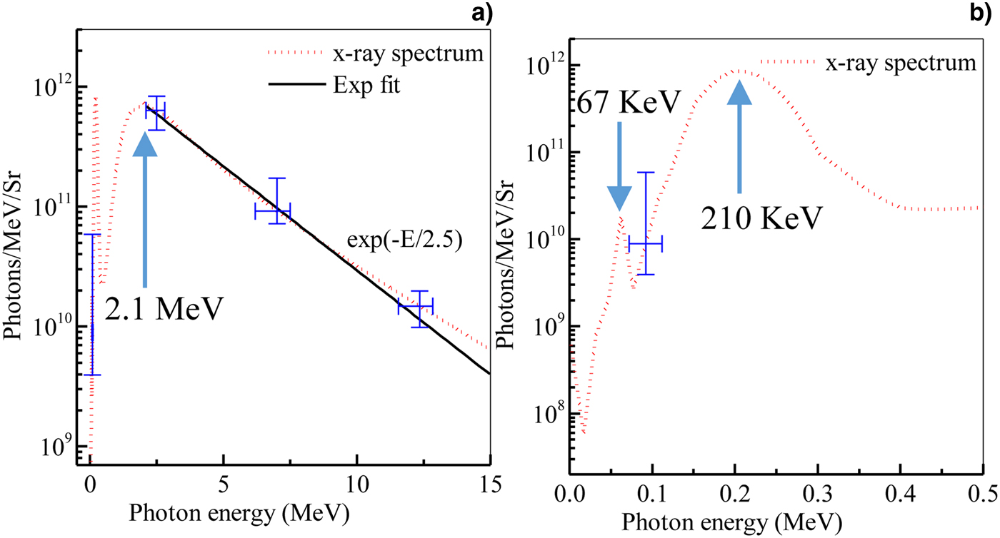
Figure 2(a) shows the X-ray spectrum retrieved from the measured X-ray transmission data when the Trident laser is incident on a 1 mm-thick tantalum foil. The X-ray spectrum peaks at 2.1 MeV with a temperature of 2.5 MeV. The X-ray spectral peak at 210 keV appears from the tantalum filter. Additionally, the X-ray spectrum shows a tantalum K-edge at 67 keV (Fig. 2b). However, the 67 and 210 keV X-ray peaks have negligible X-ray energy content compared with the broader 2.1 MeV peak. The integrated X-ray spectrum yields 2.5 × 10¹² photons/sr per shot. The yield varied typically by less than a factor of two from shot-to-shot. For the X-ray beam FWHM divergence of 0.1 sr (10° half-opening angle), we get 0.8 J of MeV X rays, which is ~1% conversion efficiency.

Fig. 2. Results from single target setup. (a) Measured X-ray spectrum from single 1 mm-thick tantalum foil. (b) Closer view of the same X-ray spectrum showing spectral peaks at 67 and 210 keV.
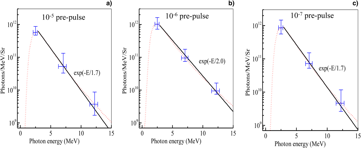
Additionally, we also tested the sensitivity of the X-ray generation from the single-foil target to the ns pre-pulse/pedestal. We added 1 ns long pre-pulses that were at the 10−5 (Fig. 3a), 10−6 (Fig. 3b), and 10−7 (Fig. 3c) level compared with the peak of the high-contrast high-power Trident laser pulse. The measured X-ray spectrum shows that the added pre-pulses do not have a significant effect on the X-ray spectra and the yield from the 1 mm-thick tantalum foil. For this setup, we added an extra 4 mm-thick tungsten filter to block X rays below 240 keV (<1% transmission below 240 keV). Hence, the 67 and 210 keV X-ray peaks are missing in Figure 3a–c. One-dimensional HELIOS rad-hydro (MacFarlane et al., Reference MacFarlane, Golovkin and Woodruff2006) simulation showed that these pre-pulses produce a pre-plasma density gradient of only a few microns at the front surface of the target due to lower sound speed of the heavier ions. Also here we use s-polarized laser where the laser E-field has no component along the pre-plasma gradient that could reduce the vacuum heating in the pre-plasma (Brunel, Reference Brunel1987).

Fig. 3. Effect of laser contrast on X-ray spectrum from single 1 mm-thick tantalum foil. A 1 ns-long pedestal at various laser intensity levels were added to the high-contrast Trident laser. Measured X-ray spectra when added (a) 10−5 pedestal, (b) 10−6 pedestal, and (c) 10−7 pedestal.
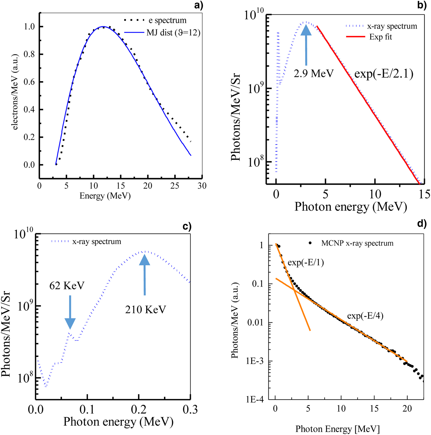
Figure 4a shows the electron spectrum (dashed red line) from the 110 nm aluminum foil measured using the magnetic spectrometer. The electron spectrum peaks around 12 MeV. The instrument had a lower (higher) energy cut-off of 0.2 (28.2) MeV. It is possible that the low-energy electrons below 3 MeV do not escape the target (Cobble et al., Reference Cobble, Palaniyappan, Johnson, Shimada, Huang, Gautier, Clark, Falk and Jung2016). The measured electron spectrum matches well with a Maxwell–Jüttner distribution f(E) = (E 2β/θK 2(1/θ))exp(-E/θ) with θ = kT/mc 2 = 12, where β = v/c, and kT is the electron temperature. Figure 4b shows the typical X-ray spectrum retrieved from the measured X-ray transmission data from the double-foil targets with ~200 µm spacing between them via Expectation-Maximization ation algorithm (Lange and Carson, Reference Lange and Carson1984; Zhang et al., Reference Zhang, Zhang, Chen, Xing, Cheng and Xiao2007). Varying the distance between foils from 50 to 400 µm did not seem to affect the X-ray spectra and yield in any consistent manner. The X-ray spectrum peaks at 210 keV and 2.9 MeV with a temperature of 2.1 MeV. Additionally, the X-ray spectrum shows a tantalum k α peak at 67 keV (Fig. 4c). However, the 67 and 210 keV X-ray peaks have negligible X-ray energy content compared with the broader 2.9 MeV peak. It is most likely that the 210 keV X-ray peak is an apparent peak due to significant X-ray filtering from the 0.5 mm-thick tantalum foil.

Fig. 4. Results from double target setup. (a) Measured electron spectrum from 100 nm-thick aluminum foil using a magnetic spectrometer. (b) Measured X-ray spectrum from double target (100 nm-thick aluminum foil ~200 µm away from 1 mm-thick tantalum converter foil). (c) Zoomed in view of the same X-ray spectrum showing spectral peaks at 67 and 210 keV. (d) Simulated X-ray spectrum using measured electron spectrum as input to Monte-Carlo algorithm MCNP.
Figure 4d shows the simulation results when an electron beam with the measured electron spectrum in Figure 4a traverses through a 1 mm-thick tantalum converter foil using the code Monte Carlo N-Particle (MCNP) (Forster and Godfrey, Reference Forster and Godfrey1985). The simulation results show a two temperature (1 and 4 MeV) X-ray spectrum that has no obvious spectral peaks. The fact that the X-ray spectrum from the MCNPX simulation (Fig. 4d) is much hotter than the measured X-ray distribution (Fig. 4b) seems to provide evidence that there may be issues in the transport of the hot electrons from the thin foil to the converter foil. We believe that better understanding of the hot electron transport from the thin foil to the converter foil could help optimize the double-foil scheme. Also, reducing the gap between the two foils down to 20 µm or lower as indicated in Sefkow et al. (Reference Sefkow, Bennett, Geissel, Schollmeier, Franke and Atherton2011) could help mitigate the hot electron transport issues. The integrated X-ray spectrum from the double-foil scheme yields 3.6 × 10¹⁰ photons/sr per shot. However, the yield varied by more than one order of magnitude from shot-to-shot.
Figure 5a shows the R2DTO object, a 10 cm × 10 cm × 6 mm tungsten object with radial slots, used for quantifying the MeV X-ray source size from the X-ray radiograph of the object using the Bayesian-Inference-Engine (BIE) analysis. Figure 5b shows R2DTO radiograph using the X rays from the double-foil source with 1:1 magnification. Figure 5c shows the same radiograph using the X rays from the single-foil source with 6.2 × magnification. The images show that the edges of the radial slots are blurred when using the double-foil X-ray source. The BIE analysis of these images shows that the MeV X-ray source size was 35–195 and 270–600 µm in the single-foil and double-foil schemes, respectively. The details of the BIE analysis is discussed elsewhere (Tobias et al., Reference Tobias, Palaniyappan, Gautier, Mendez, Burris-Mog, Huang, Favalli, Hunter, Espy, Schmidt, Nelson, Sefkow, Shimada, Johnson and Fernandez2017). The larger source size in the double-foil scheme could come form the hot electron transport issues that could increase the size of the electron beam reaching the converter foil.
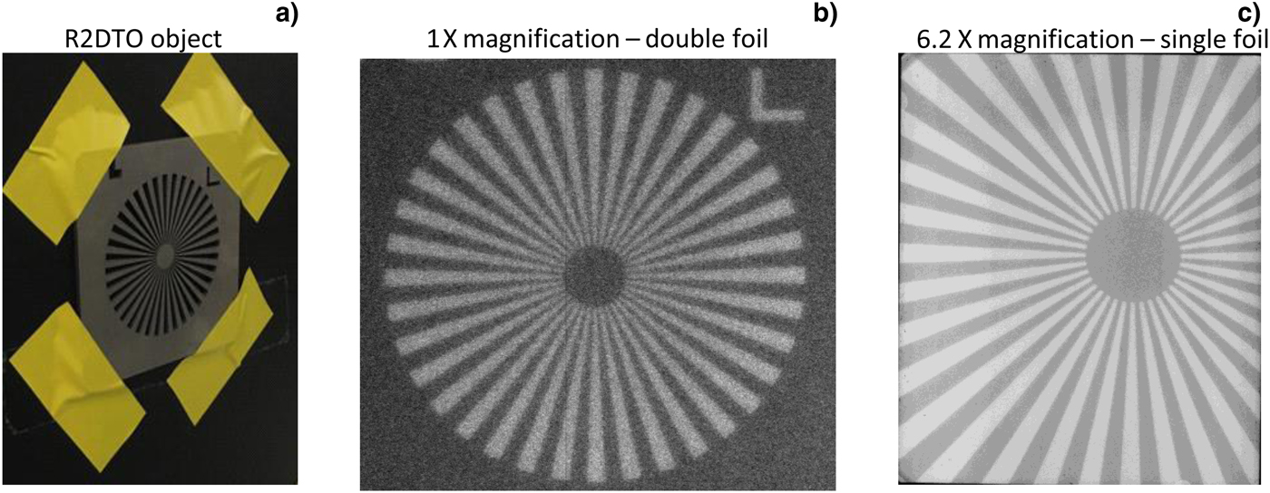
Fig. 5. MeV X-ray radiograph. (a) 10 cm × 10 cm × 6 mm tungsten object called “R2DTO” with radial slots used for measuring the MeV X-ray source size, (b) radiograph of R2DTO taken using MeV X rays from the double-foil scheme with 1:1 magnification, (c) radiograph of the same object using MeV X rays from the single-foil scheme with 6.2 × magnification.
Conclusions
In conclusion, we have characterized MeV bremsstrahlung X-ray source from 120 TW (80 J, 650 fs) Trident laser interaction with a 1 mm-thick tantalum foil. Our measurements show X-ray temperature of 2.5 MeV, flux of 3 × 10¹² photons/sr/shot, beam divergence of ~0.1 sr, conversion efficiency of ~1%, that is, ~1 J of MeV X rays out of 80 J incident laser, and source size of 80 µm. Our measurement also shows that MeV X-ray yield and temperature is largely insensitive to nanosecond laser contrast up to 10−5. Also, preliminary measurements of similar MeV X-ray source using a double-foil scheme, where the laser-driven hot electrons from a thin foil undergoing relativistic transparency impinging onto a second high-Z converter foil separated by 50–400 µm, show MeV X-ray yield more than an order of magnitude lower compared with the single-foil results. Despite the interest, further optimization and complete understanding of the double-foil scheme using comprehensive Particle-In-Cell (PIC) simulations is beyond the scope of the work presented here.
Author ORCIDs
S. Palaniyappan 0000-0001-6377-1206.
Acknowledgments
This work was performed under the auspices of the National Nuclear Security Administration Inertial Confinement Fusion Program John Kline, LANL ICF Program Manager along with the support of the Los Alamos National Laboratory LDRD (Laboratory Directed Research and Development) project 20170573ECR. This work was funded in part by DOE-NNSA.