


Introduction
Haemogregarines of the genus Hepatozoon Miller, 1908 (Apicomplexa, Hepatozoidae) comprise a ubiquitous group of intraerythrocytic parasites that are remarkably diverse among snakes (Smith, Reference Smith1996; Úngari et al., Reference Úngari, Santos, O'Dwyer, Da Silva, Santos, Da Cunha and Cury2018). For many years, species nomination in this taxon was based on classical taxonomic methods, using morphological and morphometric data (Morrison, Reference Morrison2009). However, such features alone do not provide a robust diagnosis for single-celled organisms such as Hepatozoon, whose paucity of morphological traits make them especially prone to cryptic species (Perkins et al., Reference Perkins, Martinsen and Falk2011; Zechmeisterová et al., Reference Zechmeisterová, Javanbakht, Kvicerová and Siroký2021). In this regard, the use of molecular markers, especially the 18S ribosomal nuclear gene (Hrazdilová et al., Reference Hrazdilová, Cervená, Blanvillain, Foronda and Modrý2021), was a decisive milestone for the taxonomy of these parasites, allowing access to genetic diversity and a better understanding of their phylogenetic relationships (Harris et al., Reference Harris, Maia and Perera2011; O'Dwyer et al., Reference O'Dwyer, Moço, Paduan, Spenassatto, Silva and Ribolla2013; Hrazdilová et al., Reference Hrazdilová, Cervená, Blanvillain, Foronda and Modrý2021).
Descriptions of Hepatozoon in snakes have been reported in literature for more than a century (Smith, Reference Smith1996), yet genetic data for this group are restricted to the last 15 years (Sloboda et al., Reference Sloboda, Kamler, Bulantová, Votýpka and Modrý2007). In Brazil, even with the high taxonomic richness recorded (ca. 40 spp.) in snakes (Pessôa et al., Reference Pessôa, De Biasi and Puorto1974; Smith, Reference Smith1996; Úngari et al., Reference Úngari, Santos, O'Dwyer, Da Silva, Santos, Da Cunha and Cury2018, Reference Úngari, Netherlands, De Alcantara, Emmerich and Da Silva2021), only the sequences of 5 species were available to date: Hepatozoon cevapii O'Dwyer et al., Reference O'Dwyer, Moço, Paduan, Spenassatto, Silva and Ribolla2013 reported in Crotalus durissus Linnaeus, 1758 (O'Dwyer et al., Reference O'Dwyer, Moço, Paduan, Spenassatto, Silva and Ribolla2013) and Thamnodynastes lanei Bailey et al., 2005 (De Paula et al., Reference De Paula, Picelli, Silva, Correa and Viana2021); Hepatozoon cuestensis O'Dwyer et al., Reference O'Dwyer, Moço, Paduan, Spenassatto, Silva and Ribolla2013 and Hepatozoon massardii O'Dwyer et al., Reference O'Dwyer, Moço, Paduan, Spenassatto, Silva and Ribolla2013 described in C. durissus (O'Dwyer et al., Reference O'Dwyer, Moço, Paduan, Spenassatto, Silva and Ribolla2013; Úngari et al., Reference Úngari, Santos, O'Dwyer, Da Silva, Santos, Da Cunha and Cury2018); Hepatozoon musa Borges-Nojosa et al., Reference Borges-Nojosa, Borges-Leite, Maia, Zanchi-Silva, da Rocha and Harris2017 from Philodryas nattereri (Steindachner, 1870) (Borges-Nojosa et al., Reference Borges-Nojosa, Borges-Leite, Maia, Zanchi-Silva, da Rocha and Harris2017), C. durissus and Epicrates crassus Cope, 1862 (Úngari et al., Reference Úngari, Santos, O'Dwyer, Da Silva, Santos, Da Cunha and Cury2018); and Hepatozoon quagliattus Úngari et al., 2021 in Dipsas mikanii (Schlegel, 1837) (Úngari et al., Reference Úngari, Netherlands, De Alcantara, Emmerich and Da Silva2021).
Helicops Wagler, 1830 (Colubridae, Dipsadinae) is a genus of neotropical snakes widely distributed in South America and comprises 19 described species, of which 15 can be found in Brazil, some of them are endemic to the country (Costa and Bérnils, Reference Costa and Bérnils2018; Nogueira et al., Reference Nogueira, Argôlo, Arzamendia, Azevedo, Barbo, Bérnils, Bolochio, Borges-Martins, Brasil-Godinho, Braz, Buononato, Cisneros-Heredia, Colli, Costa, Franco, Giraudo, Gonzalez, Guedes, Hoogmoed, Marques, Montingelli, Passos, Prudente, Rivas, Sanchez, Serrano, Silva, Strüssmann, Vieira-Alencar, Zaher, Sawaya and Martins2019; Schoneberg and Kohler, Reference Schoneberg and Kohler2021). These snakes are strictly aquatic, with nocturnal habits, and feed mainly on fish and anurans (Aguiar and Di-Bernardo, Reference Aguiar and Di-Bernardo2004; Ávila et al., Reference Ávila, Ferreira and Arruda2006; Carvalho et al., Reference Carvalho, De Assis Montag and Dos Santos-Costa2017). They can be viviparous or oviparous, with the exception of Helicops angulatus (Linnaeus, 1758) that possesses both reproductive modes (Braz et al., Reference Braz, Scartozzoni and Almeida-Santos2016). In terms of the occurrence of haemoparasites, 2 species of hepatozoids have been reported in these snakes (Pessôa and Cavalheiro, Reference Pessôa and Cavalheiro1969a, Reference Pessôa and Cavalheiro1969b): Hepatozoon modesta Pessôa and Cavalheiro, 1969 from Helicops modestus Gunther, 1861 and Hepatozoon carinicauda Pessôa and Cavalheiro, 1969 in Helicops carinicaudus (Wied-Neuwied, 1824).
Therefore, the present study aimed to investigate the presence of haemoparasites in the brown-banded water snake He. angulatus. As a result, through the use of an integrative taxonomy approach (morphology, morphometry and molecular data), H. carinicauda, a species named more than 50 years ago, was rediscovered and redescribed.
Materials and methods
Blood sampling and parasite morphological identification
Blood samples from 3 individuals of He. angulatus were collected via venepuncture of the tail (Sykes and Klaphake, Reference Sykes and Klaphake2008). The snakes were manually captured in 2 flooding areas in the municipal region of Macapá, in the state of Amapá, Brazil (0°01′04.29″S, 51°05′11.17″W; 0°00′40.7″N, 51°05′49.5″W) (Fig. 1). A portion of this blood was used to make blood smears, which were fixed with absolute methanol for 3 min and stained with Giemsa 10% for 30 min, while the rest of the sample was preserved in 96% ethanol (Hull and Camin, Reference Hull and Camin1960; Telford et al., Reference Telford, Wozniak and Butler2001). The slides were examined under a light microscope at ×400 and ×1000 magnification and the parasite forms were recorded with a 5.1 MP digital camera attached to the biological microscope DI – 136T. The images and measurements of the parasites were processed using Image View® software. Morphometric characterization was given in micrometres (μm) and variables such as the length, width and area of the parasite and erythrocytes were presented as mean, range and standard deviation. Parasitaemia was estimated by counting the number of parasites visualized in 2000 erythrocytes, in 20 fields of 100 examined erythrocytes (Godfrey et al., Reference Godfrey, Fedynich and Pence1987).

Fig. 1. Map of the study area, Macapá, Amapá, Brazil, containing the location of the analysed samples, indicated by red circles.
DNA extraction, amplification and sequencing
Total DNA was extracted from a sample using the DNeasy Blood & Tissue Kit (QIAGEN, Valencia, CA, USA), according to the manufacturer's instructions. The detection of the parasite DNA by polymerase chain reaction (PCR) was performed using the Hep300 and Hep900 primers, which amplified a fragment of 590 base pairs (bp) of the 18S rRNA gene for sequencing and phylogenetic analysis (Ujvari et al., Reference Ujvari, Madsen and Olsson2004). The PCR consisted of a pre-PCR step at 94°C for 3 min, followed by 45 cycles of 94°C for 45 s, 1 cycle at 56°C for 1 min, an extension at 72°C for 40 s and a final extension at 72°C for 10 min. The amplicon was purified following the manufacturer's protocol using Wizard® SV Gel and the PCR Clean-Up System. PCR products were sequenced using the BigDye™ Terminator v.3.1 Cycle Sequencing Ready Reaction Kit (Applied Biosystems, Foster City, CA, USA) and the ABI 3100 Genetic Analyser (Applied Biosystems) (Sanger et al., Reference Sanger, Nicklen and Coulson1977).
Phylogenetic analyses
The amplified sequence was edited and the consensus sequence was built using the BioEdit software package, v7.2.5 (Hall, Reference Hall1999). The identity, query coverage and E-values were assessed by the BLASTn tool (using default parameters), available in the NCBI GenBank database (Altschul et al., Reference Altschul, Gish, Miller, Myers and Lipman1990). The obtained sequence was aligned with other sequences retrieved from GenBank using MAFFT software, version 7 (Katoh et al., Reference Katoh, Rozewicki and Yamada2019). Sequences used for phylogenetic inferences were selected from the BLAST results and other studies performed in Brazil and other countries. First, the ‘best of fit’ model was selected by the IQ-TREE software package (Trifinopoulos et al., Reference Trifinopoulos, Nguyen, Von Haeseler and Minh2016), under the Akaike information criterion (Darriba et al., Reference Darriba, Taboada, Doallo and Posada2012). A Bayesian inference analysis was performed with MrBayes 3.1.2. (Ronquist and Huelsenbeck, Reference Ronquist and Huelsenbeck2003). Markov chain Monte Carlo simulations were run for 106 generations with a sampling frequency of every 100 generations and a burn-in of 25% using the CIPRES Science Gateway (Miller et al., Reference Miller, Pfeiffer and Schwartz2010). The number of generations was selected based on the value of the average standard deviation of split frequencies (<0.02, MrBayes version 3.2 Manual) (Ronquist et al., Reference Ronquist, Huelsenbeck and Teslenko2011). Maximum likelihood (ML) tree inference was performed with the IQ-TREE software package (Trifinopoulos et al., Reference Trifinopoulos, Nguyen, Von Haeseler and Minh2016). Phylogenetic tree edition and rooting (outgroup) were performed using the Treegraph 2.0 beta software.
Genetic diversity of Hepatozoon spp.
Nineteen sequences retrieved from GenBank (MN833641, MF435047, MF435048, KX880079, MF497763, MF497764, MF497765, MF497766, MF497767, KC342524, MF497769, MF497770, KC342526, MW241134, MW241135, KC342525, MF322538, MF322539, MW591599) and aligned using the MAFFT software package (version 7) (Katoh et al., Reference Katoh, Rozewicki and Yamada2019), resulting in an alignment of 461 bp, were used to evaluate the genetic diversity of the 18S rRNA gene from Hepatozoon spp. detected in the snake in the present study and in reptiles from previous studies in Brazil (O'Dwyer et al., Reference O'Dwyer, Moço, Paduan, Spenassatto, Silva and Ribolla2013, Borges-Nojosa et al., Reference Borges-Nojosa, Borges-Leite, Maia, Zanchi-Silva, da Rocha and Harris2017; Bouer et al., Reference Bouer, André, Gonçalves, Luzzi, Oliveira, Rodrigues, Varani, Miranda, Perles, Werther and Machado2017; Úngari et al., Reference Úngari, Santos, O'Dwyer, Da Silva, Santos, Da Cunha and Cury2018, Reference Úngari, Netherlands, De Alcantara, Emmerich and Da Silva2021; Picelli et al., Reference Picelli, Da Silva, Ramires, Da Silva, Pessoa, Viana and Kaefer2020). Nucleotide diversity (Pi), haplotype diversity (Hd), number of haplotypes (h), total number of mutations (Eta) and average number of nucleotide differences (k), using DnaSP v5 software (Librado and Rozas, Reference Librado and Rozas2009), were calculated. Additionally, a haplotype network was constructed using Median Joining Network parameters (Bandelt et al., Reference Bandelt, Forster and Rohl1999) using PopArt software (http://popart.otago.ac.nz). Finally, an even-distance matrix among sequences detected in reptiles in Brazil was estimated using the Mega-X software package version 10.1.8 (Kumar et al., Reference Kumar, Stecher, Li, Knyaz and Tamura2018).
Results
Microscopic analysis
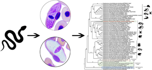
All 3 He. angulatus snakes were positive for Hepatozoon parasites, with an average parasitaemia of 7/2000 erythrocytes in the blood (0.37%; ±4.51), ranging from 3 to 12 parasites among the specimens. Gamonts (Fig. 2a–f) observed in the peripheral blood of the 3 snakes had extremely wide and elongated bodies, which induced deformities in the host cells, and had large eccentric nuclei. These characteristics agreed with H. carinicauda, a species previously described in the snake He. carinicaudus (Pessôa and Cavalheiro, Reference Pessôa and Cavalheiro1969b). Details of the morphometric and morphological traits are shown in Table 1 and in the species redescription section below.

Fig. 2. Gamonts of Hepatozoon carinicauda infecting erythrocytes of the aquatic snakes Helicops angulatus from the Eastern Amazonia, Brazil (a–f). Arrows indicate parasites; asterisks (*) indicate gamont nuclei; and ‘n’ indicates host cell nucleus. Micrographs are from Giemsa-stained thin blood films. Scale bar = 10 μm.
Table 1. Comparative morphometry of Hepatozoon carinicauda gamonts and their host cells between this study, Pessôa and Cavalheiro (Reference Pessôa and Cavalheiro1969b) and Telford (Reference Telford2009)

Measurements are in micrometres and presented as mean ± standard deviation (s.d.) and/or intervals (minimum and maximum values).
Molecular and phylogenetic analyses
The 18S rRNA sequence detected in He. angulatus exhibited 100% of query coverage, 98.47% of identity and 0.0 of E-value with Hepatozoon spp. detected in Amblyomma fimbriatum Koch, 1844 from Varanus panoptes Storr, 1980, sampled in Australia (EU430234; Vilcins et al., Reference Vilcins, Ujvari, Old and Deane2009), and 98.14% of identity with Hepatozoon ophisauri (Tartakovskii, 1913) from Iran, detected in the lizard Pseudopus apodus (Pallas, 1775) (MN723845; Zechmeisterová et al., Reference Zechmeisterová, Javanbakht, Kvicerová and Siroký2021). The sequence also exhibited 99% of query coverage, 98.63% of identity and 0.0 of E-value with Hepatozoon spp. detected in Amblyomma varanense (Supino, 1897) from the King cobra Ophiophagus hannah (Cantor, 1836) from Thailand (JQ670908; Sumrandee et al., Reference Sumrandee, Baimai, Trinachartvanit and Ahantarig2015).
Phylogenetic analysis of a 615 bp alignment using evolutionary model GTR (general-time reversible model) + F + I + G4 showed 87% bootstrap in the maximum likelihood (Ml) tree (Fig. 3) and 91% posterior probability in the Bayesian tree (Supplementary material). The topology of both trees was similar. The newly generated sequence formed a new branch, inserted into a large clade composed of Hepatozoon strains isolated from herpetofauna and mites from South America. Sequences previously detected in snakes (H. cevapii, H. massardii, H. musa and H. cuestensis) from Brazil were inserted (O'Dwyer et al., Reference O'Dwyer, Moço, Paduan, Spenassatto, Silva and Ribolla2013; Borges-Nojosa et al., Reference Borges-Nojosa, Borges-Leite, Maia, Zanchi-Silva, da Rocha and Harris2017), and H. cevapii and H. massardii were positioned in a different clade (Fig. 3). The sequences of H. cuestensis and H. musa shared the same ancestor with the sequence detected in the present study (Fig. 3). Furthermore, the sequences of Hepatozoon spp. (EU430234) and H. ophisauri (MN723845) that presented >98% of identity with our sequence were clustered in other clades.

Fig. 3. Phylogenetic tree based on an alignment of 615 bp fragment of Hepatozoon spp. 18S rRNA sequences, using ML method and GTR evolutionary model. Numbers beside nodes correspond to bootstrap. Accession numbers are indicated in the sequences. The sequence of Hepatozoon carinicauda detected in the present study is highlighted in bold red.
The distance matrix showed that the divergence (P value) between the Hepatozoon 18S rRNA sequences obtained herein and in reptiles from previous studies carried out in Brazil varied from 1.86 to 6.5% (Table 2). The Hepatozoon ameivae (Carini and Rudolph, 1912) sequence obtained from the lizard Ameiva ameiva (Linnaeus, 1758) (Picelli et al., Reference Picelli, Da Silva, Ramires, Da Silva, Pessoa, Viana and Kaefer2020) was the closest, with a distance of 1.86% (Table 2). Among the Hepatozoon sequences obtained from snakes in Brazil, the closest were those from H. musa, which had a divergence from 1.88 to 2.2%, followed by H. cuestensis, which had a divergence from 2.1 to 2.3%, H. massardii, with a distance of 2.5%, and H. cevapii, which had a divergence of distance ranging from 2.3 to 2.7% (Table 2). The sequences of H. quagliattus were the most distant of all, with a genetic distance from 6.2 to 6.7% (Table 2).
Table 2. Divergence scores among the different Hepatozoon spp. detected in reptiles from Brazil

The pairwise distance matrix was estimated using the Mega-X software version 10.1.8.
Genetic diversity
The analysis of nucleotide polymorphisms of the 18S rRNA sequences isolated in reptiles in Brazil revealed 10 different haplotypes (Fig. 4), with 54 variable sites, with haplotype diversity (Hd) = 0.9 and a nucleotide diversity (Pi) = 0.02724. As a result, most Hepatozoon species from Brazilian reptiles were represented by individual haplotypes, with the exception of the Hepatozoon caimani (Carini, 1909) and H. musa species, both of which had 2 haplotypes. Nucleotide sequence genealogy shows that the sequence of H. carinicauda originated from median vectors, which represent hypothetical haplotypes generated by the software to connect the other haplotypes, but which were not contemplated in the present study. The comparison between H. carinicauda and its closest sequence, H. ameivae, demonstrates the presence of 3 median vectors with 10 mutational events between them. These results corroborate the phylogenetic tree, and 5 groups were found: (i) one comprising H. cuestensis, H. musa and H. ameivae; (ii) another group composed of H. carinicauda; (iii) a third composed of H. massardii and H. cevapii; (iv) a fourth composed of H. quagliattus; and (v) a fifth composed of haplotypes of H. caimani (Fig. 4).

Fig. 4. Median Joining Network of 18S rRNA Hepatozoon sequences (461 bp) detected in snakes from Brazil in the present study and in previous studies. While the lines between haplotypes represent mutational steps, the black circles indicate median vectors (hypothetical haplotypes generated by the software to connect all the haplotypes).
Phylogenetic position and genetic divergence showed that the novel sequence obtained in this study is a distinct species of Hepatozoon. Therefore, based on these data, the morphological and morphometric similarities, and the congeneric host (Helicops), we believe that the parasite found herein belongs to the previously described species H. carinicauda.
Species redescription
Taxonomic summary
Phylum: Apicomplexa Levine, 1970
Class: Coccidia Leuckart, 1879
Order: Eucoccidiorida Léger and Duboscq, 1910
Suborder: Adeleorina Léger, 1911
Family: Hepatozoidae Wenyon, 1926
Genus: Hepatozoon Miller, 1908
Hepatozoon carinicauda Pessôa and Cavalheiro, 1969
Type host: Helicops carinicaudus (Wied-Neuwied, 1824) (Serpentes: Colubridae), Wied's Keelback, Cobra-D’água-Preta.
Other hosts: Helicops angulatus (Linnaeus, 1758) (Serpentes: Colubridae), Brown-banded water snake, Cobra-D’água – current study.
Vector: Unknown.
Type locality: Votuporanga, São Paulo state, Brazil (Pessôa and Cavalheiro, Reference Pessôa and Cavalheiro1969b).
Other localities: Urban flooding area (0°01′04.29″S, 51°05′11.17″W; 0°00′40.7″N, 51°05′49.5″W), municipal region of Macapá, state of Amapá, Brazil (present study).
Prevalence: One of He. carinicaudus (Pessôa and Cavalheiro, Reference Pessôa and Cavalheiro1969b); and all 3 specimens of He. angulatus (this study).
Site of infection: Gamonts in blood erythrocytes (see Pessôa and Cavalheiro, Reference Pessôa and Cavalheiro1969b; current study); meronts observed in liver, intestine and lungs (Pessôa and Cavalheiro, Reference Pessôa and Cavalheiro1969b).
Parasitaemia (this study): Mean of 7 parasites/2000 blood erythrocytes (0.37%; ±4.51), ranging from 3 to 12 parasites.
Type material: Six blood slides (hapantotypes) from He. angulatus were deposited at the Institute for Scientific and Technological Research of the State of Amapá (IEPA), Amapá, Brazil (no IEPA0001, IEPA0002, IEPA0003, IEPA0004, IEPA0005, IEPA0006).
DNA sequences: The 18S ribosomal gene sequence (590 bp) was deposited in GenBank® (accession number MT561455).
Redescription
This haemogregarine was described by Pessôa and Cavalheiro (Reference Pessôa and Cavalheiro1969b) in the colubrid snake He. carinicaudus from the southeast region of Brazil. In the original description, large gamonts were reported infecting erythrocytes and meronts, with an abundant number of merozoites in the liver, intestine and lung smears. Those authors also made an attempt at vectorial incrimination and recorded the development of sporulated oocysts in the body cavity of the leech Haementeria lutzi Pinto, 1920. In the present study, intraerythrocytic gamonts were the forms of H. carinicauda investigated in the peripheral blood of a new vertebrated host, the brown-banded water snake He. angulatus from the Eastern Amazonia, Brazil (Fig. 2a–f).
Diagnosis: Gamonts (Fig. 2a–f; Table 1) – extremely elongated, slightly curved and sometimes pyriform; both ends rounded; basophilic cytoplasm with granules; parasitophorous vacuole not evident. Wide and elliptical nucleus; eccentric and slightly oriented towards one end; position usually marked by the enlargement of the body of the parasite; with condensed dark-stained chromatin filaments or more dispersed chromatin. Body dimensions: 25.3 ± 1.9 × 8.6 ± 1.3 μm; and area 174.1 ± 17.2 μm2. Nucleus dimensions: 5.2 ± 1.6 × 6.9 ± 1.2 μm; and area 30.2 ± 14.1 μm2. The cytopathological effects caused by parasites on their host cells were remarkable when compared with non-parasitized erythrocytes. The infected erythrocytes were hypertrophied (27.3 ± 2.0 μm vs 16.9 ± 0.9 μm) with gamonts occupying almost the entire area (250.8 ± 34.2 μm2) of these cells. The parasitized erythrocyte nucleus was flattened and displaced laterally to the host cell margin [8.3 ± 0.9 × 3.9 ± 1.0 μm vs 6.9 ± 0.5 × 4.4 ± 0.4 μm (Table 1)], which was generally irregular (Fig. 2e–f).
Remarks: Hepatozoon carinicauda gamonts observed in the blood of He. angulatus were slightly smaller than those described in the type host, He. carinicaudus (Pessôa and Cavalheiro, Reference Pessôa and Cavalheiro1969b) (29.4–21.5 × 11.8–6.8 μm vs 30–23 × 13–5 μm, respectively). When compared to H. modesta (Pessôa and Cavalheiro, Reference Pessôa and Cavalheiro1969a), another species described in Helicops snakes, H. carinicauda is larger (15–13 × 3–2 μm vs 29.4–21.5 × 11.8–6.8 μm, respectively). Moreover, unlike H. carinicauda, there is nothing unique about the morphology of H. modesta, and it is considered a ‘hepatozoic’ type – a former term used for parasites that did not deform or displace the host cell nucleus (Pessôa and Cavalheiro, Reference Pessôa and Cavalheiro1969a). Morphometric and morphological differences can also be found among H. carinicauda and phylogenetically close haemogregarine species infecting lizards and snakes, such as H. ameivae, H. cuestensis and H. musa. Gamonts of H. ameivae, in addition to being smaller (14.28 × 4.50 μm), possess the outstanding feature of overlapping the host cell nucleus (Picelli et al., Reference Picelli, Da Silva, Ramires, Da Silva, Pessoa, Viana and Kaefer2020), a trait not observed in H. carinicauda. Hepatozoon cuestensis forms have arched ends, uniform cytoplasm and are smaller in size (17.07 × 3.6 μm) (O'Dwyer et al., Reference O'Dwyer, Moço, Paduan, Spenassatto, Silva and Ribolla2013). Finally, H. musa has both curved ends and bodies which, although elongated (18.9 × 3.8 μm), are smaller than H. carinicauda (Borges-Nojosa et al., Reference Borges-Nojosa, Borges-Leite, Maia, Zanchi-Silva, da Rocha and Harris2017).
Discussion
Taken together, our genetic, morphological and morphometric data allowed us to redescribe H. carinicauda in a new snake host, He. angulatus, and to place this parasite species in a current phylogenetic context. This was mainly possible because H. carinicauda has very distinct and unique morphological and morphometric characteristics (i.e. large dimensions that considerably deform the erythrocytes). Other studies that employed integrative taxonomy started from the same assumption, such as H. ameivae in the lizard A. ameiva, whose gamonts overlap the host cell nucleus (Picelli et al., Reference Picelli, Da Silva, Ramires, Da Silva, Pessoa, Viana and Kaefer2020), and the redescription of H. ophisauri in the lizard P. apodus, which has unusual pink inclusions, and Hepatozoon colubri (Borner, 1901) in the snake Zamenis longissimus (Laurenti, 1768), which presents a degeneration zone in the cytoplasm, and connection to the nucleus of the host cell (Zechmeisterová et al., Reference Zechmeisterová, Javanbakht, Kvicerová and Siroký2021).
Hepatozoon carinicauda was first observed 52 years ago infecting another Helicops snake, the Wied's Keelback He. carinicaudus. This host species and He. angulatus shared the main ecological features associated with this snake genus, namely inhabiting freshwater environments, having nocturnal habits and feeding mainly on fish and frogs (Aguiar and Di-Bernardo, Reference Aguiar and Di-Bernardo2004; Ávila et al., Reference Ávila, Ferreira and Arruda2006; Carvalho et al., Reference Carvalho, De Assis Montag and Dos Santos-Costa2017). Nevertheless, in addition to having specific differentiations, these species exhibit distinct geographic distribution patterns. Although there are no data on the sympatric occurrence of these host species, the distribution of both species suggests an area of sympatry in the state of Bahia, northeast of Brazil (Freitas, Reference Freitas2003; Costa and Bérnils, Reference Costa and Bérnils2018). In fact, studies have shown that He. carinicaudus is endemic to the coastal Atlantic Forest at low elevations, restricted to the southeast and south regions of the country (Yuki and Lema, Reference Yuki and Lema2005; Nogueira et al., Reference Nogueira, Argôlo, Arzamendia, Azevedo, Barbo, Bérnils, Bolochio, Borges-Martins, Brasil-Godinho, Braz, Buononato, Cisneros-Heredia, Colli, Costa, Franco, Giraudo, Gonzalez, Guedes, Hoogmoed, Marques, Montingelli, Passos, Prudente, Rivas, Sanchez, Serrano, Silva, Strüssmann, Vieira-Alencar, Zaher, Sawaya and Martins2019). In contrast, He. angulatus represents a cryptic species complex, which is most widespread in northern South America (Nogueira et al., Reference Nogueira, Argôlo, Arzamendia, Azevedo, Barbo, Bérnils, Bolochio, Borges-Martins, Brasil-Godinho, Braz, Buononato, Cisneros-Heredia, Colli, Costa, Franco, Giraudo, Gonzalez, Guedes, Hoogmoed, Marques, Montingelli, Passos, Prudente, Rivas, Sanchez, Serrano, Silva, Strüssmann, Vieira-Alencar, Zaher, Sawaya and Martins2019; Murphy et al., Reference Murphy, Muñoz-Mérida, Auguste, Lasso-Alcala, Rivas and Jowers2020), with records in the Brazilian biomes of the Amazonia, the Chiquitano dry forest, the Cerrado, the Caatinga and the northern portion of the Atlantic Forest (Nogueira et al., Reference Nogueira, Argôlo, Arzamendia, Azevedo, Barbo, Bérnils, Bolochio, Borges-Martins, Brasil-Godinho, Braz, Buononato, Cisneros-Heredia, Colli, Costa, Franco, Giraudo, Gonzalez, Guedes, Hoogmoed, Marques, Montingelli, Passos, Prudente, Rivas, Sanchez, Serrano, Silva, Strüssmann, Vieira-Alencar, Zaher, Sawaya and Martins2019; Murphy et al., Reference Murphy, Muñoz-Mérida, Auguste, Lasso-Alcala, Rivas and Jowers2020). Generally, the sharing of parasites by different host species can be explained by the understanding that there is a low specificity to the vertebrate host among the haemogregarines (Maia et al., Reference Maia, Perera and Harris2012; De Paula et al., Reference De Paula, Picelli, Silva, Correa and Viana2021). However, we cannot fully assume that this is the case here, due to the evolutionary and ecological history of the hosts. The presence of H. carinicauda in these snakes would be through a vector and/or an intermediate host related to both, given the similarity of the ecological niche they use (Poulin and Morand, Reference Poulin and Morand2004; Morand, Reference Morand2015). It is also noteworthy that since these snakes are congeneric hosts, the parasite may have been inherited by these species through a common ancestor during the speciation process (Poulin and Morand, Reference Poulin and Morand2004; Hay et al., Reference Hay, Poulin and Jorge2020). In this sense, it would be useful to investigate other Helicops species, as well as other aquatic snake taxa, to test these hypotheses.
With respect to morphological aspects, we observed that H. carinicauda gamonts found in He. angulatus, although slightly smaller, remain within the dimensions of the species (Pessôa and Cavalheiro, Reference Pessôa and Cavalheiro1969b). In the original description, Pessôa and Cavalheiro (Reference Pessôa and Cavalheiro1969b) measured gamonts twice, with an interval of 1 month between each measurement, and noticed an increase in the size of the parasitic forms. Nonetheless, Telford (Reference Telford2009) calculated the measurements of H. carinicauda from the original description photos, finding that larger parasites, identified during the second smear examination, had smaller dimensions (26–25 × 13–10 μm) than those presented by Pessôa and Cavalheiro (Reference Pessôa and Cavalheiro1969b). Despite the fact that both measures were consistent with one another, the author concluded that the original measurements of H. carinicauda were unsuitable. In fact, as in other older haemoprotozoan descriptions, not all the traits in H. carinicauda and the host cells were evaluated, with some discrepancies between the first and second measurements (Pessôa and Cavalheiro, Reference Pessôa and Cavalheiro1969b). For example, the parasite nuclei were considered in the first analysis only, whereas the infected erythrocytes were assessed in the second examination only, where just length was recorded. Therefore, we believe that these methodological inconsistencies are likely to have led to the morphological differences between our study and that of Pessôa and Cavalheiro (Reference Pessôa and Cavalheiro1969b).
To our knowledge, this is the first molecular characterization of a haemogregarine species isolated from a Helicops snake. It should be noted that Hepatozoon spp. DNA was recently detected in a tick (Amblyomma rotundatum Koch, 1844) collected from a He. carinicaudus snake (Fonseca et al., Reference Fonseca, Bahiense, Silva, Onofrio, Barral, Souza, Lira-da-Silva, Biondi, Meyer and Portela2020). However, in this study, infection in snakes was not confirmed, and so it was not clear whether the tick acts as a vector of Hepatozoon for this aquatic snake host. Furthermore, as the authors did not perform morphological characterization or molecular sequencing, it was not possible to compare this parasite with H. carinicauda found herein. Furthermore, it is important to emphasize that available molecular data for Hepatozoon infecting snakes remain scarce in all countries within the Amazonian domain. So far, there has been only 1 molecular characterization in the entire region, H. cevapii in T. lanei (De Paula et al., Reference De Paula, Picelli, Silva, Correa and Viana2021), a parasite previously described in C. durissus in the southeast of Brazil (O'Dwyer et al., Reference O'Dwyer, Moço, Paduan, Spenassatto, Silva and Ribolla2013).
The 18S rRNA nuclear marker is very popular, and it is used almost exclusively, in phylogenetic analyses of haemogregarines (Hrazdilová et al., Reference Hrazdilová, Cervená, Blanvillain, Foronda and Modrý2021). It has proven useful for molecular screening of Hepatozoon and species descriptions in snakes (O'Dwyer et al., Reference O'Dwyer, Moço, Paduan, Spenassatto, Silva and Ribolla2013; Borges-Nojosa et al., Reference Borges-Nojosa, Borges-Leite, Maia, Zanchi-Silva, da Rocha and Harris2017; Ùngari et al., Reference Úngari, Santos, O'Dwyer, Da Silva, Santos, Da Cunha and Cury2018, Reference Úngari, Netherlands, De Alcantara, Emmerich and Da Silva2021), including in our study. Yet, in recent years the need for the improvement and employment of other genetic markers capable of responding more clearly to the phylogenetic relationships of the group has been discussed (Abdel-Baki et al., Reference Abdel-Baki, Al-Quraishy and Zhang2014; Maia et al., Reference Maia, Carranza and Harris2016; Cook et al., Reference Cook, Netherlands, Smit and Van As2018; Gutiérrez-Liberato et al., Reference Gutiérrez-Liberato, Lotta-Arévalo, Rodríguez-Almonacid, Vargas-Ramírez and Matta2021; Hrazdilová et al., Reference Hrazdilová, Cervená, Blanvillain, Foronda and Modrý2021). In the recent differentiation of Hepatozoon catesbianae (Stebbins, 1903), Hepatozoon clamatae (Stebbins, 1905) and a third Hepatozoon genotype infecting frogs, the 18S rRNA marker identified low molecular divergence, and was found to be inadequate for the differentiation of these species (Léveillé et al., Reference Léveillé, Zeldenrust and Barta2021). Hrazdilová et al. (Reference Hrazdilová, Cervená, Blanvillain, Foronda and Modrý2021) argue that the solution to the taxonomic puzzle of historical roots requires combinations of nuclear and mitochondrial markers. This approach may resolve deeper issues permeating the phylogenetic position of Hepatozoon lineages vis-à-vis other haemogregarines, such as the problematic proposal of a new genus (‘Bartazoon’) for hepatozoids of non-carnivorous vertebrates (reptiles, amphibians, bats, rodents and marsupials) (Karadjian et al., Reference Karadjian, Chavatte and Landau2015; Maia et al., Reference Maia, Carranza and Harris2016; Hrazdilová et al., Reference Hrazdilová, Cervená, Blanvillain, Foronda and Modrý2021). In the present work, there were attempts to analyse long fragments of the 18S gene of H. carinicauda, which could contribute to a more robust database. However, the stored genetic material samples degrade, making DNA extraction and subsequent sequencing impossible.
Overall, the phylogeny recovered in our study maintained the known paraphyletic pattern for Hepatozoon (Maia et al., Reference Maia, Perera and Harris2012; Karadjian et al., Reference Karadjian, Chavatte and Landau2015), where sequences obtained from amphibians, reptiles, rodents and mites tend to fall within the same major clade (Zechmeisterová et al., Reference Zechmeisterová, Javanbakht, Kvicerová and Siroký2021). In this respect, most likely due to the amount of sequences analysed here, our phylogeny reveals clearer relationships with regard to the positioning of Hepatozoon lineages in the herpetofauna, resulting in 3 well-structured clades. Furthermore, the phylogenetic relationships in our study were quite similar to those obtained by Úngari et al. (Reference Úngari, Netherlands, De Alcantara, Emmerich and Da Silva2021) and Zechmeisterová et al. (Reference Zechmeisterová, Javanbakht, Kvicerová and Siroký2021), but diverged from the positioning of some lineages in the study by Guitiérrez-Liberato et al. (Reference Gutiérrez-Liberato, Lotta-Arévalo, Rodríguez-Almonacid, Vargas-Ramírez and Matta2021). In this case, our phylogenetic tree reveals that Hepatozoon simidi Guitiérrez-Liberato et al., Reference Gutiérrez-Liberato, Lotta-Arévalo, Rodríguez-Almonacid, Vargas-Ramírez and Matta2021 is most closely related to H. massardii and H. cevapii, 2 sequences that were absent in the analyses performed by Guitiérrez-Liberato et al. (Reference Gutiérrez-Liberato, Lotta-Arévalo, Rodríguez-Almonacid, Vargas-Ramírez and Matta2021).
In terms of the phylogenetic positioning of H. carinicauda, this new lineage appears as a sister taxon to a clade composed of Hepatozoon sequences from anurans, lizards, snakes and a trombiculid mite, from different locations in South America. Interestingly, H. carinicauda was placed as isolated and distant from the other sequences obtained in aquatic hosts. Hepatozoon sipedon Smith et al., Reference Smith, Desser and Martin1994 from the aquatic snake, Nerodia sipedon (Linnaeus, 1758) (JN181157; Barta et al., Reference Barta, Ogedengbe, Martin and Smith2012), and H. simidi from the Colombian wood turtle, Rhinoclemmys melanosterna (Gray, 1861) (MT754271; Guitiérrez-Liberato et al., Reference Gutiérrez-Liberato, Lotta-Arévalo, Rodríguez-Almonacid, Vargas-Ramírez and Matta2021) possess closer proximity and are inserted into a large clade. Haplotypes of H. caimani (MF435048–MF322539; Bouer et al., Reference Bouer, André, Gonçalves, Luzzi, Oliveira, Rodrigues, Varani, Miranda, Perles, Werther and Machado2017) from caimans are clustered in another clade, distantly related to other aquatic haemogregarine lineages, including H. carinicauda. Therefore, Hepatozoon lineages isolated from hosts associated with aquatic environments are paraphyletic, with at least 3 evolutionary origins.
It is assumed that for Hepatozoon, as for many other groups of heteroxenous parasites (Votýpka et al., Reference Votýpka, Modrý, Oborník, Slapeta, Lukes, Archibald, Simpson and Slamovits2017), evolutionary history can be better told through vectors (Barta et al., Reference Barta, Ogedengbe, Martin and Smith2012), and that the unique position of H. carinicauda in the phylogeny may reflect this relationship. Unfortunately, the vectors of H. carinicauda are still to be confirmed. Pessôa and Cavalheiro (Reference Pessôa and Cavalheiro1969b) carried out an experimental transmission of H. carinicauda to H. lutzi leeches, by haematophagy on an infected He. carinicaudus snake, and observed the formation of sporulated oocysts after 40 days. However, as the authors did not provide information about whether snakes and/or possible paratenic hosts were fed leeches containing developed oocysts, and were later examined for infection, it cannot be concluded whether these invertebrates are vectors or not. Furthermore, there is no proof to date that leeches are involved in the transmission of Hepatozoon (Telford, Reference Telford2009). Indeed, in aquatic vertebrates, the recognized vectors for these haemoparasites are mosquitoes, such as: Culex pipiens Linnaeus, 1758 and Culex territans Walker, 1856 which can transmit H. sipedon to Lithobates pipiens (Schreber, 1782), a paratenic host subsequently preyed upon by N. sipedon (Smith et al., Reference Smith, Desser and Martin1994); Culex fatigans Wiedemann, 1828, in which the complete sporogony of H. caimani was observed after haematophagy in infected caimans (Lainson et al., Reference Lainson, Paperna and Naiff2003); and Culex mosquitoes belonging to the subgenus Melanoconion, mainly Culex theobaldi (Lutz, 1904), have also been reported as natural vectors for H. caimani (Viana et al., Reference Viana, Soares, Paiva and Lourenço-de-Oliveira2010). We therefore emphasize the importance of conducting studies with vectors, especially in the natural environment, to explore the relationships that may exist between Hepatozoon from terrestrial and aquatic hosts, as well as to elucidate the life cycles of these parasites, a research field that remains underexplored.
The evolutionary relationships obtained from the phylogenetic tree and haplotype network were similar. The trend of isolation of H. carinicauda is repeated and the haplotypes from reptiles remain evenly distributed, as they are in the clades of the phylogenetic tree, an organization pattern already observed in haplotypes of distinct Hepatozoon species, such as the wild cat Felis silvestris (Schreber, 1775) and domestic cats from South Africa (Hodzic et al., Reference Hodzic, Alic, Prasovic, Otranto, Baneth and Duscher2017; Harris et al., Reference Harris, Santos, Rampedi, Halajian and Xavier2019). In the network, the H. carinicauda haplotype is located in a central position, which suggests that it is an ancestral haplotype, in comparison with more external haplotypes which represent species that have diverged more recently (Flanley et al., Reference Flanley, Ramalho-Ortigao, Coutinho-Abreu, Mukbel, Hanafi, El-Hossary, Fawaz, Hoel, Bray, Stayback, Shoue, Kamhawi, Karakus, Jaouadi, Yaghoobie-Ershadi, Krüger, Amro, Kenawy, Dokhan, Warburg, Hamarsheh and McDowell2018). However, as the relationships in the network have only recently been studied, they can be unstable and can change as more sequences are incorporated. Furthermore, many mutational events are visualized throughout the network, enabling the visualization of 5 haplogroups. The presence of median vectors indicate that these are Hepatozoon species that have not been sampled yet, so more reptilian hepatozoid sequences need to be analysed in haplotype networks to better understand the relationships among these groups.
In summary, our study presented the first redescription, with the addition of molecular data, of a Hepatozoon species infecting snakes in the Brazilian Amazonia. We also provide the first genetic record of haemogregarines in neotropical aquatic snakes and expand knowledge of the geographic distribution of H. carinicauda in Brazil. Furthermore, our phylogenetic results showed that sequences isolated from aquatic hosts are not monophyletic, which may indicate a lower evolutionary relationship between these parasites and the environment used by their vertebrate hosts. Finally, we stress that there is a need for more studies, especially involving vector aspects, as there is still a large gap in knowledge about the taxonomy, natural history and evolution of parasites in wild hosts, especially in the Amazonia region.
Supplementary material
The supplementary material for this article can be found at https://doi.org/10.1017/S0031182022000919.
Data availability
All sequences used here are available at GenBank.
Acknowledgements
We would like to acknowledge Brazilian CAPES (the Coordination for the Improvement of Higher Education Personnel) for the Masters Scholarship to F. P. R.; the Postgraduate Development Program (PDPG – Amazônia Legal/CAPES) and the One Health in Areas of Urban and Peri-Urban Streams Project of Porto Velho (SUIg_PVH/UNIR/CAPES) for the Postdoctoral Fellowship to A. M. P.; FAPESP (Process#2019/15150-4) for the PhD Fellowship to L. P.; and Adriane C. Ramires for editing the figures. We are also grateful to CNPq (the National Council for Scientific and Technological Development) for the Productivity Grant awarded to M. R. A. (CNPq Process #302420/2017-7).
Author contributions
F. R. P. and L. A. V. conceived and designed the study. F. R. P. performed the fieldwork and the microscopic analysis. L. P. and M. R. A. performed the molecular analysis. A. M. P. and F. R. P. processed the data, interpreted the results and worked on the manuscript. All authors took part in the preparation, revised and approved the final version of the manuscript.
Financial support
This study was financed in part by CAPES (Finance Code 001), and was also supported by the Brazilian National Council for Scientific and Technological Development (CNPq Universal 429.132/2016-6 to LAV).
Conflict of interest
None.
Ethical standards
All procedures performed in this study involving animals were approved by the ethics committee on animal use from UNIFAP (protocol number 02/2020), and snake sampling and access to the genetic data were authorized by the Brazilian Ministry of the Environment (SISBIO number 74153 and SISGEN AB23235, respectively).


