


Introduction
The most recent taxonomic overview of the Monorchiidae Odhner, 1911 characterizes the family by the possession of a spined tegument, complex and spined terminal genitalia, and restricted fields of vitelline follicles (Madhavi, Reference Madhavi, Bray, Gibson and Jones2008). However, members of the family exhibit wide variation in the number of testes, body shape, structure of the terminal genitalia and distribution of vitelline follicles, which has led the position of some presently-recognized monorchiid genus concepts to be controversial. Helicometroides Yamaguti, 1934 was originally placed in the Allocreadiidae Looss, 1902. Yamaguti (Reference Yamaguti1971) transferred it to the Opecoelidae Ozaki, 1925, before Gibson and Bray (Reference Gibson and Bray1982) transferred it to the Enenteridae Yamaguti, 1958. Bray and Cribb (Reference Bray and Cribb2001) commented that Helicometroides likely belonged in the Monorchiidae, but did not formally transfer it. Madhavi (Reference Madhavi, Bray, Gibson and Jones2008) finally transferred Helicometroides to the Monorchiidae, and synonymized another monorchiid genus, Hysterorchis Durio and Manter, 1968, with the concept. Of the five currently recognized species of Helicometroides, only H. longicollis Yamaguti, Reference Yamaguti1934 is represented by genetic sequence data. Recent phylogenetic analyses have reported it as sister to all other monorchiid taxa (Wee et al., Reference Wee, Cribb, Cutmore and Martin2020a, Reference Wee, Crouch, Cutmore and Cribb2020b, Reference Wee, Cutmore, Pérez-del-Olmo and Cribb2020c), or to all except Cableia pudica Bray et al., 1996 (Panyi et al., Reference Panyi, Curran and Overstreet2020; Wee et al., Reference Wee, Cutmore and Cribb2021b).
In this study, new collections from haemulid fishes from the waters off Japan and Australia revealed what we interpret as five species of Helicometroides, three of which are described as new here. The other two species are morphologically cryptic with respect to each other. Their relationship to the type-species, H. longicollis, is explored.
Materials and methods
Host and parasite collection
Fishes were collected from multiple localities across the Indo-west Pacific (see Table 1) via spear or line fishing, or purchased fresh from local fisheries. Live fish were euthanized via cranial pithing or an overdose of anaesthetic [AQUI-S® (AQUI-S, Melling, Lower Hutt, New Zealand)]. The gastrointestinal tract was removed and examined for parasites using the gut wash method described by Cribb and Bray (Reference Cribb and Bray2010). Trematodes were washed in vertebrate saline, fixed without pressure in near-boiling saline, and preserved in 80% ethanol for parallel morphological and molecular characterization. Hologenophores (sensu Pleijel et al., Reference Pleijel, Jondelius, Norlinder, Nygren, Oxelman, Schander, Sundberg and Thollesson2008) were prepared for several specimens of each species.
Table 1. Haemulid fishes examined from the Indo-west Pacific during this study

Number of examined specimens for fish species that harbour species of Helicometroides in bold, with number of infected specimens in parentheses.
Morphological analysis
Specimens were washed in fresh water, stained in Mayer's haematoxylin, destained in dilute HCl (1.0%), neutralized in dilute ammonium hydroxide (1.0%), dehydrated in a graded ethanol series, cleared in methyl salicylate and mounted in Canada balsam. Measurements were made using an Olympus SC50 digital camera mounted on an Olympus BX-53 compound microscope with cellSens Standard imaging software (Olympus, Tokyo, Japan). Measurements are in micrometres (μm) and presented as the range followed by the mean in parentheses. Drawings were made using the same Olympus BX-53 compound microscope fitted with a drawing tube and digitized using Adobe Illustrator CC 2018 software (Adobe Corporation, San Jose, California, USA). Type- and voucher specimens collected from fishes from Australian waters are lodged in the Queensland Museum (QM), Brisbane, and those from fishes from Japanese waters are lodged in the Meguro Parasitological Museum, Japan (MPM). To comply with the regulations set out in article 8.5 of the amended 2012 version of the International Code of Zoological Nomenclature (ICZN, 2012), details of all new taxa have been submitted to ZooBank; the Life Science Identifier (LSID) is reported in the taxonomic summary for each species.
Molecular sequencing
Total genomic DNA was extracted using standard phenol/chloroform extraction techniques (Sambrook and Russell, Reference Sambrook and Russell2001). Molecular sequence data were generated for three ribosomal DNA (rDNA) markers: the second internal transcribed spacer region (ITS2), the partial D1–D3 region of the large ribosomal subunit (28S) rDNA coding region and the partial small ribosomal 18S rDNA region; and one mitochondrial DNA (mtDNA) region: the partial cytochrome c oxidase subunit (cox1) region. Amplification of the ITS2 and 28S regions was performed as per the protocols of Wee et al. (Reference Wee, Cutmore, Yong and Cribb2017b) using the primers 3S (5′-GGT ACC GGT GGA TCA CGT GGC TAG TG-3′; Morgan and Blair, Reference Morgan and Blair1995) and ITS2.2 (5′-CCT GGT TAG TTT CTT TTC CTC CGC-3′; Cribb et al., Reference Cribb, Anderson, Adlard and Bray1998) for the ITS2 amplification, and LSU5 (5′-TAG GTC GAC CCG CTG AAY TTA AGC A-3′; Littlewood et al., Reference Littlewood, Curini-Galletti and Herniou2000) and 1500R (5′-GCT ATC CTG AGG GAA ACT TCG-3′; Snyder and Tkach, Reference Snyder and Tkach2001) or 1200R (5′-GGG CAT CAC AGA CCT G-3′; Lockyer et al., Reference Lockyer, Olson and Littlewood2003) for the 28S amplification. Amplification of the 18S region was performed as per the protocols of Martin et al. (Reference Martin, Cutmore and Cribb2017) using the primers WormA (5′-GCG AAT GGC TCA TTA AAT CAG-3′; Littlewood and Olson, Reference Littlewood, Olson, Littlewood and Bray2001) and WormB (5′-CCT GTT ACG ACT TTT ACT TCC-3′; Littlewood and Olson, Reference Littlewood, Olson, Littlewood and Bray2001). Amplification of the cox1 region was performed following the protocols of Wee et al. (Reference Wee, Cribb, Bray and Cutmore2017a) using the primers Dig_cox1Fa (5′-ATG ATW TTY TTY TTY YTD ATG CC-3′; Wee et al., Reference Wee, Cribb, Bray and Cutmore2017a) and Dig_cox1R (5′-TCN GGR TGH CCR AAR AAY CAA AA-3′; Wee et al., Reference Wee, Cribb, Bray and Cutmore2017a). Amplifications were conducted on a Takara TP-650 PCR Thermocycler (Takara Bio, Shiga, Japan). Sanger sequencing of the amplified DNA was conducted at the Australian Genome Research Facility. Sequencing of the ITS2 and cox1 regions was conducted with the same primers used for amplification. For the 28S region, the primers used for amplification were also used in sequencing, in addition to two internal primers: 300F (5′-CAA GTA CCG TGA GGG AAA GTT G-3′; Littlewood et al., Reference Littlewood, Curini-Galletti and Herniou2000) and ECD2 (5′-CCT TGG TCC GTG TTT CAA GAC GGG-3′; Littlewood et al., Reference Littlewood, Rohde and Clough1997). For the 18S rDNA region, four internal primers were used: 300F_18S (5′-AGG GTT CGA TTC CGG AG-3′; Elwood et al., Reference Elwood, Olson and Sogin1985), 600R (5′-ACC GCG GCK GCT GGC ACC-3′; Littlewood and Olson, Reference Littlewood, Olson, Littlewood and Bray2001), 1100F (CAG AGG TTC GAA GAC GAT C-3′; Littlewood and Olson, Reference Littlewood, Olson, Littlewood and Bray2001) and 1270R (5′-CCG TCA ATT CCT TTA AGT-3′; Littlewood and Olson, Reference Littlewood, Olson, Littlewood and Bray2001). Geneious® version 11.0.5 (Kearse et al., Reference Kearse, Moir, Wilson, Stones-Havas, Cheung, Sturrock, Buxton, Cooper, Markowitz, Duran, Thierer, Ashton, Mentjies and Drummond2012) was used for the assembly and editing of contiguous sequences. Collection data for taxa sequenced in the current study are presented in the taxonomic section of each species description.
Molecular analyses
Phylogenetic analyses were performed with the ITS2, 28S and cox1 sequences only, ITS2 and cox1 to determine species boundaries and 28S for testing relationships between the monorchiid taxa. 18S sequence data were generated specifically for future analyses as few comparable sequences are available, although analyses were still conducted on the sequences to determine species boundaries.
ITS2, cox1 and 18S sequences were aligned separately, with each dataset comprising only the species of Helicometroides; alignments were conducted in MEGA version X (Kumar et al., Reference Kumar, Stecher, Li, Knyaz and Tamura2018) with the MUSCLE algorithm and UPGMA clustering for iterations 1 and 2. To determine the correct reading frame, the cox1 alignment was translated with the echinoderm/flatworm mitochondrial code and inspected for internal stop codons in MESQUITE v.3.6 (Maddison and Maddison, Reference Maddison and Maddison2019). Once the correct reading frame was identified, the alignment was trimmed so that the reading frame began on position one; the final alignment comprised 464 base pairs. All codons in the alignment were then tested for non-stationarity and substitution saturation with a χ 2 test run on PAUP* (Swofford, Reference Swofford2002) and Xia's test as implemented in DAMBE7 (Xia et al., Reference Xia, Xie, Salemi, Chen and Wang2003; Xia and Lemey, Reference Xia, Lemey, Lemey, Salemi and Vandamme2009; Xia, Reference Xia2018), respectively; no significant non-stationarity or substitution saturation was detected. Neighbour-joining (NJ) and pairwise distance analyses were conducted on all three datasets to explore the number of base pair differences, and to determine species boundaries, respectively. The parameters used for the NJ analyses were: ‘test of phylogeny = bootstrap’, ‘no. of bootstrap replications = 10 000’, ‘model/method = no. of differences’, ‘substitutions to include = d: Transitions + Transversions’ and ‘rates among sites = Uniform rates’. The pairwise distance analyses used the ‘no. of differences’ model and gaps were treated as a character state.
Newly generated partial 28S sequences were aligned with comparable monorchiid sequences available on GenBank (Table 2) using MUSCLE version 3.7 (Edgar, Reference Edgar2004) with ClustalW sequence weighting and UPGMA clustering for iterations 1 and 2. The resulting alignment was trimmed manually, and indels three bases and larger, and affecting >5% of sequences were removed; the removed bases amounted to <2% of the original alignment. Maximum likelihood (ML) and Bayesian inference (BI) analyses were conducted using the implementations of RAxML version 8.2.6 (Stamatakis, Reference Stamatakis2014) and MrBayes version 3.2.7a (Ronquist et al., Reference Ronquist, Teslenko, van der Mark, Ayres, Darling, Höhna, Larget, Liu, Suchard and Huelsenbeck2012), respectively, in the CIPRES portal (Miller et al., Reference Miller, Pfeiler and Schwartz2010). Both analyses were run with the closest estimation of the GTR + I + Г model of evolution, based on implementations of both the Akaike Information Criterion (AIC) and Bayesian Information Criterion (BIC) in jModelTest version 2.1.10 (Darriba et al., Reference Darriba, Taboada, Doallo and Posada2012). Nodal support for the ML analysis was estimated by performing 1000 bootstrap pseudoreplicates. The BI analysis was run over 10 000 000 generations (ngen = 10 000 000) with two runs each containing four simultaneous Markov chain Monte Carlo (MCMC) chains (nchains = 4) and every 1000th tree saved (samplefreq = 1000). BI analysis used the following parameters: nst = 6, rates = invgamma, ngammacat = 4, and the prior parameters of the combined dataset were set to ratepr = variable. Samples of substitution model parameters are ‘sump burnin’ = 3000 and ‘sumt burnin’ = 3000. Sequence data for the Lissorchiidae, the sister family to the Monorchiidae, were included, and Skrjabinopsolus nudidorsalis Sokolov et al., Reference Sokolov, Voropaeva and Atopkin2020, of the Deropristidae, was designated as the outgroup taxon, based on the phylogenetic findings of Sokolov et al. (Reference Sokolov, Voropaeva and Atopkin2020).
Table 2. Collection data for 28S sequences from GenBank analysed in this study

Results
We examined 105 specimens of haemulids (17 species, four genera) from multiple Indo-west Pacific localities. Specimens consistent with the concept of Helicometroides were identified from five haemulid taxa – Diagramma pictum pictum (Thunberg), D. pictum labiosum Macleay, Plectorhinchus chrysotaenia (Bleeker), P. flavomaculatus (Cuvier) and P. multivittatus (Macleay) (Table 1). Based on the morphology, we can easily distinguish four species – Helicometroides longicollis and three new species. However, based on the genetic sequence data, we recognize five species; the specimens morphologically consistent with H. longicollis from Japan represent two distinct species, but which of the two truly represents H. longicollis cannot presently be determined given an absence of any basis of association. Specimens of one of the two cryptic Japanese species are genetically identical in the ITS2 region to specimens previously identified as H. longicollis infecting D. pictum labiosum from off Heron and Lizard Islands in Australia. The samples differ slightly in the 28S region, and significantly in the cox1 region, but at levels we consider best interpreted as representing intraspecific variation.
Species discrimination based on the molecular sequence data
The phylogram generated from the NJ analysis of the ITS2 dataset (Fig. 1A) suggests the presence of five operational taxonomic units (OTUs) which differ by 19–53 base pairs. Three of the OTUs correspond to morphologically distinctive forms that are formally described as new species below. The other two OTUs (represented by 41 sequences) correspond to specimens with morphology consistent with that of H. longicollis. Of the 41 sequences, 37 are nearly identical and relate to specimens from D. pictum pictum and D. pictum labiosum from Japan and Australia, respectively; the remaining four sequences differ from these by 19–21 base pairs and relate to samples from D. pictum pictum from Japan only.

Fig. 1. Unrooted neighbour-joining analyses for the species of Helicometroides. (A) Phylogram generated for the ITS2 rDNA region; (B) phylogram generated for the cox1 mtDNA region. Scale bars indicate the number of base pair differences. Numbers in bold and brackets in (A) represent the number of identical replicates for each genotype. Aus, Australia; HI, Heron Island; LI, Lizard Island.
The phylogram generated from the NJ analysis of the cox1 dataset (Fig. 1B) suggests the presence of six OTUs which differ by 51–81 base pairs. As with the NJ analysis of the ITS2 dataset, three of the OTUs correspond to new species. The remaining three OTUs (represented by 13 sequences) correspond to specimens morphologically consistent with H. longicollis. Of the 13 sequences, nine relate to specimens from D. pictum labiosum from Australia, and the remaining four specimens from D. pictum pictum from Japan (two sequences for each Japanese OTU). Sequences of the two OTUs from D. pictum pictum differ by 52 base pairs; sequences of these OTUs differ from those from D. pictum labiosum by 51–53 and 33–36 base pairs, respectively.
Phylograms generated from the ML and BI analyses of the 28S dataset (Fig. 2) suggest the presence of five OTUs which differ by 10–70 base pairs. As with the ITS2 and cox1 phylograms, three OTUs correspond to new species and the remaining OTUs (represented by three sequences) correspond to specimens morphologically consistent with H. longicollis. Of these three sequences, two (which differ by three base pairs) belong to an OTU that relates to specimens from D. pictum pictum and D. pictum labiosum from Japan and Australia, respectively; the third differs from the other two by 10–13 base pairs and relates to specimens from D. pictum pictum from Japan.

Fig. 2. Relationships of monorchiid taxa based on Bayesian inference analysis of the 28S rDNA dataset. Subfamilies are marked with a grey box, with the Hurleytrematinae in light grey and the Monorchiinae in dark grey; Cableia pudica and Monorchiidae sp. are unassigned. Posterior probabilities and bootstrap values are shown above and below the nodes. Nodal support below 0.80/80 not shown. Scale bar indicates expected number of substitutions per site. H, Hurleytrematinae; O, outgroup taxon.
The phylogram generated from the NJ analysis of the 18S dataset (not shown) suggests the presence of five OTUs which differ by 11–37 base pairs. As with the ITS, cox1 and 28S phylograms, three OTUs correspond to new species and the remaining OTUs (represented by four sequences) correspond to specimens morphologically consistent with H. longicollis. Of these four sequences, three (which differ by 1–3 base pairs) relate to an OTU with specimens from D. pictum pictum and D. pictum labiosum from Japan and Australia, respectively; the fourth differs from the other three by 11–12 base pairs and relates to specimens from D. pictum pictum from Japan only.
Based on the combined morphological and molecular data, we interpret the sequences to represent five species. These five putative species are characterized below. Three species are morphologically and genetically distinct, and are formally described as new below. In contrast, the H. longicollis morphotype is interpreted as a complex of two cryptic species which occur together in Japan. One of these two species has been found in Australia where it shows intraspecific variation relative to the Japanese specimens. As it is not presently possible to determine which of the two species relates to the true H. longicollis, we distinguish them below as H. longicollis lineage 1 and H. longicollis lineage 2.
Genus concept and species descriptions
Some morphological features exhibited by the species of Helicometroides in this study mean that the existing diagnosis of the genus no longer captures its full variability; an amended diagnosis is presented to reflect these features.
Family Monorchiidae Odhner, 1911
Genus Helicometroides Yamaguti, 1934
Amended diagnosis
Body pyriform to elongate. Forebody long. Tegument with minute spines throughout. Oral sucker unspecialized. Pharynx spherical or ellipsoidal. Ventral sucker unspecialized. Intestine bifurcates in forebody; caeca terminate in hindbody. Testes two, spherical to elongate, opposite or diagonal. Cirrus-sac contains bipartite seminal vesicle, tubular pars prostatica and spined cirrus. Genital pore median or slightly sinistral, anterior to or level with ventral sucker. Ovary pre-testicular, with three to four distinct lobes. Uterus mainly in hindbody, distinctly helical. Metraterm present, may contain spines. Terminal organ absent or present; if present, unipartite, with robust spines. Vitellarium dense, in two lateral fields of follicles, reaches anteriorly and posteriorly beyond ventral sucker. Eggs with single polar filament at anopercular end. Excretory vesicle variable, tubular, saccular or Y-shaped. In digestive tract of marine fishes; Indo-west Pacific and Atlantic Oceans.
Type-species: Helicometroides longicollis Yamaguti, Reference Yamaguti1934.
Other species: Helicometroides atlanticus (Gaevskaya and Aleshkina, Reference Gaevskaya and Aleshkina1983) Searle et al., Reference Searle, Cutmore and Cribb2014 (syn: Hysterorchis atlanticus Gaevskaya and Aleshkina, Reference Gaevskaya and Aleshkina1983); Helicometroides gabrieli n. sp. (present study); H. leiperi (Ahmad, Reference Ahmad1982) Madhavi, Reference Madhavi, Bray, Gibson and Jones2008 (syn: Hysterorchis leiperi Ahmad, Reference Ahmad1982); Helicometroides murakamii n. sp. (present study); H. pseudovitellosus (Madhavi, Reference Madhavi1974) Searle et al., Reference Searle, Cutmore and Cribb2014 (syn: Hysterorchis pseudovitellosus Madhavi, Reference Madhavi1974); H. vitellosus (Durio and Manter, Reference Durio and Manter1968) Madhavi, Reference Madhavi, Bray, Gibson and Jones2008 (syn: Hysterorchis vitellosus Durio and Manter, Reference Durio and Manter1968); Helicometroides wardae n. sp. (present study)
Remarks: The use of the terms ‘metraterm’ and ‘terminal organ’ in the taxonomic descriptions of monorchiids is confused. The two terms have been used interchangeably in the descriptions of Helicometroides species, with some studies reporting a metraterm with no mention of a terminal organ (Yamaguti, Reference Yamaguti1934; Madhavi, Reference Madhavi1974; Gaevskaya and Aleshkina, Reference Gaevskaya and Aleshkina1983), others reporting a terminal organ with no mention of a metraterm (Ahmad, Reference Ahmad1982; Searle et al., Reference Searle, Cutmore and Cribb2014), and in one, the terminal organ being described as undifferentiated from the metraterm (Durio and Manter, Reference Durio and Manter1968). In each of these cases, a metraterm is the best descriptor, as the structure typically appears to be no more than a thickening of the walls of the anterior end of the uterus and is a direct continuation of the uterus. We consider a terminal organ to be a more specialized structure, strongly differentiated from the uterus (and metraterm). Under this interpretation, four of the five species reported here, H. longicollis lineage 1 and 2, H. murakamii, and H. gabrieli, have a simple metraterm and the fifth, H. wardae, has a metraterm and a terminal organ.
New species
Helicometroides murakamii n. sp. (Fig. 3A–C)

Fig. 3. Helicometroides murakamii n. sp. from Diagramma pictum pictum from Japan and Helicometroides gabrieli n. sp. from Plectorhinchus chrysotaenia from Australia. (A) Ventral view of H. murakamii n. sp.; (B) ventral view of H. murakamii n. sp., uterus not shown; (C) terminal genitalia of H. murakamii n. sp.; (D) ventral view of H. gabrieli n. sp.; (E) ventral view of H. gabrieli n. sp., uterus not shown; (F) terminal genitalia of H. gabrieli n. sp. Scale bars: A, B, D and E: 200 μm; C: 100 μm; F: 50 μm.
Type-host: Diagramma pictum pictum (Thunberg), painted sweetlips (Perciformes: Haemulidae).
Type-locality: off Minabe, Wakayama Prefecture, Japan (33°44′N, 135°19′E).
Site of infection: Intestine.
Prevalence: 3/4 (75%).
Deposition of specimens: Holotype and 11 paratypes, including hologenophores (MPM Coll. No. 21817a & 21817b).
Molecular sequence data: ITS2 rDNA, seven identical replicates, one submitted to GenBank (GB OM230119); 28S rDNA, seven sequences, six identical replicates, other sequence differs by one base position, one of the six submitted to GenBank (GB OM230128); 18S rDNA, three identical replicates, one submitted to GenBank (GB OM230113); cox1 mtDNA, five identical replicates, one submitted to GenBank (GB OM156426).
ZooBank registration: To comply with the regulations set out in article 8.5 of the amended 2012 version of the International Code of Zoological Nomenclature (ICZN, 2012), details of the new species have been submitted to ZooBank. The LSID for H. murakamii n. sp. is urn:lsid:zoobank.org:act:2D0DB23C-2D33-4FBB-875A-A31C838997F0.
Etymology: This species is named in honour of the famous Japanese author, Haruki Murakami, in recognition of his many inspirational books.
Description (Based on 12 gravid, unflattened specimens; measurements in Table 3). Body fusiform, widest just behind mid-hindbody. Forebody long. Tegument thin, covered with minute spines throughout, with prominence decreasing slightly in hindbody. Eye-spot pigment granules obvious, restricted to forebody. Oral sucker terminal, roughly round, with opening subterminal. Ventral sucker round, approximately same size as oral sucker. Prepharynx distinct. Pharynx almost round to oval, smaller than oral sucker. Oesophagus simple, long. Intestinal bifurcation in posterior forebody, distinctly anterior to ventral sucker; caeca with thick gastrodermis, terminate in mid-hindbody just anterior to testes.
Table 3. Measurements of three new species of Helicometroides

B, body; L, length; W, width; FB, forebody; OS, oral sucker; VS, ventral sucker; RT, right testis; LT, left testis; Post T, post-testicular region; CS, cirrus-sac; ASV, anterior seminal vesicle; PSV, posterior seminal vesicle; PP, pars prostatica; Egg cap, egg capsule.
Measurements are in micrometres or percentages, means in parentheses.
Testes subspherical to slightly ellipsoidal, in posterior hindbody, slightly oblique, contiguous; left testis overlaps right testis ventrally, both overlap excretory vesicle ventrally. Post-testicular region short. Vas deferens not detected. Cirrus-sac in mid-body, reaches anteriorly and posteriorly beyond ventral sucker; anterior third of cirrus-sac recurves postero-sinistrally. Seminal vesicle bipartite; posterior chamber much larger than anterior chamber. Pars prostatica short, distinct from seminal vesicle and cirrus, surrounded by distinct prostatic cells. Cirrus follows recurved portion of cirrus-sac, armed with minute spines. Genital atrium distinct, unspined. Common genital pore small, immediately anterior to and sinistro-lateral to ventral sucker.
Ovary with three or four distinct lobes, just postero-dextral to ventral sucker, ventrally overlaps cirrus-sac, excretory vesicle, right caecum, and one or both testes in some specimens. Egg-forming complex not traced. Uterine seminal receptacle present. Vitellarium comprising dense lateral follicles extending from level of intestinal bifurcation to level of anterior margin of testes, confluent only in forebody, dorsally overlaps ovary; posterior follicles dorsally and ventrally overlap anterior margins of one or both testes in some specimens. Uterine coils mostly in hindbody, never in post-testicular space, extending from anterior half of testes to just anterior to ventral sucker, usually with two broadly spiral coils, ventrally overlap ovary, posterior end of cirrus-sac and testes in some specimens; ascending loop always partially overlaps ventral sucker, opens into genital atrium. Metraterm distinct, thick-walled, at anterior end of ascending loop of uterus. Terminal organ absent. Egg capsules with single long filament at anopercular end.
Excretory vesicle I-shaped, saccular anteriorly, tapering into long tube posteriorly, extends to level of anterior margin of testes, dorsal to uterus and testes. Excretory pore terminal.
Helicometroides gabrieli n. sp. (Fig. 3D–F)
Type-host: Plectorhinchus chrysotaenia (Bleeker), yellow-striped sweetlips (Perciformes: Haemulidae).
Type-locality: off Lizard Island, northern Great Barrier Reef, Queensland, Australia (14°40′S, 145°27′E).
Site of infection: Intestine.
Prevalence: 7/18 (39%).
Deposition of specimens: Holotype and 19 paratypes, including hologenophores (QM G239463–G239482).
Molecular sequence data: ITS2 rDNA, three identical replicates, one submitted to GenBank (GB OM230120); 28S rDNA, three sequences, one submitted to Genbank (GB OM230129); 18S rDNA, two identical replicates, one submitted to GenBank (GB OM230114); cox1 mtDNA, three replicates representing two genotypes, one of each genotype submitted to GenBank (GB OM156427–OM156428).
ZooBank registration: To comply with the regulations set out in article 8.5 of the amended 2012 version of the International Code of Zoological Nomenclature (ICZN, 2012), details of the new species have been submitted to ZooBank. The LSID for H. gabrieli n. sp. is urn:lsid:zoobank.org:act:0BE05236-59ED-4C12-B7F0-716802EFD14A.
Etymology: This species is named in memory of Gabriel Debono, brother-in-law of NW.
Description (Measurements based on 20 gravid, unflattened specimens; measurements in Table 3). Body elongate, weakly fusiform, widest approximately at level of ventral sucker. Forebody long. Tegument thin, covered with minute spines, with prominence decreasing slightly in hindbody. Eye-spot pigment granules obvious, restricted to forebody. Oral sucker terminal, roughly round, with opening immediately subterminal. Ventral sucker round, roughly same size as oral sucker. Prepharynx distinct. Pharynx oval, smaller than oral sucker. Oesophagus simple, long. Intestinal bifurcation in posterior forebody, distinctly anterior to ventral sucker; caeca lack thick gastrodermis, terminate in mid-hindbody at level of or just anterior to testes.
Testes typically ellipsoidal, almost globular in some specimens, in posterior hindbody, oblique, separated by excretory vesicle in some specimens. Post-testicular region short. Vas deferens not detected. Cirrus-sac in mid-body, reaches anteriorly and posteriorly beyond ventral sucker; anterior third of cirrus-sac recurves postero-sinistrally. Seminal vesicle bipartite; chambers typically approximately same size. Pars prostatica short, distinct from seminal vesicle and cirrus, surrounded by distinct prostatic cells. Cirrus follows recurved portion of cirrus-sac, armed with minute spines. Genital atrium distinct, unspined. Common genital pore small, immediately anterior to and sinistro-lateral to ventral sucker.
Ovary with three or four distinct lobes, just postero-dextral to ventral sucker, ventrally overlaps cirrus-sac, excretory vesicle, and right caecum in most specimens. Egg-forming complex not traced. Uterine seminal receptacle present. Vitellarium comprising dense lateral follicles extending from level of middle of oesophagus to level of anterior margin of testes, confluent only at anterior end, dorsally and ventrally overlap ovary, dorsally or ventrally overlap one or both testes. Uterine coils mainly in hindbody, never in post-testicular space, extending from middle of testes to just anterior to ventral sucker, usually with two broadly spiral coils, ventrally overlap ovary, testes and posterior end of cirrus-sac; ascending loop always partially overlaps ventral sucker, opens into genital atrium. Metraterm distinct, thick-walled, at anterior end of ascending loop of uterus. Terminal organ absent. Egg capsules with single long filament at anopercular end.
Excretory vesicle I-shaped, extends anteriorly between testes, reaches just posterior to posterior margin of cirrus-sac; anterior margin indiscernible in most specimens. Excretory pore terminal.
Helicometroides wardae n. sp. (Fig. 4A–C)

Fig. 4. Helicometroides wardae n. sp. from Plectorhinchus flavomaculatus from Australia. (A) Ventral view of H. wardae n. sp.; (B) ventral view of H. wardae n. sp., ovary and uterus not shown; (C) terminal genitalia of H. wardae n. sp. Scale bars: A and B: 300 μm; C: 100 μm.
Type-host: Plectorhinchus flavomaculatus (Cuvier), goldspotted sweetlips (Perciformes: Haemulidae).
Other hosts: Plectorhinchus multivittatus (MacLeay), many-lined sweetlips (Perciformes: Haemulidae).
Type-locality: off Heron Island, southern Great Barrier Reef, Queensland, Australia (23°27′S, 151°55′E).
Site of infection: Intestine.
Prevalence: 4/15 (26%) ex P. flavomaculatus; 1/5 (20%) ex P. multivittatus.
Deposition of specimens: Holotype and 18 paratypes, including hologenophores (QM G239483–G239501).
Molecular sequence data: ITS2 rDNA, four identical replicates, three from P. flavomaculatus and one from P. multivittatus, one from each host submitted to GenBank (GB OM230121–OM230122); 28S rDNA, three identical replicates, two from P. flavomaculatus and one from P. multivittatus, one from each host submitted to Genbank (GB OM230130 & OM260310); 18S rDNA, four identical replicates, all from P. flavomaculatus, one submitted to GenBank (GB OM230115); cox1 mtDNA, two identical replicates, both from P. flavomaculatus, one submitted to GenBank (GB OM156429).
ZooBank registration: To comply with the regulations set out in article 8.5 of the amended 2012 version of the International Code of Zoological Nomenclature (ICZN, 2012), details of the new species have been submitted to ZooBank. The LSID for H. wardae n. sp. is urn:lsid:zoobank.org:act:11BEBFDF-F34B-4BC6-A87B-DCE677E70F57.
Etymology: The species is named in honour of Dr Selina Ward, in recognition of her constant encouragement, support and guidance.
Description (Measurements based on 19 gravid, unflattened specimens; measurements in Table 3). Body fusiform, widest in middle third of body. Forebody long. Tegument thin, covered with minute spines, with prominence decreasing slightly in hindbody. Eye-spot pigment granules obvious, restricted to forebody. Oral sucker terminal, roughly round, with opening immediately subterminal. Ventral sucker round, slightly larger than oral sucker. Prepharynx distinct. Pharynx oval, smaller than oral sucker. Oesophagus simple, long. Intestinal bifurcation in posterior forebody, distinctly anterior to ventral sucker; caeca lack thick gastrodermis, terminate in mid-hindbody just anterior to testes.
Testes noticeably elongate, in posterior hindbody, slightly oblique or symmetrical, never contiguous or overlapping, slightly overlap excretory vesicle dorsally. Post-testicular region short. Vas deferens not detected. Cirrus-sac in mid-body, reaches anteriorly and posteriorly beyond ventral sucker; anterior third not recurving posteriorly. Seminal vesicle bipartite; chambers approximately same size. Pars prostatica short, distinct from seminal vesicle and cirrus, surrounded by distinct prostatic cells. Cirrus armed with robust spines. Genital atrium distinct, unspined. Common genital pore small, immediately anterior to and sinistro-lateral to ventral sucker, or level with middle of and sinistro-lateral to ventral sucker.
Ovary with three or four distinct lobes, just postero-dextral to ventral sucker, ventrally overlaps cirrus-sac, dorsally overlaps ventral sucker and right caecum. Egg-forming complex not traced. Uterine seminal receptacle present. Vitellarium comprising dense lateral follicles extending from level of anterior margin of testes to just anterior to intestinal bifurcation, confluent only anteriorly, separate in middle and posterior sections, dorsally overlaps ovary and testes. Uterine coils restricted to hindbody, never in post-testicular space, extending from middle of testes to ventral sucker, usually with two broadly spiral coils, ventrally overlap ovary, testes and posterior end of cirrus-sac; ascending loop partially overlaps ventral sucker in some specimens (not in drawn specimen), opens into posterior end of terminal organ. Metraterm distinct, thick-walled, at anterior end of ascending loop of uterus; contains large spines in some specimens (not in drawn specimen). Terminal organ unipartite, clearly differentiated from metraterm, short, with robust spines. Egg capsules with single long filament at anopercular end.
Excretory vesicle I-shaped, extends anteriorly between testes; anterior termination indiscernible. Excretory pore terminal.
Helicometroides longicollis species complex
Helicometroides longicollis Yamaguti, Reference Yamaguti1934
Type-host: Listed as ‘Plectorhinchus pictus’ (authority not given).
Type-locality: off Tarumi, Japan.
Helicometroides longicollis lineage 1
Previous reports [see Searle et al. (Reference Searle, Cutmore and Cribb2014)]
Host: Diagramma pictum labiosum Macleay (Perciformes: Haemulidae).
Locality: off Heron Island, southern Great Barrier Reef, Queensland, Australia.
Representative DNA sequences: ITS2 rDNA (KJ658288); 28S rDNA (KJ658287).
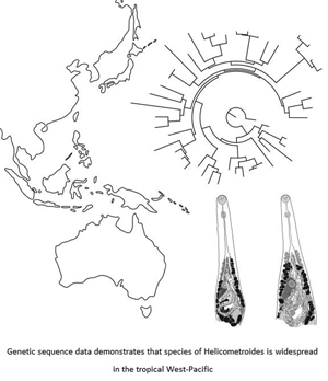
New material from Japan (Fig. 5A and B)

Fig. 5. Hologenophores of Helicometroides longicollis lineage 1 and 2 from Japan and Australia, illustrating the morphological similarity and variation between and within each species; variation can be exaggerated by the mounted position of a specimen (fully dorsoventral or semi-lateral). (A and B) H. longicollis lineage 1 from Japan; (C–E) H. longicollis lineage 1 from Australia; (F and G) H. longicollis lineage 2 from Japan. Scale bars: A–G: 200 μm.
Host: Diagramma pictum pictum (Thunberg).
Locality: off Minabe, Wakayama Prefecture, Japan (33°44′N, 135°19′E).
Site of infection: Intestine.
Prevalence: Unknown; sequences derived from three of four individual D. pictum pictum examined.
Deposition of specimens: Six vouchers, all hologenophores (MPM Coll. No. 21818).
Representative DNA sequences: ITS2 rDNA, six identical replicates, one submitted to GenBank (GB OM230123); 28S rDNA, three identical replicates, one submitted to GenBank (GB OM230131); 18S rDNA, two sequences, both submitted to GenBank (GB OM230116 & OM260311); cox1 mtDNA, two replicates, both submitted to GenBank (GB OM156430–OM156431).
Remarks: The specimens from Japan were collected from near the type-locality of H. longicollis (distance between the localities, <100 km), and broadly conform to the concept of the species. However, the true identity of these specimens remains uncertain, as a second morphologically indistinguishable species of Helicometroides infected the same individual fish. Measurements for these specimens are given in Table 4. The differentiation between the two species is considered in the discussion.
Table 4. Measurements of Helicometroides longicollis from present (both lineages) and previous studies

B, body; L, length; W, width; FB, forebody; OS, oral sucker; VS, ventral sucker; RT, right testis; LT, left testis; Post T, post-testicular region; CS, cirrus-sac; ASV, anterior seminal vesicle; PSV, posterior seminal vesicle; PP, pars prostatica; Egg cap, egg capsule.
Measurements are in micrometres or percentages, means in parentheses
New material from Australia (Fig. 5C–E)
Host: Diagramma pictum labiosum Macleay.
Locality: off Heron Island, southern Great Barrier Reef, Queensland, Australia (23°27′S, 151°55′E); off Lizard Island, northern Great Barrier Reef, Queensland, Australia (14°40′S, 145°27′E).
Site of infection: Intestine.
Prevalence: 4/5 (80%), Heron Island; 4/30 (13%), Lizard Island.
New DNA sequences: ITS2 rDNA, 30 replicates, 29 identical, 25 from Heron Island, four from Lizard Island, one sequence from Lizard Island differs by two base pairs, an identical sequence from each location and the other sequence submitted to GenBank (identical sequences: GB OM230125–OM230126, other sequence: GB OM230124); 28S rDNA, 11 replicates, six from Heron Island, five from Lizard Island, one from each location submitted to GenBank (GB OM230132–OM230133); 18S rDNA, two identical replicates, both from Heron Island, one submitted to GenBank (GB OM230117); cox1 mtDNA, nine replicates, six from Heron Island, three from Lizard Island, a replicate of each genotype (four replicates) submitted to GenBank (GB OM156432–OM156435).
Remarks: All but one of the 30 ITS2 sequences of this species generated in this study (from both Australia and Japan) are identical to the sequence generated by Searle et al. (Reference Searle, Cutmore and Cribb2014), with one sequence (from Lizard Island) differing at a single base position. Similarly, all but two 28S sequences generated in this study are identical to that generated by Searle et al. (Reference Searle, Cutmore and Cribb2014), with two sequences (from Heron Island) differing at three base positions. Morphological measurements for 17 of these specimens, including 10 hologenophores, are given in Table 4.
Helicometroides longicollis lineage 2 (Fig. 5F and G)
Host: Diagramma pictum pictum (Thunberg).
Locality: off Minabe, Wakayama Prefecture, Japan (33°44′N, 135°19′E).
Site of infection: Intestine.
Prevalence: Unknown; sequences derived from two of four individual D. pictum pictum examined.
Deposition of specimens: Three vouchers, all hologenophores (MPM Coll. No. 21819).
Representative DNA sequences: ITS2 rDNA, four identical replicates, one submitted to GenBank (GB OM230127); 28S rDNA, one sequence, submitted to GenBank (GB OM230134); 18S rDNA, one sequence, submitted to GenBank (GB OM230118); cox1 mtDNA, two sequences, both submitted to GenBank (GB OM156436–OM156437).
Remarks: Hologenophores morphologically indistinguishable from those of H. longicollis lineage 1. Measurements for these specimens are given in Table 4.
Molecular phylogenetic analyses
ML and BI analyses of the 28S dataset produced phylograms with similar topologies (Fig. 2). The species of Helicometroides form a well-supported clade with identical strongly supported internal nodes in both analyses. There are three key differences between the two analyses. First, the relative phylogenetic positions of C. pudica and Helicometroides spp. are swapped in the two analyses, with the most basal monorchiid in the BI analysis being C. pudica, and in the ML analysis, Helicometroides spp. This instability has been observed previously in recent phylogenetic analyses (Wee et al., Reference Wee, Cutmore and Cribb2021b). The second difference involves the phylogenetic position of Postmonorchis orthopristis Hopkins, 1941. In the BI analysis, it resolves as sister to Genolopa vesca Panyi et al., Reference Panyi, Curran and Overstreet2020, and this clade is sister to the other represented species of Genolopa Linton, 1910; the position of P. orthopristis in this analysis is weakly supported. In the ML analysis, P. orthopristis resolves sister to all the species of Genolopa with strong nodal support. Finally, in the BI analysis, Retroporomonorchis pansho Wee et al., 2020 is sister to a clade comprising Monorchis monorchis (Stossich, 1890) Looss, 1902, species of Sinistroporomonorchis Wee et al., 2020, and a species identified only as ‘Lasiotocus sp’. In the ML analysis, R. pansho resolves sister to a clade comprising the aforementioned species, as well as Infundiburictus arrhichostoma (Searle et al., Reference Searle, Cutmore and Cribb2014) Wee et al., 2020, Monorchis lewisi Cribb et al., 2018, and the species of Allobacciger Hafeezullah and Siddiqi, 1970. The position of R. pansho in both arrangements is weakly supported.
Discussion
Genus concept
In both analyses of the 28S dataset presented in this study, the five species of Helicometroides recognized form a strongly-supported clade. Given the strength of this clade, it is inferred that the presence of a spined terminal organ is a labile character for Helicometroides. Four species, H. longicollis lineage 1 and 2, H. murakamii n. sp., and H. gabrieli n. sp., lack terminal organs but possess metraterms, which conforms to the previous generic diagnosis of Helicometroides. However, H. wardae n. sp. possesses a distinct terminal organ that has robust spines. In this, it is consistent with typical monorchiids; species from 49 of 54 other monorchiid genera are reported to possess a spined terminal organ. Of the five remaining genera, the species of Cableia Sogandares-Bernal, 1959, Opisthomonorchis Yamaguti, 1952 and Pseudopisthomonorchis Madhavi, 1974 do not possess a terminal organ, while some species of Hurleytrematoides and all species of Pseudoproctotrema Yamaguti, 1942 have unspined terminal organs. Given that Helicometroides comprises species with or without a spined terminal organ, and Hurleytrematoides species have spined or unspined terminal organs, the form of the terminal organ is clearly labile for some groups of monorchiids. It will be necessary to establish the phylogenetic position of the species of Pseudoproctotrema, Opisthomonorchis and Pseudopisthomonorchis to comprehend the evolution of terminal organ forms within the Monorchiidae. Notably, symmetrical testes have been previously used as a uniting morphological feature for Helicometroides (see Madhavi, Reference Madhavi, Bray, Gibson and Jones2008), despite H. atlanticus possessing somewhat oblique testes (Gaevskaya and Aleshkina, Reference Gaevskaya and Aleshkina1983); H. murakamii n. sp. and H. gabrieli n. sp. characterized here also possess distinctly oblique testes, as do some specimens of H. wardae n. sp. and H. longicollis. With the loss of a lack of a terminal organ and symmetrical testes as features common to all Helicometroides species, the genus is now best united by the possession of a distinctly helical uterus, filamented eggs, and well-developed and extensive vitelline follicles. The distinctly helical uterus easily distinguishes Helicometroides from all other monorchiid genera. Filamented eggs are relatively common in the Monorchiidae, with species from 10 genera (of the Anamonorchiinae Yamaguti, 1970, Hurleytrematinae Yamaguti, 1958 and Opisthomonorchiinae Yamaguti, 1952) reported to possess filamented eggs, but notably, the feature is labile in some genera (see Provitellus in Wee et al., Reference Wee, Cutmore and Cribb2019). Extensive vitelline follicles are uncommon in the Monorchiidae. Only species of three other genera, Cableia, Hurleytrema Srivastava, 1939, and Octotestis Yamaguti, 1951, have a comparable vitelline follicle range (ranging from at least the level of the ventral sucker to the testes).
Recognition of species
The conclusion is that the present samples represent four morphospecies, of which one, H. longicollis, is a complex of two cryptic species. Below, the validity of the three morphologically distinct forms, and the issues associated with H. longicollis, is considered.
Helicometroides murakamii n. sp.
Helicometroides murakamii n. sp. most closely resembles H. pseudovitellosus (from Lutjanus sp. [Lutjanidae], Bay of Bengal, India) in body size, range of the vitelline follicles and position of the ovary, cirrus-sac and testes. However, it differs in the possession of a much longer oesophagus (at least twice as long as the prepharynx vs about as long as the prepharynx), and oblique, contiguous testes (vs symmetrical, non-contiguous testes). It also resembles H. vitellosus (from Plectorhinchus sp. [Haemulidae], New Caledonia) in body shape and size, cirrus-sac shape, position and size, and shape and position of the ovary. The two species differ in the anterior extent of the vitelline follicles (level of intestinal bifurcation in H. murakamii n. sp. vs distinctly anterior to the intestinal bifurcation in H. vitellosus), the shape of the testes (globular in H. murakamii n. sp. vs distinctly elongate in H. vitellosus), the posterior extent of the testes (distinctly anterior to the posterior margin of the body in H. murakamii n. sp. vs immediately anterior to the posterior margin of the body in H. vitellosus) and cirral spines (spines lining the entire length of the cirrus in H. murakamii n. sp. vs absent from the middle third of the cirrus in H. vitellosus). Helicometroides murakamii n. sp. is easily differentiated from H. atlanticus [from Parapristipoma octolineatum (Valenciennes) [Haemulidae], from the coast of Angola (see Gaevskaya and Aleshkina, Reference Gaevskaya and Aleshkina1983), and from Dakar (see Diagne et al., Reference Diagne, Quilichini, Bâ, Ndiaye, Dione and Marchand2015), east Atlantic Ocean] by the presence of a long, distinct prepharynx (vs very short), the anterior extent of the vitelline follicles (reaches the level of the intestinal bifurcation vs reaches distinctly anterior to the intestinal bifurcation) and the position of the ovary (anterior to the right testis vs anterior to the left testis). It differs from H. longicollis in the morphology of the vitelline follicles (not contiguous at the posterior end vs contiguous at the posterior end), length of prepharynx (28–65 vs 85–294), position of the ovary (just anterior to the left testis vs distinctly anterior to the left testis) and shape of the testis (globular vs elongate). It is most easily differentiated from H. leiperi [from Otolithes ruber (Bloch and Schneider) [Sciaenidae], Puri Coast, Bay of Bengal, India] in the range and extent of the vitelline follicles (from the level of the intestinal bifurcation to the level of the testes vs from just posterior to the intestinal bifurcation to the anterior region of ovary), position of the ovary (just anterior to the right testis vs in the middle of the body, distinctly pre-testicular), extent of caeca (terminate at the middle of the hindbody vs terminate near the posterior end of the hindbody) and size of the eggs (31–38 × 14–21 vs 55–61 × 26–29).
Helicometroides gabrieli n. sp.
In the possession of vitelline follicles that extend well-anteriorly beyond the intestinal bifurcation, reaching approximately the mid-oesophagus level, H. gabrieli n. sp. closely resembles H. atlanticus and H. vitellosus; all other species have a much more restricted vitelline distribution, with the follicles not extending anterior to the intestinal bifurcation, or only just anterior to it. Helicometroides gabrieli n. sp. is easily differentiated from H. atlanticus in the possession of a bipartite seminal vesicle (vs unipartite seminal vesicle) as well as a distinctly longer prepharynx, and from H. vitellosus in the lack of a gap in the cirral spines (vs middle third lacking spines) and oblique testes (vs symmetrical testes).
Helicometroides wardae n. sp.
Helicometroides wardae n. sp. is easily differentiated from all other species of Helicometroides in the possession of a spined terminal organ. It should be noted that it is also the only species in which some specimens have been observed to possess large spines in the metraterm; the drivers behind this variation remain unknown. Additionally, it has an ovoid and wide cirrus with large, robust spines, which further separates it from all other species of Helicometroides; H. atlanticus, H. longicollis and H. vitellosus each has a thin, elongate cirrus with small spines, and H. leiperi has an elongate cirrus with both long and short spines. While the description of H. pseudovitellosus lacks detail about the cirrus and cirral spines, the illustration of an elongated cirrus leads us to think that the cirral spines are small and thin. The other new species, H. murakamii n sp. and H. gabrieli n. sp., each also possesses a narrow cirrus with small spines.
Helicometroides longicollis lineage 1 and lineage 2
The interpretation of genetic variation in marine fish trematodes for the recognition of species is receiving substantial attention, particularly in the light of ITS2 and cox1 sequence data (Bray et al., Reference Bray, Cutmore and Cribb2021). Recent studies in the tropical Indo-west Pacific using the cox1 region have frequently identified morphologically indistinguishable trematodes in the same or comparable fishes forming distinct clades based on the geographical distribution (allopatric populations) as reflecting intraspecific variation rather than distinct species. Such findings have been reported for six species of the Aporocotylidae Odhner, 1912 (Cutmore et al., Reference Cutmore, Yong, Reimer, Shirakashi, Nolan and Cribb2021), three species of the Gorgocephalidae Manter, 1966 (Huston et al., Reference Huston, Cutmore, Miller, Sasal, Smit and Cribb2021), three species of the Lepocreadiidae Odhner, 1905 (Bray et al., Reference Bray, Cutmore and Cribb2018, Reference Bray, Cutmore and Cribb2021) and seven species of the Monorchiidae Odhner, 1911 (McNamara et al., Reference McNamara, Miller and Cribb2014; Wee et al., Reference Wee, Cribb, Corner, Ward and Cutmore2021a). These intraspecific variations are much reduced in the ITS2 region (in some cases, the differences in the ITS2 region disappear entirely), resulting in the interpretation of a single population (one clade) rather than multiple populations (distinct clades). For example, Preptetos paracaballeroi Bray et al., Reference Bray, Cutmore and Cribb2021 occurs at Heron and Lizard Islands where the populations differ by 27–31 base pairs in the cox1 region while there are no differences at all for the ITS2 region. Such dramatic conflict in genetic variation between cox1 and ITS2 sequences has presented a significant obstacle in the interpretation of the status of some populations. These issues led Bray et al. (Reference Bray, Cutmore and Cribb2021) to propose a model for the recognition of species. Specifically, they proposed that, where appropriate data are available, a species should be recognized as distinct only if it comprises a monophyletic lineage in at least the most discriminating available molecular marker (in most recent cases, cox1), and has consistent morphological differences relative to other species or a host distribution distinct from that of closely related species. By these criteria, all the cases mentioned above can be interpreted as involving single, widespread species showing intraspecific geographical population structures. However, the nature of the H. longicollis complex presents components not accounted for by the criteria proposed by Bray et al. (Reference Bray, Cutmore and Cribb2021). Specifically in this system, cryptic morphology is paired with sympatric lineages identified from a recombinant region (ITS2) in addition to a non-recombining region (cox1). In none of the cases described above was there sympatric co-occurrence of morphologically indistinguishable ITS2 rDNA lineages. The analyses of McNamara et al. (Reference McNamara, Miller and Cribb2014) hint at such a possibility for Hurleytrematoides loi McNamara and Cribb, Reference McNamara and Cribb2011 and H. sasali McNamara and Cribb, Reference McNamara and Cribb2011, but there is insufficient replication for the result to be compelling.
Sequencing of specimens collected from D. pictum pictum from Japan and D. pictum labiosum from Australia that corresponded to the morphotype of H. longicollis revealed two genotypes in the ITS2 and 28S datasets, and three genotypes in the cox1 dataset. In the first two datasets, the genotypes were separated into two forms, one that occurred in both D. pictum pictum and D. pictum labiosum from Japan and Australia, respectively, and another restricted to D. pictum pictum from Japan. In the cox1 dataset, the genotypes were separated into three forms, one from Australia, and two from Japan, with the genotype occurring in both Japan and Australia in the ITS2 and 28S datasets separated into distinct genotypes. Importantly, the genetic differences between the Japanese and Australian genotypes that were the same genotype in the ITS2 and 28S datasets (33–36 base pairs) are significantly less than their differences from the other genotype (51–53 base pairs). Considering the condition of the ITS2 and 28S datasets, the two genotypes with smaller genetic difference in the cox1 dataset are interpreted as representing a single species, and the other genotype as a second species. The key aspect that affects the interpretation of these data is that the two distinct genotypes from Japan occur sympatrically, including within the same individual host fish. As such, the two genotypes are interpreted as representing two truly cryptic species that correspond morphologically to H. longicollis. These circumstances contrast to the findings of Bray et al. (Reference Bray, Cutmore and Cribb2021) who showed that Preptetos laguncula Bray and Cribb, 1996 and P. zebravaranus Bray et al., Reference Bray, Cutmore and Cribb2021 are each represented by two cox1 clades which may co-occur in the same fish individual. However, those cox1 clades had identical ITS2 sequences, consistent with the interpretation of one species.
At present, there is no way to determine which of the two species is the true H. longicollis, given that no genetic sequence data exist for the original collection of the species. Thus, the two species can only be recognized as H. longicollis lineage 1 and lineage 2, with the former occurring in both Japan and Australia, and the latter so far restricted to Japan. It is hoped that new morphological material may reveal a reliable morphological basis for the distinction of these species and that the distinction will be recognizable in the holotype of H. longicollis.
Key to the species of Helicometroides
The inclusion of the three new species brings the number of formally characterized Helicometroides spp. to eight and introduces some important morphological differences between them. A dichotomous key is provided here to differentiate them.
1a. Terminal organ present, armed with robust spines ……… Helicometroides wardae n. sp.
1b. Terminal organ absent ……… 2
2a. Distinct gap in cirral spines, with middle third of cirrus lacking spines ……… H. vitellosus
2b. No gap in cirral spines ……… 3
3a. Vitelline follicles extend posteriorly only to middle of body, well anterior to testes; follicles not confluent anteriorly or posteriorly ……… H. leiperi
3b. Vitelline follicles extend posteriorly to level of testes, does not extend into post-testicular zone; follicles confluent at one or both ends ……… 4
4a. Oesophagus shorter than prepharynx ……… H. pseudovitellosus
4b. Oesophagus distinctly longer than prepharynx ……… 5
5a. Seminal vesicle unipartite ……… H. atlanticus
5b. Seminal vesicle bipartite ……… 6
6a. Vitelline follicles extend well anterior to intestinal bifurcation ……… Helicometroides gabrieli n. sp.
6b. Vitelline follicles extend to level of or just posterior to intestinal bifurcation ……… 7
7a. Testes globular; oesophagus <4× pharynx length ……… Helicometroides murakamii n. sp.
7b. Testes elongate; oesophagus >4× pharynx length ……… H. longicollis (species complex)
Host exploitation
Species of Helicometroides have been reported from three families of marine bony fishes: Haemulidae, Lutjanidae and Sciaenidae. The haemulids are the most heavily parasitized group; four species from three genera harbour six species of Helicometroides. Haemulid fishes as a major host group for Helicometroides is consistent with the Monorchiidae as a whole; to our knowledge, 47 species of monorchiids from 18 genera infect a wide range of haemulids. Sciaenid fishes are significantly less parasitized by monorchiids; to our knowledge, 10 species of monorchiids from five genera have been reported from the family, with a single species of Helicometroides, H. leiperi, reported only from O. ruber (see Ahmad, Reference Ahmad1982). Wee et al. (Reference Wee, Cribb, Cutmore and Martin2020a) observed that most reports of monorchiid infections in lutjanid fishes are unconvincing; just two are well-supported with evidence. Wee et al. (Reference Wee, Cribb, Cutmore and Martin2020a) suggested that the two lutjanids reported as hosts of H. pseudovitellosus (see Madhavi, Reference Madhavi1974) and H. vitellosus (see Durio and Manter, Reference Durio and Manter1968) were likely haemulids as neither host was identified to species, lutjanids are easily confusable with haemulids, and as species of Helicometroides are known overwhelmingly from haemulids.
Acknowledgements
We thank the directors and staff of both Heron Island Research Station and Lizard Island Research Station for their continued support of our field activities. We thank our colleagues, Dr Daniel Huston and Dr Storm Martin for their assistance in collecting fish and members of the Marine Parasitology Laboratory for their continued support. We also sincerely thank the staff and students of Kindai University, Wakayama for their hospitality in accommodating and assisting us in our collecting efforts in Japan.
Author contribution
N.W. and T.H.C. conceived and designed the study. N.W., S.C.C. and T.H.C. collected samples from Australia, and S.C.C., S.S. and T.H.C. collected samples from Japan. N.W. performed morphological and genetic analyses, and wrote the article with critical input from S.C.C. and T.H.C.
Financial support
N.W. is supported by The Holsworth Wildlife Research Endowment and The Ecological Society of Australia, and the PADI Foundation. S.C.C. and T.H.C. are supported by the Australian Biological Resources Study (ABRS National Taxonomy Research Grant RG19-37). Collecting in Japan was supported by an Australian Society for Parasitology (ASP) Network Researcher Exchange, Training and Travel Award grant to SCC. N.W. is also supported by a PhD scholarship from the University of Queensland [Research Training Program (RTP) Scholarship].
Conflict of interest
None.
Ethical standards
All fish were caught and handled according to the University of Queensland Animal Ethics Approval Certificate SBS/248/15/ABRS/ARC.


