


INTRODUCTION
Technological advancements in the field of radiotherapy, largely play their part in driving cancer treatment to enhance local tumour control.Reference Deng, Feng, Ma and Yin 1 In the present era, linear accelerators have successfully superseded other treatment units for treating various forms of malignancies. They perform an immensely complicated job of shaping high-dose energy beams to the target volume along with excluding normal tissues and delivering radiation beams both with uniform and modulated intensities.
Before the clinical implementation of linear accelerators, they undergo acceptance testing and commissioning. Radiotherapy is connected to the accurate delivery of radiation doses to the malignant cells, which in turn depends on the accuracy of the dosimetric data utilised in creating optimum treatment plans.Reference Sahoo and Mukharjee 2
Acceptance testing conducted on linear accelerators, involves the consideration of the characteristics of absorbed doses at various depths, radiation beam uniformity determined by homogeneity and symmetry of beam profiles, systems for monitoring radiation doses, penumbra, isocentre and the movement of treatment table. The details of all of these characteristics are documented in the literature.Reference Fogliata, Garcia and Knoos 3 – Reference Nath, Biggs and Bova 5
Commissioning follows acceptance testing and consists of checking functions of the system, documentation of various capabilities, measurement of parameters of absorbed doses, which are essential for the verification of the treatment planning system to choose the optimum treatment modality and verification of the potential of dose calculation algorithm, for the purpose of reproducing measured dose calculations. During the commissioning of linear accelerator, it is vital that the radiation beam profile matches with its specification.Reference Pathak, Mishra, Singh and Mishra 6 In order to achieve accuracy and reproducibility in dose delivery, a consistent beam profile is required.Reference Klein, Hanley and Bayouth 7 The baseline values obtained during acceptance testing and commissioning are helpful in assuring the characteristics of treatment machine that they do not deviate from these values and this is the main objective of the quality assurance programme.Reference Khan and Gibbons 8
Several authors determined the dosimetric characteristics of photon beams emerging from the linear accelerator. Clivio et al.Reference Clivio, Nicolini, Vanetti, Fogliata and Cozzi 9 analysed the dosimetric properties of the 6 MV beam from linear accelerator UNIQUE by Varian Medical System (Palo Alto, CA, USA) and beam profile characteristics like homogeneity and symmetry. Xhafa et al.Reference Xhafa, Mulaj, Hodolli and Nafezi 10 determined the dosimetric parameters of the photon beam emanating from linear accelerator, Siemens, Primus for various wedge angles. Shende et al.Reference Shende, Gupta, Patel and Kumar 11 gave detailed analysis of dosimetric characteristics of 6, 15 and 10 MV flattening filter photon beam and 6 and 10 MV flattening filter free photon beam coming from the true beam linear accelerator.
The primary objective of this exploration is to assess the characteristics of photon beam profiles generated by 6 and 15 MV photon energies produced by Varian Clinac DHX (Varian Medical Systems Inc.) at various depths and field sizes and analysing the variations in these characteristics with increasing depths and field sizes in order to assure the quality and improve the accuracy of radiotherapy plans.
METHODS AND MATERIALS
We used a high-energy linear accelerator Varian Clinac DHX with two photon beam energies 6 and 15 MV and it was equipped with millennium 80 multi-leaf collimator. Cylindrical ionisation chamber N30001/PTW FREIBURG was used for absorbed dose measurements. MP3-M water tank (PTW, Freiberg, Germany) was used for radiation dosimetry and MEPHYSTO (PTW) version 7·3 software for data processing and analysing the parameters of beam profiles. CAX Dev (central axis deviation), left and right penumbras, field 50%, D max, D min, symmetry, homogeneity were analysed and compared for both beam energies (6 and 15 MV), at depths of 5, 10 and 20 cm and for squared field sizes with various dimensions of 5×5, 10×10, 15×15, 20×20, 25×25 and 30×30 cm2 by keeping the source to surface distance at 100 cm.
RESULTS
We have analysed beam profile characteristics for 6 and 15 MV photon beam, shown their variation with increasing depth, energy and field sizes and also found their mean values, standard deviations of the particular beam data generated by linear accelerator Varian Clinac for both energy beams in the tables presented below.
In Table 1, for 6 MV energy beam at 5 cm, it is obvious that both left and right penumbras increased with increasing field sizes at the depth of 5 cm. The average values of maximum and minimum doses are 101·61 and 99·04%, respectively. The concept of homogeneity presented here actually depicts inhomogeneity of the beam doses.Reference Hussain and Rhoades 12 Homogeneity increased with the increasing field sizes and its average value is 0·93%.
Table 1 6 MV photon beam characteristics at 5 cm

In Table 2, both left and right penumbras at 10 cm depth were larger than those at the smaller depth of 5 cm. Their averages were gone to the value of 8·51 and 8·38. At the higher depth of 10 cm, these doses also increased with increasing field sizes but this increase is lesser than that at 5 cm depth as it is evident from their mean values. Homogeneity showed an average of 0·92% in Table 2. Symmetry showed the similar behavior as in Table 1. The dosimetric characteristics of 6 MV photon beam at the reference depth of 10 cm and for 10×10 cm2 field size are shown in the Figure 1.
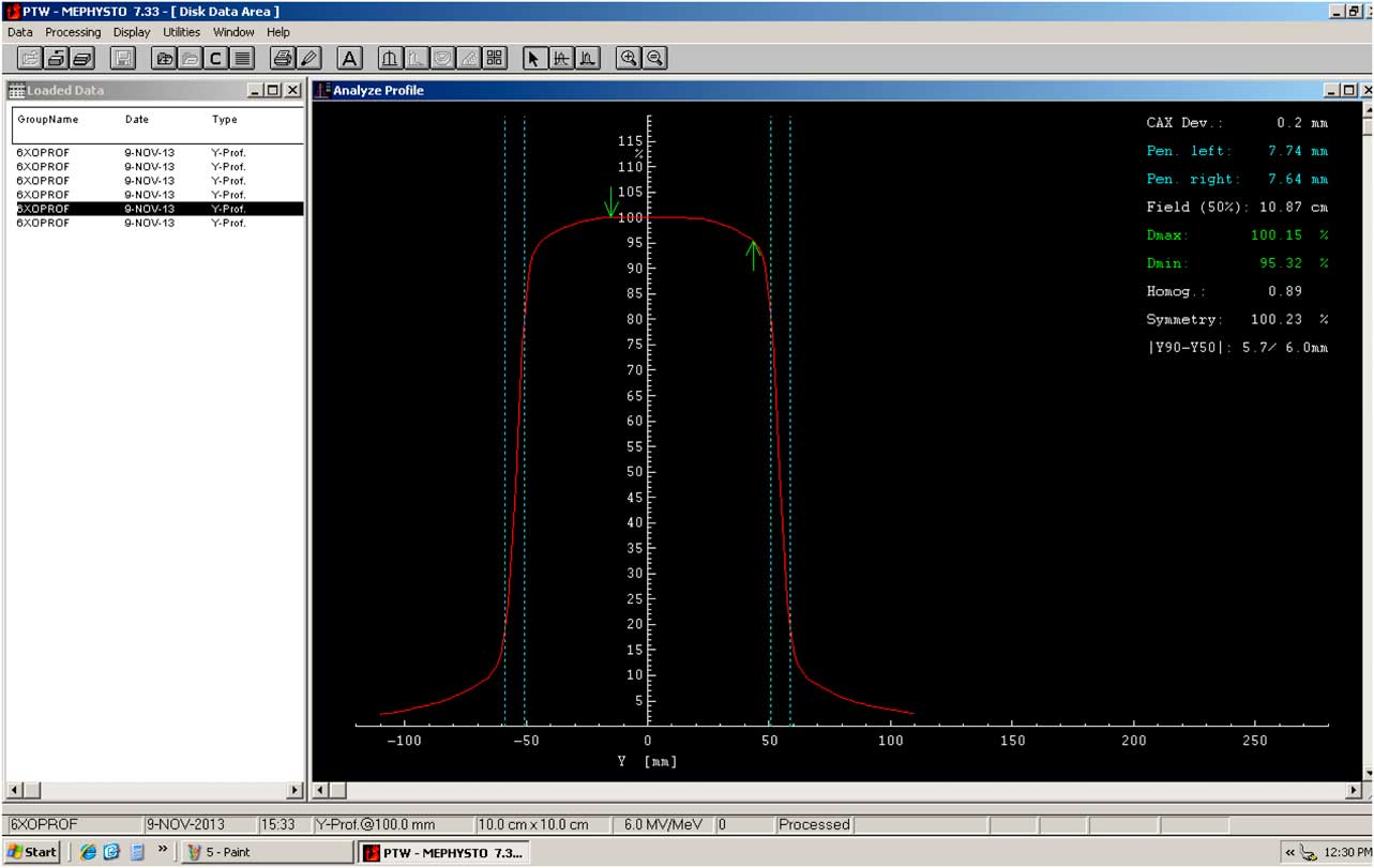
Figure 1 6 MV beam profile at 10 cm depth for 10×10 cm2 field size.
Table 2 6 MV photon beam characteristics at 10 cm

In Table 3, both penumbras at 20 cm depth were greater than that at 5 and 10 cm depth. The average of D max and D min was observed to be 100·29 and 93·09%, respectively and smaller than that at 5 and 10 cm. At 20 cm depth, homogeneity decreased with increasing field size and gave an average of 0·86%. It decreased at higher depth due to the reduction in photon energy with increasing depth. Again at this depth, symmetry remained independent of energy, field size and depth.
Table 3 6 MV photon beam characteristic at 20 cm

In Table 4, for 15 MV beam at 5 cm depth, the left and right penumbras increased with increasing field sizes. Their average had gone to 7·66 and 7·51. D max and D min values at 5 cm depth increased with increasing field sizes. They showed an average of 103·33 and 99·37%, respectively. Homogeneity for 15 MV beam also increased with increasing field sizes with an average of 0·92%. It showed dependence on energy, depth and field size in the same way as for 6 MV beam. Symmetry did not vary considerably with varying depth, energy and field size.
Table 4 15 MV photon beam characteristics at 5 cm

Left and right penumbras at the depth of 10 cm in Table 5, showed an average of 8·55 and 8·4, respectively, which is higher than that presented in Table 4. It was also seen that both left and right penumbras were higher for 15 MV beam as compared with 6 MV beam. D max and D min at 10 cm depth increased with field size and they have average values of 102·69 and 98·91%. It is seen from their average values that these doses decreased with increasing depth for the same field sizes. Homogeneity for 15 MV beam at 10 cm depth gave average of 0·91%. Symmetry remained independent of depth, energy and field size. Dosimetric characteristics of 15 MV photon beam profiles at 10 cm depth and for 10×10 cm2 field size are presented in Figure 2.
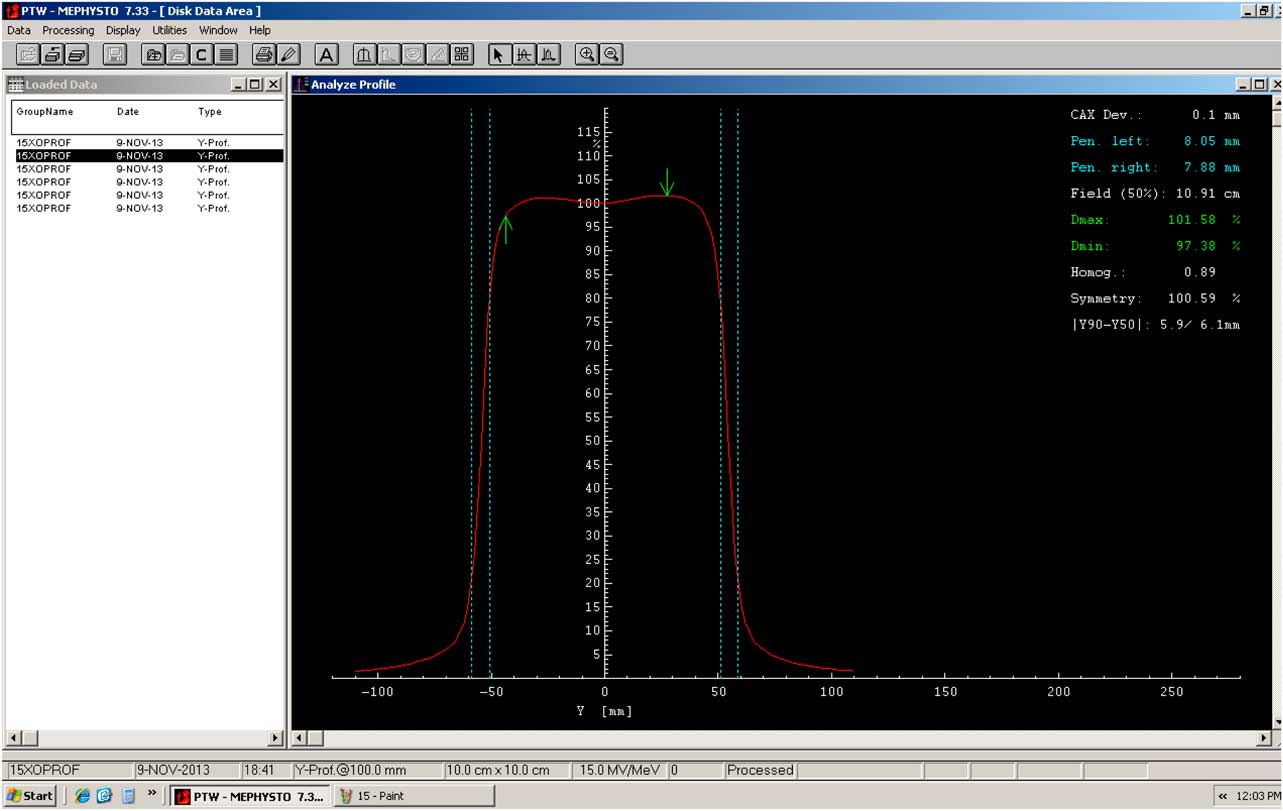
Figure 2 15 MV beam profile at 10 cm depth for 10×10 cm2 field size.
Table 5 15 MV photon beam characteristics at 10 cm

Table 6 shows left and right penumbras at 20 cm depth which gave higher average values than that at 5 and 10 cm. D max and D min decreased with depth in the similar way as in Tables 5 and 6. Homogeneity increased with the field size and has an average of 0·9%. Symmetry at 20 cm depth showed the same average value of 100·78% as that at 5 cm depth.
Table 6 15 MV photon beam characteristics at 20 cm

DISCUSSION
According to the European Society for Therapeutic Radiology and Oncology and American Association of Physicists in Medicine codes of practice, the tolerance level for photon beam homogeneity is recommended not to exceed 3% and the symmetry of the photon beam must remain within 103% acceptance level.Reference Das, Cheng and Watts 13 , Reference Aletti 14 All the values of homogeneity were found within the recommended tolerance level and the largest value of homogeneity went to 0·97% and among all the above given values, the largest observed value of symmetry was 101·01%. Homogeneity increases with increasing field sizes but decreases with depth owing to the fact that it depends on the energy of the incident beam which changes with depth. The increase in homogeneity with increasing field size is due to scattering which becomes larger for larger field sizes.Reference Nath, Biggs and Bova 5 The symmetry values do not show any significant dependence on the depth, energy of the photon beams and the field sizes.Reference Pathak, Mishra, Singh and Mishra 6
Kouloulias et al.Reference Kouloulias, Poortmans, Antypas, Kappas and Sandilos 15 stated that both homogeneity and symmetry are considered as the quality determining factor of the photon beam delivered from linear accelerators. Galiano et al.Reference Galiano, Joly and Wiebe 16 proposed the notions for symmetry and homogeneity of beam dose, which were an extension of the description of both of these tools of dosimetry and these are given in the literature.
In this exploration, it was also seen that both left and right penumbras were greater for higher energy beam like 15 MV beam as compared with lower energy beam, 6 MV beam, this is because higher energy radiation beam produces secondary electrons of larger range that deliver more lethal doses to the normal tissues as compared with the target volumes.Reference Zhang, Feng, Ming and Deng 17 The maximum and minimum doses (D max and D min) increased with increasing field sizes. This is because larger field sizes allow more photons to penetrate through them as well as they generate more scattered photons, thus leads to greater dose at the specified point.Reference Lopez, Rajan and Podgorsak 18
CONCLUSION
Linear accelerators are accomplishing the most cumbersome task of hitting target volumes with electron beam and high-energy photon beams with greater accuracy. This exploration concentrated on the analysis and comparison of dosimetric characteristics of the photon beam profile generated from the linear accelerator Varian Clinac with dual photon energies (6 and 15 MV). All the dosimetric characteristics of the beam profiles were obtained in a water phantom, by using an ionisation chamber. Their variation with the treatment parameters like energy, depth and field size was examined. It was scrutinised that the beam profile characteristics like homogeneity and symmetry precisely agree with the acceptance level of 3 and 103%, respectively, as recommended by the radiotherapy protocols. These findings should be widened to the electron beam and other energies of the photon beam at various depths and field sizes.
Acknowledgements
The authors thank the department of clinical and radiation oncology, Shaukat Khanum Memorial Cancer Hospital and Research Centre, Lahore, Pakistan.
Financial support
This research did not receive any specific grant from funding agencies in the public, commercial, or not-for-profit sectors.


