


Introduction
The still actively forming bat guano assemblage at depth (125 feet) in the Rowley mine near Theba, Arizona has already proven to be a remarkable source of new minerals. Among these are the first minerals that contain both oxalate and phosphate groups: phoxite, (NH4)2Mg2(C2O4)(PO3OH)2(H2O)4 (Kampf et al. Reference Kampf, Celestian, Nash and Marty2019a) and davidbrownite-(NH4), (NH4,K)5(V4+O)2(C2O4)[PO2.75(OH)1.25]4⋅3H2O (Kampf et al. Reference Kampf, Cooper, Rossman, Nash, Hawthorne and Marty2019b). The new mineral described herein, thebaite-(NH4), is the third known mineral containing both of these anionic groups, and several others are currently under study.
The mineral is named for Theba, Arizona, a small settlement and railroad depot ~20 km SE of the Rowley mine. At times in the past, the mine has been referred to as the Theba mine and, according to Wilson (Reference Wilson2020), old labels on specimens from the mine often say “near Theba”. For naming and species definition, the total occupancy of the three large cation sites in the structure is employed; thereby, the ‘-(NH4)’ suffix in the name reflects the fact that NH4+ > K+. If an analogue with K+ > NH4+ were found, it would be named thebaite-(K).
The new mineral and name were approved by the Commission on New Minerals, Nomenclature and Classification of the International Mineralogical Association (IMA2020-072, Kampf et al. Reference Kampf, Cooper, Celestian, Nash and Marty2021). The holotype specimen of thebaite-(NH4) is deposited in the collections of the Natural History Museum of Los Angeles County, Los Angeles, California, USA, with catalogue number 75082.
Occurrence
Thebaite-(NH4) was found on the 125-foot level of the Rowley mine (33°2′57″N, 113°1′49.59″W), ~20 km NW of Theba (small settlement and railroad depot), Maricopa County, Arizona, USA. The Rowley mine is on the western slope of the Painted Rock Mountains (in the Painted Rock mining district) and overlooks the Dendora Valley, immediately to the west. It is a former Cu–Pb–Au–Ag–Mo–V–baryte–fluorspar mine that exploited veins presumed to be related to the intrusion of an andesite porphyry dyke into Tertiary volcanic rocks. Although the mine has not been operated for ore since 1923, collectors took notice of the mine as a source of fine wulfenite crystals in ~1945. An up-to-date account of the history, geology and mineralogy of the mine was published recently by Wilson (Reference Wilson2020).
The new mineral was found in a hot and humid area of the mine (see figure 26 in Wilson, Reference Wilson2020) in an unusual bat-guano-related, post-mining assemblage of phases that include a variety of vanadates, phosphates, oxalates and chlorides, some containing NH4+. This secondary mineral assemblage is found growing on baryte-quartz-rich matrix and, besides thebaite-(NH4) includes allantoin (Kampf et al., 2020), ammineite, antipinite, aphthitalite, bassanite, biphosphammite, cerussite, davidbrownite-(NH4) (Kampf et al., Reference Kampf, Celestian, Nash and Marty2019a), fluorite, halite, hydroglauberite, mimetite, mottramite, natrosulfatourea (Kampf et al., 2020), perite, phoxite (Kampf et al., Reference Kampf, Cooper, Rossman, Nash, Hawthorne and Marty2019b), quartz, rowleyite (Kampf et al., Reference Kampf, Cooper, Nash, Cerling, Marty, Hummer, Celestian, Rose and Trebisky2017), salammoniac, struvite, thénardite, urea, vanadinite, weddellite, willemite, wulfenite and several other potentially new minerals. Thebaite-(NH4) was found in intimate association with antipinite, vanadinite and at least one other new mineral.
Physical and optical properties
Crystals of thebaite-(NH4) are colourless blades, up to ~0.1 mm in length, often growing in sprays (Fig. 1). The blades are elongate on [010], flattened on {001} and exhibit the crystal forms {100}, {001} and {320} (Fig. 2). Possible twinning on {100} was observed under crossed polars. The streak is white, the lustre is vitreous, the Mohs hardness is between 1½ and 2, the tenacity is brittle and the fracture is splintery. There are two good cleavages in the [010] zone, probably {100} and {10$\bar{2}$ }. The tiny crystals are virtually invisible in density liquids making the measurement of their density impossible. The calculated density is 2.093 g⋅cm–3 using the empirical formula and 1.991 g⋅cm–3 using the ideal (NH4 end-member) formula. Thebaite-(NH4) is non-fluorescent in long- and short-wave ultraviolet light. The mineral is insoluble at room temperature in H2O, but easily soluble in dilute HCl.

Fig. 1. Sprays of thebaite-(NH4) blades; field of view 0.6 mm across. Holotype, catalogue number 75082.
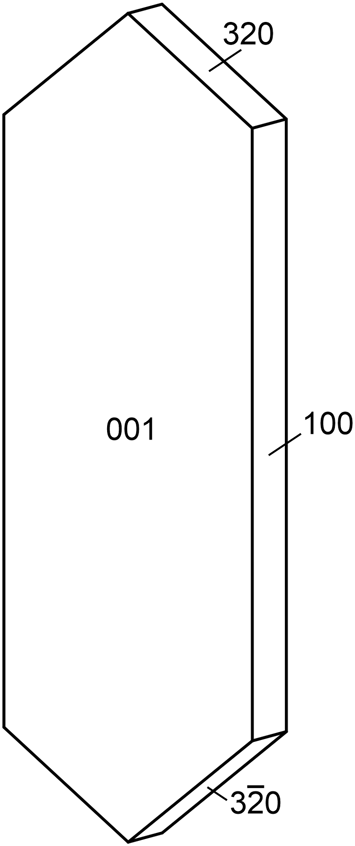
Fig. 2. Crystal drawing of thebaite-(NH4); clinographic projection in non-standard orientation, b vertical.
Thebaite-(NH4) is optically biaxial (–) with α = 1.490(2), β = 1.534(2) and γ = 1.570(2) determined in white light. The 2V measured using extinction data with EXCALIBR (Gunter et al., Reference Gunter, Bandli, Bloss, Evans, Su and Weaver2004) is 82.7(5)° and the calculated 2V is 82.0°. Slight r > v dispersion was observed. The optical orientation is X = b, Y ^ c = 13° in the obtuse angle β. The mineral is non-pleochroic.
Raman spectroscopy
Raman spectroscopy was conducted on a Horiba XploRA PLUS using a 532 nm diode laser, 100 μm slit and 2400 gr/mm diffraction grating and a 100× (0.9 NA) objective. The spectrum from 4000 to 60 cm–1 is shown in Fig. 3. Full pattern peak fitting was performed using a least-squares approach using Gaussian peak shapes to minimise the difference between measured and calculated profiles, and cubic-spline was used for base-line modelling.
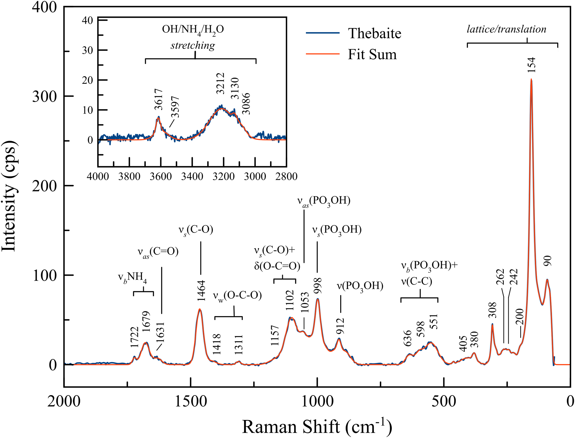
Fig. 3. Raman spectrum of thebaite-(NH4).
Both O–H and N–H stretching are manifest as bands in the 3600–2800 cm–1 range (Colmenero, Reference Colmenero2019) and definitive distinction between them in this and other regions is not possible. The assignment of most of the other bands can be made by reference to the spectrum of phoxite, (NH4)2Mg2(C2O4)(PO3OH)2(H2O)4 (Kampf et al., 2019) and other oxalate minerals (e.g. weddellite, wheatleyite, moolooite, natroxalate, oxammite and glushinskite) (Frost, Reference Frost2004, Reference Frost, Locke and Martens2008) (Peterson and Pullman, Reference Peterson and Pullman2016) as described below.
The Raman bands at 1722 cm–1 and 1679 cm–1 are related to H–N–H bending, while the lower wavenumber shoulder at 1631 cm–1 is related to the asymmetric stretching of C=O. The moderately strong Raman band at 1464 cm–1 is related to a combination of C–O symmetric stretch, and very weak bands at 1418 cm–1 and 1311 cm–1 may be related to O–C–O wagging in the oxalate group, although it also occurs in a region in which O–H and H–N–H bending is manifest. The bands at 1157 cm–1 and 1102 cm–1 are C–O stretching plus out-of-plane deformation bending of O–C=O. The PO3OH asymmetric stretch and the PO3OH symmetric stretch were observed at 1053 cm–1 and 998 cm–1, respectively; the 912 cm–1 band is probably also related to PO3OH stretching. The origin of the broad band between 636 cm–1 and 551 cm–1 is uncertain, but it could be related to O–P–O bending combined with C–C bending. Bands between 405 cm–1 and 60 cm–1 are assigned as lattice modes.
Chemical analysis
Analyses of thebaite-(NH4) (3 points) were done at the University of Utah on a Cameca SX-50 electron microprobe with four wavelength dispersive spectrometers using Probe for EPMA software. Analytical conditions were 15 kV accelerating voltage, 10 nA beam current and a beam diameter of 5 μm. Raw X-ray intensities were corrected for matrix effects with a ϕρ(z) algorithm (Pouchou and Pichoir, Reference Pouchou, Pichoir, Heinrich and Newbury1991). Na and K intensities were corrected for time-dependent decreases in intensity during analyses. Severe damage from the electron beam was observed. Attempts to analyse N (syn. Cr2N standard) provided values much lower than those predicted by the structure refinement. The low values for N [(NH4)2O] by EPMA are probably due to loss of most of the NH4 under vacuum and especially resulting from beam damage. Because insufficient material is available for CHN analyses, C2O3, H2O and (NH4)2O were calculated based upon the structure determination. Note that the structure indicates NH4 + K + Na = 3 atoms per formula unit (apfu), and this is the basis for the calculated (NH4)2O value. The high analytical total is probably due to the partial loss of NH4 and H2O under vacuum and due to beam damage, which results in higher concentrations for the remaining constituents. Analytical data are given in Table 1.
Table 1. Analytical data (wt.%) for thebaite-(NH4).

* (NH4)2O, C2O3 and H2O values in the Normalised column are based on the structure.
S.D. – standard deviation
The empirical formula (based on P + Si = 2 and O = 13 apfu) is [(NH4)2.12K0.69Na0.20]Σ3.01(Al0.84Fe3+0.11V3+0.04)Σ0.99(C2O4)[(P0.98Si0.02)O3OH]2(H2O) (+0.05 H for charge balance. The simplified formula is (NH4,K,Na)3(Al,Fe3+,V3+)(C2O4)(PO3OH)2(H2O) and the ideal (NH4 end-member) formula is (NH4)3Al(C2O4)(PO3OH)2(H2O), which requires (NH4)2O 20.61, Al2O3 13.45, P2O5 37.44, C2O3 19.00, H2O 9.50, total 100 wt.%. The Gladstone–Dale compatibility (Mandarino, Reference Mandarino2007) 1 – (K p/K c) is 0.029 in the range of excellent compatibility for the empirical formula.
X-ray crystallography and structure determination
Powder X-ray studies were done using a Rigaku R-Axis Rapid II curved imaging plate microdiffractometer with monochromatised MoKα radiation. A Gandolfi-like motion on the φ and ω axes was used to randomise the sample. Observed d values and intensities were derived by profile fitting using JADE Pro software (Materials Data, Inc.). The powder data are presented in Supplementary Table S1. Unit-cell parameters refined from the powder data using JADE Pro with whole pattern fitting are a = 11.12(3), b = 6.27(3), c = 18.74(2) Å, β = 103.54(9)° and V = 1270(7) Å3.
Single-crystal X-ray studies were done using a Bruker D8 three-circle diffractometer equipped with a rotating anode generator (MoKα X-radiation), multilayer optics and an APEX-II CCD area detector. A total of 23,373 reflections (6268 in the Ewald sphere) were integrated using 64s frames with a 0.3° frame width. The unit-cell dimensions were obtained by least-squares refinement of 1521 reflections with I o > 7σI. Systematically absent reflections are consistent with the space group P21/c. Careful visual inspection of frames confirmed that all reflections were consistent with this cell and space group. Empirical absorption corrections (SADABS, Bruker AXS) were applied and equivalent reflections were merged. The structure was solved by direct methods using SHELXS-2013 and the structure was refined using SHELXL-2016 (Sheldrick, Reference Sheldrick2015). All atoms were refined with anisotropic displacement parameters. H atom sites could not be reliably extracted from the difference-Fourier map.
Of the three large cation sites, K has the smallest average bond length along with three distinctively shorter (~2.68 Å) individual bonds, and Na is presumed to order at this site; hence, the refined scattering was modelled with coupled K and Na scattering factors (site K is K dominant, in agreement with the chemical analysis, Tables 1, 4). The other two large cation sites (N1 and N2) have significantly less relative scattering and were modelled with coupled K and N scattering factors (both are NH4 dominant, Table 4). The Al site exhibited greater scattering than expected for Al alone, so it was modelled with coupled Al and Fe; refined site-scattering in close agreement with minor Fe and V measured chemically. There are two P sites; The P1 site and its coordinated O sites are fully occupied; however, the P2 site-scattering reveals a significant deficiency from full occupation by P. Of the four O sites coordinated to P2 (OH5, O6, O7 and O8), O6 and O8 also exhibit scattering deficiency, while OH5 and O7 appear to be fully occupied; in the final refinement cycles, the occupancies of the P2, O6 and O8 sites were jointly refined to 0.879(9) occupancy. An H2O site was located, but because of the highly prolate shape of its ellipsoid when refined anisotropically, it was refined as a split site (OWA and OWB separated by 0.66(4) Å and refined isotropically) with a total occupancy of 1.0. The occupancy deficient P2, O6 and O8 sites and the split H2O site are discussed below. Data collection and refinement details are given in Table 3, atom coordinates and displacement parameters in Table 4, selected bond distances in Table 5 and a bond-valence analysis in Table 6. The crystallographic information file has been deposited with the Principal Editor of Mineralogical Magazine and is available as Supplementary material (see below).
Table 2. Data collection and structure refinement details for thebaite-(NH4).

*R int = Σ|F o2–F o2(mean)|/Σ[F o2]. GoF = S = {Σ[w(F o2–F c2)2]/(n–p)}½. R 1 = Σ||F o|–|F c||/Σ|F o|. wR 2 = {Σ[w(F o2–F c2)2]/Σ[w(F o2)2]}½; w = 1/[σ2(F o2) + (aP)2 + bP] where a is 0.081, b is 0 and P is [2F c2 + Max(F o2,0)]/3.
Table 3. Atom positions, occupancy and displacement parameters (Å2) for thebaite-(NH4).

Table 4. Selected bond lengths (Å) for thebaite-(NH4).

Subscripts in brackets are site occupancies.
Table 5. Bond-valences analysis for thebaite-(NH4). Values are in valence units (vu).

Bond-valence parameters for NH4+–O are from Garcia-Rodriguez et al. (Reference García-Rodríguez, Rute-Pérez, Piñero and González-Silgo2000); all others are from Gagné and Hawthorne (Reference Gagné and Hawthorne2015). Hydrogen bond valences based on OD⋅⋅⋅OA distances using the relation of Ferraris and Ivaldi (Reference Ferraris and Ivaldi1988). The refined occupancies of all sites have been considered.
The refinement clearly establishes the structure type, but suffers to some degree in precision due to poorer general data observation; mean I/σ for reflections ranges from ~18 (low 2θ) to ~1.5 (at resolution cut-off of 45°2θ). This poorer data observation is due to a combination of very low scattering (light element structure) and very small single-crystal volume. The very bright rotating anode X-ray source, combined with the long frame exposure, allowed for modest data observation out to an imposed resolution cut-off of 45°2θ. We note that about half of the unique data collected is observed (I o > 2σI) and that statistical measures (R values, merging of reflections) are somewhat elevated as a result.
Discussion of the structure
The structure of thebaite-(NH4) includes three large cation sites coordinated by O (O, OH and/or H2O) sites: K (nine-fold coordinated), N1 (eight-fold coordinated) and N2 (seven-fold coordinated). There is one Al site (octahedrally coordinated by O) and two P sites, P1 and P2, both tetrahedrally coordinated by three O and one OH. One oxalate (C2O4) group includes two independent C sites, C1 and C2 and four independent O sites.
The structural unit is a double-strand chain of corner-sharing AlO6 octahedra and P1O3OH tetrahedra decorated by P2O3OH tetrahedra and C2O4 groups (Fig. 4). As noted above, the tetrahedral coordination surrounding the P2 site is formed by the OH5, O6, O7 and O8 sites; the occupancies of the P2, O6 and O8 sites refined jointly to an occupancy of 0.879. This appears to indicate a real deficiency in the P2O3(OH) tetrahedron. When this group is missing (12% of the time), the OH5 site would be an H2O group and the O7 site would be an OH site. Furthermore, the aforementioned splitting of the H2O site into OWA and OWB is probably related to the deficiency in the P2O3(OH) tetrahedron. It should be noted that the normalised EPMA data are consistent with full occupancy of the P sites; however, we do not regard the EPMA data to be of sufficient quality to draw definitive inferences regarding occupancies.

Fig. 4. The chain in the thebaite-(NH4) structure along [010]. The O sites are numbered. [100] is vertical; [010] is canted down 20° from right to left.
Without the decorating P2O3OH tetrahedra and C2O4 groups, the octahedral–tetrahedral double-strand chain in the structure of thebaite-NH4 is topologically similar to chain components in several structures (Fig. 5). It is topologically identical to the chain in hannayite, Mg3(NH4)2(PO3OH)4⋅8H2O (Catti and Franchini-Angela, Reference Catti and Franchini-Angela1976), while the chains in galliskiite, Ca4Al2(PO4)2F8⋅5H2O (Kampf et al., Reference Kampf, Colombo, Simmons, Falster and Nizamoff2010), kapundaite, (Na,Ca)2Fe3+4(PO4)4(OH)3⋅5H2O (Mills et al., Reference Mills, Birch, Kampf, Christy, Pluth, Pring, Raudsepp and Chen2010a) and synthetic Pb3Fe2(PO4)4(H2O) (Mills et al., Reference Mills, Kolitsch, Miyawaki, Hatert, Porier, Kampf, Matsubara and Tillmanns2010b) are geometrical isomers. The octahedral–tetrahedral double-strand chains in synthetic PbIn(AsO4)(AsO3OH) (Kolitsch and Schwendtner, Reference Kolitsch and Schwendtner2005) and synthetic Na3Fe(PO4)2 (Hatert, Reference Hatert2007) differ topologically from those in the aforementioned structures in that their octahedra share three corners of the same octahedral face with tetrahedra, while in all of the other chains, the octahedra share three equatorial corners with tetrahedra.
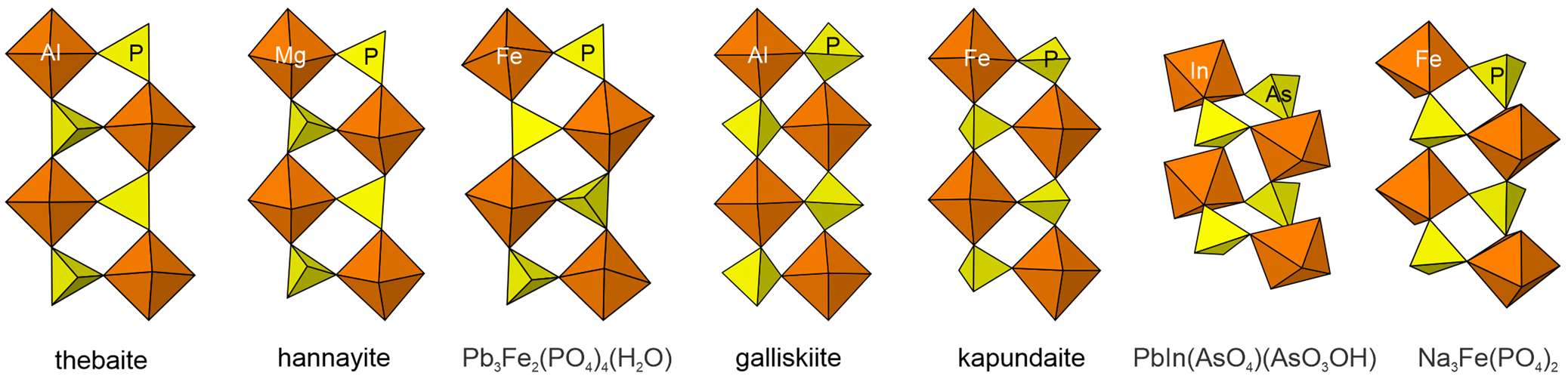
Fig. 5. Octahedral–tetrahedral double-strand chains in the structures of thebaite-(NH4), hannayite, Pb3Fe2(PO4)4(H2O), galliskiite, kapundaite, PbIn(AsO4)(AsO3OH) and Na3Fe(PO4)2.
In thebaite-(NH4), the chains, including the decorating components, connect to one another through bonds to the K site and the two NH4 sites (N1 and N2) and through hydrogen bonds (Fig. 6). In contrast, the chains in hannayite connect to one another through decorating PO4 tetrahedra corner-linked to intermediary FeO6 octahedra to form sheets. The chains in kapundaite link directly to edge-sharing chains of FeO6 octahedra, as well as to additional PO4 tetrahedra to form a framework. In Pb3Fe2(PO4)4(H2O), two sets of interpenetrating chains are corner-linked into a framework. Other than thebaite-(NH4), the only one of these structures with double-strand chain structural units is that of galliskiite in which the chains are undecorated and connect to one another via bonds to Ca cations.

Fig. 6. The thebaite-(NH4) structure viewed down [001] (left) and [010] (right). The unit-cell outline is shown with dashed lines.
Synthetic phases containing both phosphate and oxalate groups are often referred to as oxalatophosphates. In many of these phases, the phosphate and oxalate groups link with octahedrally coordinated cations to form porous frameworks with potential technological applications: catalysis, adsorption, ion exchange, gas storage, separation and sensing (Luan et al., Reference Luan, Li, Chen, Lin and Huang2015). These structures take advantage of the oxalate group's ability to form strong bidentate linkages between octahedra. Such strong bidentate linkages between octahedra also occur in the structures of the other two oxalatophosphate minerals, phoxite, (NH4)2Mg2(C2O4)(PO3OH)2(H2O)4 (Kampf et al., Reference Kampf, Celestian, Nash and Marty2019a) and davidbrownite-(NH4), (NH4,K)5(V4+O)2(C2O4)[PO2.75(OH)1.25]4⋅3H2O (Kampf et al., Reference Kampf, Cooper, Rossman, Nash, Hawthorne and Marty2019b), which both are found in the Rowley-mine guano mineral assemblage. The oxalate group in the thebaite-(NH4) structure forms a bidentate linkage to a single AlO6 octahedron, thereby decorating the double-strand chain rather than strongly linking it to other structural components. We are not aware of any synthetic phases with similarly decorated double-strand chains; however, another potentially new mineral in the Rowley-mine guano has a structural unit with an identical topology.
Acknowledgements
Structural Editor Pete Leverett and two anonymous reviewers are thanked for constructive comments, which improved the manuscript. Keith Wentz, claim holder of the Rowley mine, is thanked for allowing underground access for the study of the occurrence and the collecting of specimens, along with Frank Hawthorne for providing access to the single-crystal instrument at the University of Manitoba. This study was funded, in part, by the John Jago Trelawney Endowment to the Mineral Sciences Department of the Natural History Museum of Los Angeles County.
Supplementary material
To view supplementary material for this article, please visit https://doi.org/10.1180/mgm.2021.26


