


Introduction
Cambrian Lagerstätten with exceptional preservation have opened unique windows into the origin and early evolution of metazoans. There are mainly two types of such Lagerstätten, which are especially influential: those of the typical Burgess Shale type (Conway Morris, Reference Conway Morris1989), including the Chengjiang or Maotianshan Shale type (Chen, Reference Chen2004), and those of the Orsten type (Maas et al., Reference Maas, Braun, Dong, Donoghue, Müller, Olempska, Repetski, Siveter, Stein and Waloszek2006). In typical Burgess Shale type Lagerstätten, mainly macroscopic metazoans (of centimeter scale) can be completely preserved, but are preserved in a flat two-dimensional manner, in some cases as carbonaceous films (Gaines et al., Reference Gaines, Kennedy and Droser2005, Reference Gaines, Briggs and Zhao2008; Butterfield et al., Reference Butterfield, Balthasar and Wilson2007). Orsten-type Lagerstätten are a special mode of taphonomy (Müller and Walossek, Reference Müller and Walossek1985, Reference Müller and Walossek1991; Butterfield, Reference Butterfield2003; Maas et al., Reference Maas, Braun, Dong, Donoghue, Müller, Olempska, Repetski, Siveter, Stein and Waloszek2006). In this case, only microscopic metazoans (at millimeter scale, generally no larger than 2 mm in size, down to 100 μm) have become three-dimensionally preserved by phosphate, better apatite, and an apparent replacement of the original surface (sometimes also secondary coating). Remarkable surface details are preserved so that the topology of body and surface structures can be directly observed in their original context and precisely reconstructed. Orsten-type preservation seems to affect mainly two taxa, both being specific chitin-cuticle-bearing animals, i.e., the cycloneuralians and panarthropods (Maas et al., Reference Maas, Braun, Dong, Donoghue, Müller, Olempska, Repetski, Siveter, Stein and Waloszek2006). Orsten-type preservation seems to preserve mainly components of the marine meiofauna, the small-scale animals living at or within the bottom layer of aquatic regimes. This is highly significant since this fauna is lacking in almost all other Lagerstätten (Maas et al., Reference Maas, Braun, Dong, Donoghue, Müller, Olempska, Repetski, Siveter, Stein and Waloszek2006). Also of significance is that Orsten-type Lagerstätten occur at the coastline of more or less all microcontinents along the northern edge of the Cambrian supercontinent Gondwana and have a geological range from the early Cambrian to the Early Ordovician (Maas et al., Reference Maas, Braun, Dong, Donoghue, Müller, Olempska, Repetski, Siveter, Stein and Waloszek2006).
Morphology-based phylogenetic analyses support monophyly of Scalidophora (including Kinorhyncha, Loricifera, and Priapulida) and Nematoida (Nematoda and Nematomorpha). Together, these constitute the monophyletic Cycloneuralia (Ahlrichs, Reference Ahlrichs1995; Nielsen, Reference Nielsen2012). According to molecular estimates, the scalidophoran stem species should have lived in the Ediacaran Period and the group should have diversified in the Cambrian Fortunian (Rota-Stabelli et al., Reference Rota-Stabelli, Daley and Pisani2013). In fact, scalidophoran material was comparatively rare in the Cambrian Fortunian Stage. In a series of recent studies of Lagerstätten in China, the oldest known scalidophoran animals were recovered from the Cambrian Fortunian Zhangjiagou section and Xinli section, represented by Eopriapulites (Liu et al., Reference Liu, Xiao, Shao, Broce and Zhang2014; Shao et al., Reference Shao, Liu, Wang, Zhang, Tang and Li2016) and Eokinorhynchus (Zhang et al., Reference Zhang, Xiao, Liu, Yuan, Wan, Muscente, Shao, Gong and Cao2015), respectively, and all specimens are preserved in Orsten-type preservation. Subsequently, continuous large-scale acetic-acid etching of rock samples recovered more scalidophoran material from the Xinli section, indicating potential high abundance and diversity. Here, we report more of the Orsten-type scalidophoran material as part of a continuing study of Orsten-type scalidophorans from the Cambrian Fortunian Xinli section of South China.
Materials and methods
The specimens considered here were liberated from the phosphatic dolostone of lower Xinli Member of Dengying Formation at Xinli section in Nanjiang County, northern Sichuan Province, South China (Fig. 1). The Xinli Member at Xinli section is about 26 m in thickness and consists of four units in ascending order. It is underlain by dolostone of the Dengying Formation and overlain by the black shale of the Guojiaba Formation (Fig. 1). The scalidophoran fossils are from the lower part of the second unit, 3 m above the base of Xinli Member. Co-occurring microfossils include a great number of small shelly fossils, such as Anabarites (Fig. 2.1–2.4), Protohertzina (Fig. 2.5, 2.6), Acanthocassis (Fig. 2.10), Xinlispina (Fig. 2.11), and embryo fossils of Olivooides (Fig. 2.7) and its hatched form ‘Punctatus’ (Fig. 2.8) and their putatively close relatives Quadrapyrgites (Fig. 2.13) and Hexaconularia (Fig. 2.9), as well as other small shelly fossils such as smooth tubes with a multilayered wall (Fig. 2.12) and abundant globular specimens with smooth surface (Fig. 2.14). The co-occurring microfossils indicate that the horizon yielding the present material is part of the Anabarites trisulcatus–Protohertzina anabarica Assemblage Zone (Steiner et al., Reference Steiner, Li, Qian, Zhu and Erdtmann2007), which belongs to the Fortunian Stage (Peng et al., Reference Peng, Babcock and Cooper2012). The age of the Fortunian Stage is considered to span between 541.0 and 529.0 Ma (Peng et al., Reference Peng, Babcock and Cooper2012), and the Anabarites trisulcatus-Protohertzina anabarica Assemblage Zone is estimated to be approximately 535 Ma (Steiner et al., Reference Steiner, Li, Qian, Zhu and Erdtmann2007, Reference Steiner, Qian, Li, Hagadorn and Zhu2014).
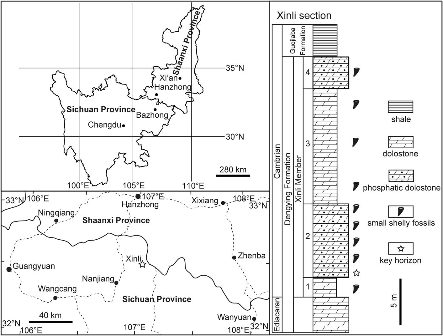
Figure 1 Location map and stratocolumn of the Xinli section in northern Sichuan Province, South China. The Xinli section and the key horizon yielding the present specimens are denoted by a star. Revised from Zhang et al. (Reference Zhang, Xiao, Liu, Yuan, Wan, Muscente, Shao, Gong and Cao2015).
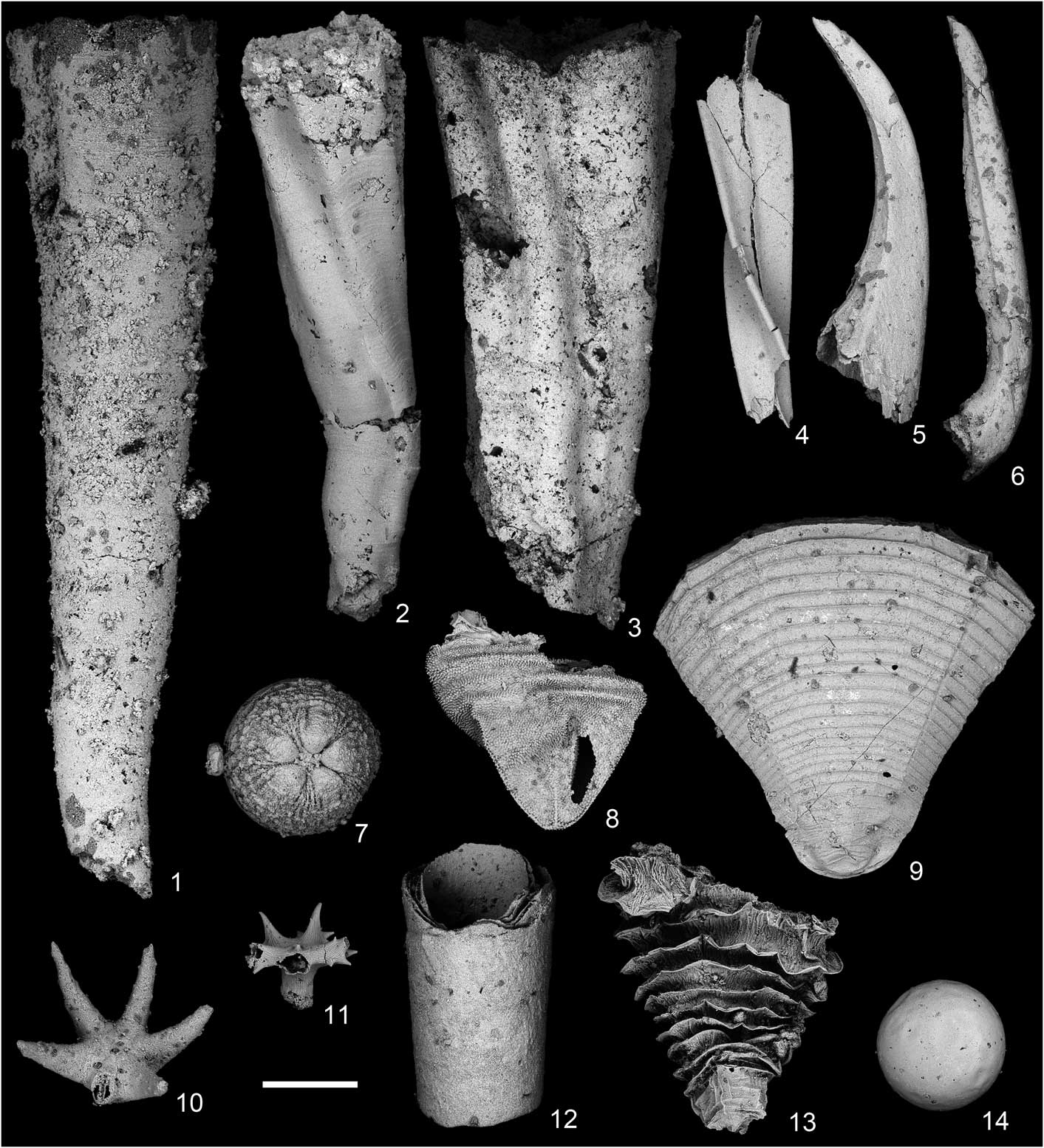
Figure 2 SEM images of representative microfossils from the lower Cambrian Xinli section. (1) Anabarites trisulcatus Missarzhevsky in Voronova and Missarzhevsky, Reference Voronova and Missarzhevsky1969, NIGP160422; (2) Anabarites isisticus Missarzhevsky, Reference Missarzhevsky1974, NIGP160423; (3) Anabarites hexasulcatus (Missarzhevsky, Reference Missarzhevsky1974), NIGP160424; (4) Anabarites ternarius Missarzhevsky in Rozanov et al., Reference Rozanov, Missarzhevsky, Volkova, Voronova, Krylov, Keller, Korolyuk, Lendzion, Michniak, Pykhova and Sidarov1969, NIGP160425; (5) Protohertzina anabarica Missarzhevsky, Reference Missarzhevsky1983, NIGP160426; (6) Protohertzina unguliformis Missarzhevsky, Reference Missarzhevsky1973, NIGP160427; (7) Olivooides multisulcatus Qian, Reference Qian1977, NIGP160428; (8) ‘Punctatus’ stage of Olivooides multisulcatus, NIGP160429; (9) Hexaconularia sichuanensis He and Yang, Reference He and Yang1986, NIGP160430; (10) Acanthocassis orthacanthus (Yang and He, Reference Yang and He1984) He and Xie, Reference He and Xie1989, NIGP160431; (11) Xinlispina spinosa Shao et al., Reference Shao, Liu, Wang and Zhang2015, NIGP160432; (12) A tube with multilayered wall, NIGP160433; (13) Quadrapyrgites quadratacris (Li, Reference Li1984) Steiner et al., Reference Steiner, Qian, Li, Hagadorn and Zhu2014, NIGP160434; (14) A globular specimen with smooth surface, NIGP160435. Scale bar=500 μm.
The rock samples collected from the lower second unit of the Xinli Member (Fig. 1) were first crushed into walnut-sized pieces (2~3 cm in diameter) and then dissolved in acetic acid following procedures described by Müller (Reference Müller1985). Rock fragments were immersed in diluted acetic acid (~10%), and residues were retrieved regularly after seven days of reaction. The residues were dried naturally, and microfossils were handpicked under a binocular microscope. The selected microfossils were mounted on aluminum stubs for scanning electron microscopy (SEM) on a LEO 1530VP field-emission environmental SEM at Nanjing Institute of Geology and Paleontology. The figures in this paper were processed using Adobe Illustrator CS5 and Adobe Photoshop CS5, respectively.
Repository and institutional abbreviation
The specimens are deposited at Nanjing Institute of Geology and Paleontology, Chinese Academy of Sciences, and the depository numbers are prefixed with NIGP.
Systematic paleontology
Scalidophora Lemburg, Reference Lemburg1995 total group
Genus Eokinorhynchus Zhang et al., Reference Zhang, Xiao, Liu, Yuan, Wan, Muscente, Shao, Gong and Cao2015
Type and only species
Eokinorhynchus rarus Zhang et al., Reference Zhang, Xiao, Liu, Yuan, Wan, Muscente, Shao, Gong and Cao2015.
Eokinorhynchus rarus Zhang et al., Reference Zhang, Xiao, Liu, Yuan, Wan, Muscente, Shao, Gong and Cao2015
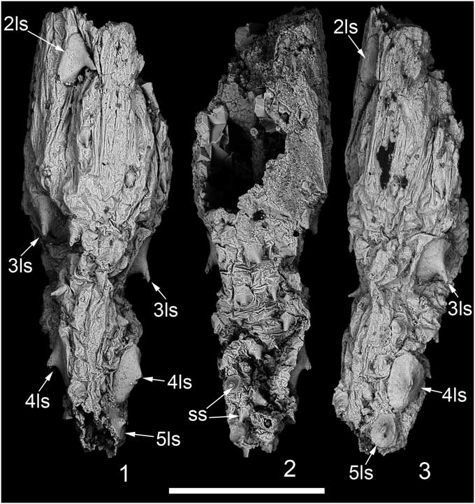
Figure 3 SEM images of Eokinorhynchus rarus Zhang et al., Reference Zhang, Xiao, Liu, Yuan, Wan, Muscente, Shao, Gong and Cao2015 from the lower Cambrian Xinli section. (1) A specimen representing part of the trunk, NIGP160436, dorsal view; (2) ventral view of (1); (3) right lateral view of (1). 2ls–5ls=2nd to 5th large spinose sclerite; ss=small spinose sclerite. Scale bar=500 μm.
2015 Eokinorhynchus rarus Reference Zhang, Xiao, Liu, Yuan, Wan, Muscente, Shao, Gong and CaoZhang et al., figs. 1, 2, 3a–g, 5, supplementary figs. S2, S3, supplementary movies S1–S4.
Holotype
Specimen NIGP160400 (Zhang et al., Reference Zhang, Xiao, Liu, Yuan, Wan, Muscente, Shao, Gong and Cao2015, fig. 1), a completely preserved specimen with partly protruded pharyngeal teeth.
Diagnosis
(Revised from Zhang et al., Reference Zhang, Xiao, Liu, Yuan, Wan, Muscente, Shao, Gong and Cao2015) Worm-like animal composed of a frontal region, a neck region, and a trunk. Frontal region consists of a pharynx with octaradially arranged teeth and an introvert with pentaradially arranged hollow scalids. Neck region covered with five circlets of neck scalids. Trunk has at least 20 annuli, and each annulus is covered with a circlet of tightly sutured small plates and armored with spinose sclerites. Five pairs of large spinose sclerites are bilaterally arranged, and a single large spinose sclerite is midventrally located. Two pairs of caudal spines are located slightly ventral to the terminal anus.
Occurrence
Xinli section, Nanjiang County, northern Sichuan Province, South China (Yang et al., Reference Yang, He and Deng1983; Zhang et al., Reference Zhang, Xiao, Liu, Yuan, Wan, Muscente, Shao, Gong and Cao2015). Small shelly fossils, Anabarites trisulcatus–Protohertzina anabarica Assemblage Zone, Xinli Member, Dengying Formation, Cambrian Fortunian Stage (Steiner et al., Reference Steiner, Li, Qian, Zhu and Erdtmann2007; Peng et al., Reference Peng, Babcock and Cooper2012).
Materials
One specimen, NIGP160436.
Measurements
Specimen NIGP160436 is about 1.4 mm long, with the second large spinose sclerite about 180 μm long.
Remarks
For a detailed description of Eokinorhynchus rarus, please refer to Zhang et al. (Reference Zhang, Xiao, Liu, Yuan, Wan, Muscente, Shao, Gong and Cao2015). Here, we revise the possible ontogenetic stages, with the originally proposed trunk part NIGP160414 (Zhang et al., Reference Zhang, Xiao, Liu, Yuan, Wan, Muscente, Shao, Gong and Cao2015, fig. 3h) replaced by a new specimen NIGP160436 (Fig. 3). Specimens NIGP160400 (Zhang et al., Reference Zhang, Xiao, Liu, Yuan, Wan, Muscente, Shao, Gong and Cao2015, fig. 1), NIGP160402 (Zhang et al., Reference Zhang, Xiao, Liu, Yuan, Wan, Muscente, Shao, Gong and Cao2015, fig. 3a), NIGP160401 (Zhang et al., Reference Zhang, Xiao, Liu, Yuan, Wan, Muscente, Shao, Gong and Cao2015, fig. 2), and NIGP160436 (Fig. 3) represent, most likely, progressively advanced ontogenetic stages. With growth of the body length, the large spinose sclerites expand their bases, and the cuticular annuli become more differentiated. The rectangular plates on the annuli are not well differentiated in NIGP160400 and NIGP160402. In NIGP160436, the annular rectangular plates are not preserved; thus, the growth trend of the rectangular plates during ontogeny could not be detected. Orsten-type preserved animals may, as adults, be much longer than 2–3 mm; however, entire or fragmented specimens larger than 2 mm in size are rare, indicative of a preservational bias (Maas et al., Reference Maas, Braun, Dong, Donoghue, Müller, Olempska, Repetski, Siveter, Stein and Waloszek2006; see also Müller and Hinz-Schallreuter, Reference Müller and Hinz-Schallreuter1993, for their palaeoscolecid material; Duan et al., Reference Duan, Dong and Donoghue2012). Specimen NIGP160436 is only part of the trunk but has a length of 1.4 mm. The original entire individual should presumably have a body length far exceeding 2 mm.
Form A
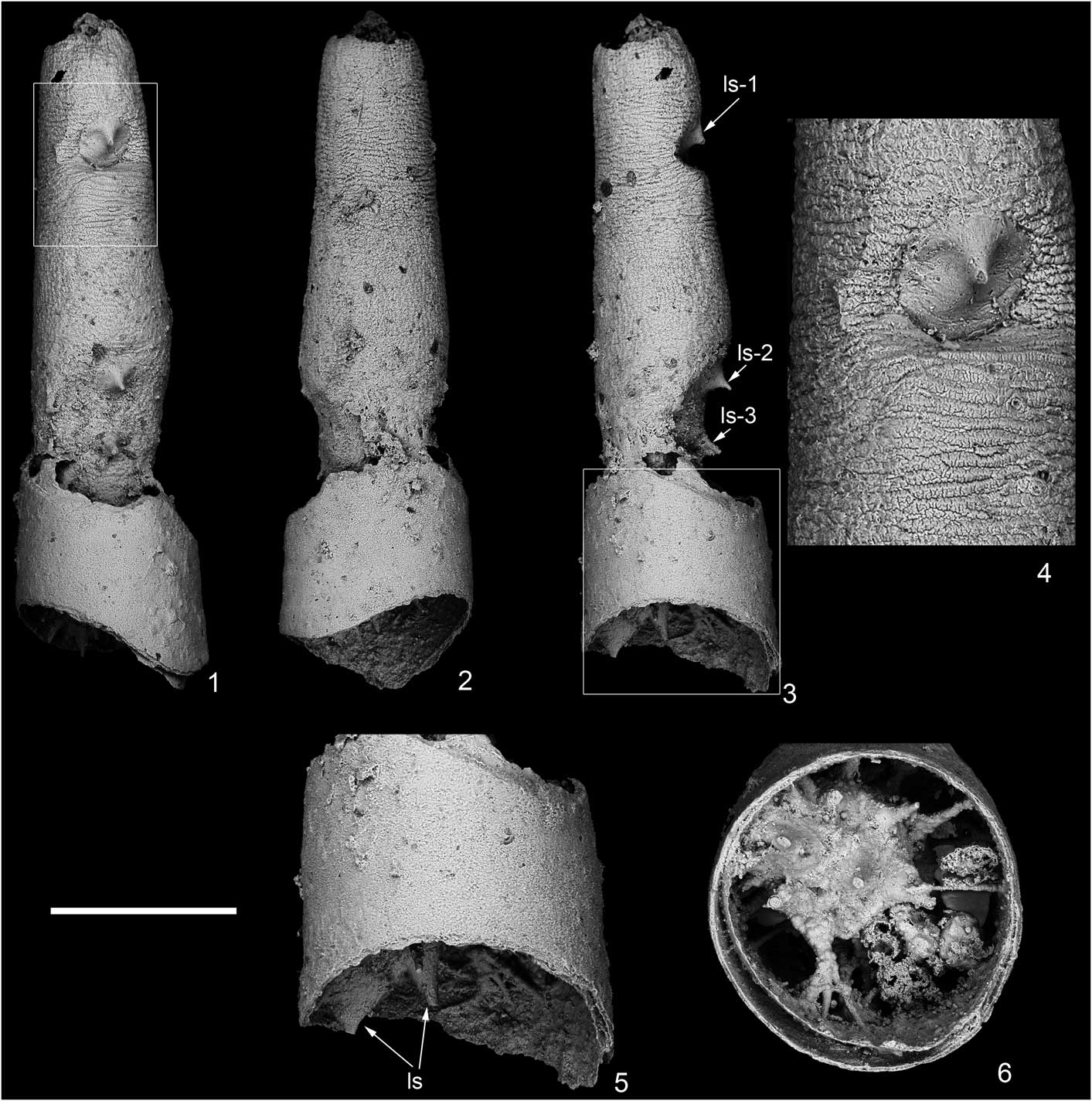
Figure 4 SEM images of Form A from the lower Cambrian Xinli section, NIGP160437. (1) Dorsal view; (2) ventral view; (3) left lateral view; (4) enlargement of area in white triangle in (1); (5) enlargement of area in white rectangle in (3); (6) posterior view. ls-1–ls-3=1st to 3rd large spinose sclerite. (1–3) Scale bar=500 μm; (4) scale bar=187 μm; (5, 6) scale bar=319 μm.
Occurrence
Xinli section, Nanjiang County, northern Sichuan Province, South China (Yang et al., Reference Yang, He and Deng1983; Zhang et al., Reference Zhang, Xiao, Liu, Yuan, Wan, Muscente, Shao, Gong and Cao2015). Small shelly fossils, Anabarites trisulcatus–Protohertzina anabarica Assemblage Zone, Xinli Member, Dengying Formation, Cambrian Fortunian Stage (Steiner et al., Reference Steiner, Li, Qian, Zhu and Erdtmann2007; Peng et al., Reference Peng, Babcock and Cooper2012).
Description
NIGP160437 represents the trunk part of a worm-like animal, and the overall morphology of this animal is currently unknown. It is cylindrical in shape and has a line of at least five large spinose sclerites on the dorsal side (ls-1, ls-2, ls-3, and two more sclerites caudally; Fig. 4.1, 4.3, 4.5, 4.6). Except the large spinose sclerites (Fig. 4.2), there are no other sclerites or spines on the trunk. The large spinose sclerites decrease in size from anterior to posterior (Fig. 4.1). The cuticle is ornamented with a large number of annuli (Fig. 4.4). The posterior end of the trunk might have a tuft of sclerites or spines, but they are embedded within secondary phosphate calcium and cannot be detected here (Fig. 4.6). The trunk is posteriorly nested within a tube, which has a smooth and multilayered wall (Fig. 4.5, 4.6).
Materials
One specimen, NIGP160437.
Measurements
Specimen NIGP160437 is about 1.8 mm in length and 370 μm in diameter at midpart, with the first large spinose sclerite (ls-1) about 140 μm long.
Remarks
The orientation of Form A follows the topology of Eokinorhynchus rarus. The large spinose sclerites of E. rarus are distributed mostly on the dorsal side, functioning as defense organs. When this topology is applied to Form A, we treat the side with the large spinose sclerites as the dorsal side. The spines of the large spinose sclerites of E. rarus are directed posteriorly; thus, when this topology is applied to Form A, we treat the end bearing the smooth tube as the posterior end.
The smooth tube has a diameter of about 522 μm at the larger end. It should be some small shelly fossil that mantled the worm trunk end during taphonomy. Such smooth tube with a multilayered wall is separately recovered in the Xinli section (Fig. 2.12); it has a diameter of about 810 μm at its larger end. As to NIGP160433 (Fig. 2.12), we treat the end with larger diameter as the upper part. When this orientation is applied to the tube mantling Form A (NIGP160437), it is evident that the upper part of the tube (with larger diameter) is opposite to the anterior part of the worm trunk. The orientations of the tube and Form A do not match up, and it supports the nonbiological connection between them.
Form A has densely spaced trunk annuli, different from the 20 annuli of Eokinorhynchus rarus. Form A also differs from Eopriapulites sphinx Liu and Xiao in Liu et al., Reference Liu, Xiao, Shao, Broce and Zhang2014 because the latter does not have any sclerites or spines on the trunk (Shao et al., Reference Shao, Liu, Wang, Zhang, Tang and Li2016). Therefore, Form A probably represents part of the body of a new species.
Form B
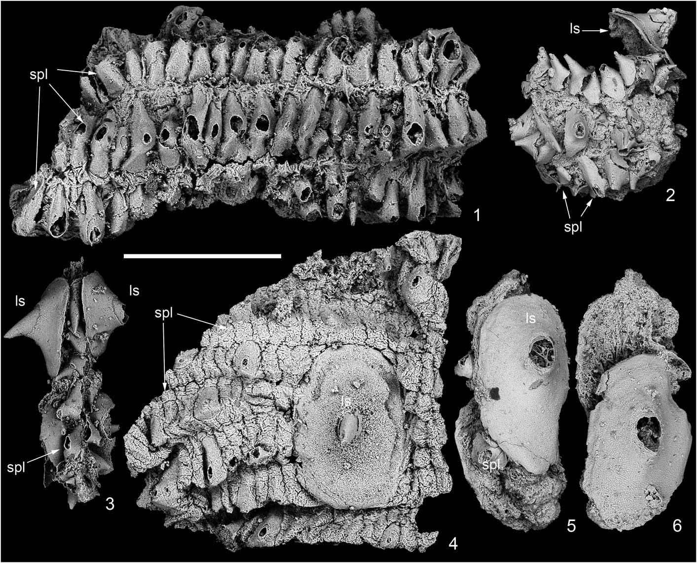
Figure 5 SEM images of Form B from the lower Cambrian Xinli section. (1) NIGP160438, with four annuli, each bearing densely sutured small plates; (2) NIGP160439, a fragment with a single large spinose sclerite and a few small plates; (3) NIGP160440, with a pair of large spinose sclerites and a few small plates; (4) NIGP160441, with eight annuli and a large spinose sclerite loosely covering 4 annuli; (5, 6) NIGP160442, opposite views, with two large spinose sclerites basally attached to each other and a few small plates in between. ls=large spinose sclerite; spl=small plate. Scale bar=500 μm.
2015 Eokinorhynchus rarus Reference Zhang, Xiao, Liu, Yuan, Wan, Muscente, Shao, Gong and CaoZhang et al., fig. 3h–i
2015 Unnamed Form I; Reference Zhang, Xiao, Liu, Yuan, Wan, Muscente, Shao, Gong and CaoZhang et al., fig. 4a, b
Occurrence
Xinli section, Nanjiang County, northern Sichuan Province, South China (Yang et al., Reference Yang, He and Deng1983; Zhang et al., Reference Zhang, Xiao, Liu, Yuan, Wan, Muscente, Shao, Gong and Cao2015). Small shelly fossils, Anabarites trisulcatus–Protohertzina anabarica Assemblage Zone, Xinli Member, Dengying Formation, Cambrian Fortunian Stage (Steiner et al., Reference Steiner, Li, Qian, Zhu and Erdtmann2007; Peng et al., Reference Peng, Babcock and Cooper2012).
Description
The illustrated specimens might represent different trunk parts of the same animals, because of the similar morphology and arrangement mode of the small plates, but in different ontogenetic stages because of the size difference of the sclerites and plates. The tightly sutured small plates are aligned in three rows in NIGP160439 (Fig. 5.2), four rows in NIGP160438 (Fig. 5.1), and eight rows in NIGP160441 (Fig. 5.4). These rows with small plates represent annuli of the trunk. The small plates are much longer than wide, have an expanded base, and protrude distally into a spine. Sometimes, the distal spines are broken, leaving a hollow hole centrally (Fig. 5.1). The large spinose sclerites have an expanded base, which may be elliptical, and in one specimen the large spinose sclerite straddles as many as four annuli (Fig. 5.4). The small plates and the large spinose sclerites are all hollow internally.
Materials
Five specimens, NIGP160438–160442.
Measurements
Specimen NIGP160438 (Fig. 5.1) is about 1.5 mm long, and each small plate is about 240 μm long. The large spinose sclerite in NIGP160442 (Fig. 5.5, 5.6) is about 580 μm long, representing the trunk part of a very large individual.
Remarks
A fragment (NIGP160414, Zhang et al., Reference Zhang, Xiao, Liu, Yuan, Wan, Muscente, Shao, Gong and Cao2015, fig. 3h) was originally assigned to Eokinorhynchus rarus and was hypothesized to represent the trunk part of a very large individual. The small plates of E. rarus are rectangular in shape and never protrude into spines, but the small plates of NIGP160414 are spinose. Thus, we move NIGP160414 to Form B, and Form B possibly represents a new scalidophoran animal, different from E. rarus and Form A. The second large spinose sclerites of E. rarus (Fig. 3.1) are about 180 μm long, and the first large spinose sclerite of Form A is about 140 μm long. By contrast, the large spinose sclerites of NIGP160442 (Fig. 5.5) are 580 μm long. If the body length is proposed to be positively correlated with the size of the large spinose sclerite, Form B probably reached a centimeter-scale body length.
Form C
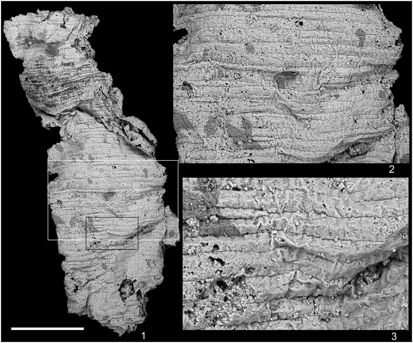
Figure 6 SEM images of Form C from the lower Cambrian Xinli section. (1) A fragment with densely spaced annuli, NIGP160493; (2) enlargement of the area in the white rectangle in (1); (3) enlargement of the area in the black rectangle in (1). (1) Scale bar=500 μm; (2) scale bar=270 μm; (3) scale bar=122 μm.
Occurrence
Xinli section, Nanjiang County, northern Sichuan Province, South China (Yang et al., Reference Yang, He and Deng1983; Zhang et al., Reference Zhang, Xiao, Liu, Yuan, Wan, Muscente, Shao, Gong and Cao2015). Small shelly fossils, Anabarites trisulcatus–Protohertzina anabarica Assemblage Zone, Xinli Member, Dengying Formation, Cambrian Fortunian Stage (Steiner et al., Reference Steiner, Li, Qian, Zhu and Erdtmann2007; Peng et al., Reference Peng, Babcock and Cooper2012).
Description
A fragmentary trunk part of a scalidophoran worm, with a length of 2.3 mm. The preserved trunk part bears densely spaced annuli, and each annulus is about 42 μm long. The annuli are counted to be about 72 for this fragment. There are no other ornaments or structures on the trunk part.
Materials
One specimen, NIGP160493.
Measurements
Specimen NIGP160493 is about 2.3 mm long and 830 μm wide. Each annulus is about 42 μm long (anterior–posterior).
Remarks
Form C might be the trunk part of Eopriapulites sphinx Liu and Xiao in Liu et al., Reference Liu, Xiao, Shao, Broce and Zhang2014, because they share a similarly unornamented trunk with densely spaced trunk annuli.
Form D
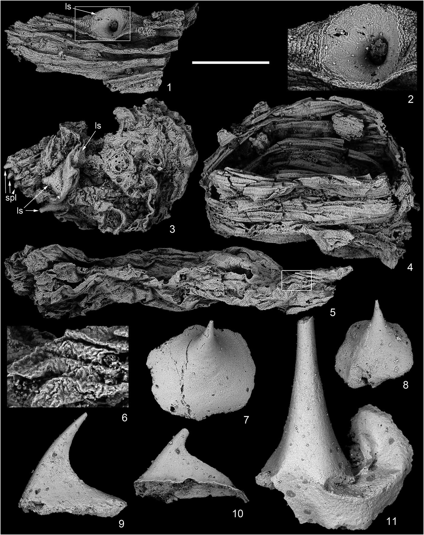
Figure 7 (1, 2) SEM images of Form D; (3–6) possible exuviae; (7–11) disassociated small shelly fossils from the lower Cambrian Xinli section. (1) NIGP160443; (2) enlargement of area in white rectangle in (1); (3) NIGP160444; (4) NIGP160445; (5) NIGP160446; (6) enlargement of area in white rectangle in (5); (7) NIGP160447; (8) NIGP160448; (9) NIGP160449; (10) NIGP160450; (11) NIGP160451. ls=large spinose sclerite; spl=small plate. (1, 3–5, 7–11) Scale bar=500 μm; (2) scale bar =194 μm; (6) scale bar=115 μm.
Occurrence
Xinli section, Nanjiang County, northern Sichuan Province, South China (Yang et al., Reference Yang, He and Deng1983; Zhang et al., Reference Zhang, Xiao, Liu, Yuan, Wan, Muscente, Shao, Gong and Cao2015). Small shelly fossils, Anabarites trisulcatus–Protohertzina anabarica Assemblage Zone, Xinli Member, Dengying Formation, Cambrian Fortunian Stage (Steiner et al., Reference Steiner, Li, Qian, Zhu and Erdtmann2007; Peng et al., Reference Peng, Babcock and Cooper2012).
Description
Part of the trunk is preserved as external mold, judged by the morphology of the only preserved large spinose sclerite and the preserved trunk annuli (Fig. 7.2) that have clear boundaries in between. The base of the large spinose sclerite is nearly circular in shape. Its outer edge is continuous into the annuli of the cuticle. The cuticle is irregularly annulated with a large number of annuli.
Materials
One specimen, NIGP160443.
Measurements
Specimen NIGP160443 is about 1.3 mm long, with the only large spinose sclerite about 200 μm long.
Remarks
It is possible that Form D represents the trunk part of Form A because they share some similarities; for example, the trunk cuticle is densely annulated with a large number of annuli, and the large spinose sclerite is sparsely distributed. Due to the lack of completely preserved specimens, we tentatively adopt open nomenclature and treat them as different forms. Deduced from the size of the large spinose sclerite, Form D might have reached a body length of more than 2 mm.
Affinities
Scalidophoran worms possess, plesiomorphically, a subectodermal double-layered body-wall musculature, i.e., made of longitudinal and circular layers (e.g., Brusca and Brusca, Reference Brusca and Brusca2003; Nielsen, Reference Nielsen2012). During locomotion, the contraction of the circular muscles would result in trunk annuli; thus, the scalidophoran worms might be superficially annulated. In the absence of cilia along the epidermal cell layer, locomotion is mainly achieved using the extrudable introvert and backwardly oriented scalids (as anchoring devices) to pull the trunk forward. From extant taxa, it is known that these scalids are internally hollow, with the cuticle limited to a thin outer covering (Merriman, Reference Merriman1981; Neuhaus et al., Reference Neuhaus, Kristensen and Lemburg1996, Reference Neuhaus, Kristensen and Peters1997; Neuhaus and Higgins, Reference Neuhaus and Higgins2002). Nematoids autapomorphically lack circular muscles, and their ‘scalids,’ if developed (e.g., in larval nematomorphs and some nematodes), are solid and composed exclusively of cuticle (Schmidt-Rhaesa, Reference Schmidt-Rhaesa1998; Nielsen, Reference Nielsen2012). Eokinorhynchus rarus has cuticular annulation and, most likely, internally hollow scalids, indicating a close relationship with Scalidophora.
Eokinorhynchus rarus has heterogenous annulation embodied by the bilaterally symmetrical arrangement of large spinose sclerites on different parts of the trunk and the randomly distributed small spines especially on the posteroventral side of the trunk. Moreover, E. rarus has 20 annuli, each annulus being comparatively long relative to trunk length. This type of annulation differs from the homonomous annulation of other Cambrian putative scalidophorans, for example, the long, macroscopic worm-like forms from the Chengjiang biota Chen, Reference Chen2004), the likewise worm-like species of Markuelia, known only from individuals close to hatching (Dong et al., Reference Dong, Donoghue, Cheng and Liu2004; Haug et al., Reference Haug, Maas, Waloszek, Donoghue and Bengtson2009) that might have as many as 60 or more annuli (Cheng et al., Reference Cheng, Peng, Duan and Dong2011), or Eopriapulites sphinx, which has a long vermiform trunk with more than 140 annuli (Shao et al., Reference Shao, Liu, Wang, Zhang, Tang and Li2016). The annulation of the middle Cambrian Shergoldana australiensis Maas et al., Reference Maas, Huang, Chen, Waloszek and Braun2007a is restricted to the neck region, while the hind body is characterized by hexagonal plates that are arranged in rings with each plate extending into a posteriorly oriented spine (Maas et al., Reference Maas, Huang, Chen, Waloszek and Braun2007a). The annulation type of E. rarus coincides with the macroannuli (= zonites) exemplified by kinorhynch worms (Budd, Reference Budd2001; Neuhaus, Reference Neuhaus2013). E. rarus also has some other features that are similar to modern kinorhynchs, for example, each trunk zonite ornamented with densely sutured plates, trunk with small spines, and trunk end with caudal spines. Despite these similarities, E. rarus differs from modern kinorhynchs in many aspects. For example, E. rarus has a trunk with 20 macroannuli, whereas modern kinorhynchs have a trunk with a consistent 11 zonites. Each zonite of modern kinorhynchs has only two to four cuticular plates (dorsally and ventrally, no rings), while each annulus of E. rarus has as many as 10 to 40 small plates. The modern kinorhynchs have one pair of lateral terminal spines (relatively long) and one pair of lateral terminal accessory spines (relatively short), and all these spines are positioned lateral to the anus; some taxa bear one single midterminal spine that is 150% longer than the lateral terminal spines (Sørensen and Pardos, Reference Sørensen and Pardos2008), whereas the caudal spines of E. rarus are positioned ventrally to the anus and relatively short. This may indicate that E. rarus represents a stem member of the taxon Kinorhyncha, with the macroannuli as a key synapomorphy. Since the Kinorhyncha is represented by about 240 extant species, with no fossil species reported yet (Neuhaus, Reference Neuhaus2013), E. rarus would be the first and only fossil record of this cycloneuralian taxon.
It has previously been argued that a long vermiform trunk with a large number of annuli might be a plesiomorphic state inherited from the last common ancestor of Scalidophora, or even Cycloneuralia; thus, most Cambrian so-called priapulid-like worms might be assigned to total-group Cycloneuralia or total-group Scalidophora as a whole, but a further assignment to an exact taxon within Scalidophora is currently not possible (e.g., Maas et al., Reference Maas, Waloszek, Haug and Müller2007b; Harvey et al., Reference Harvey, Dong and Donoghue2010; Wills et al., Reference Wills, Gerber, Ruta and Hughes2012). In addition, Eopriapulites sphinx from the lower Cambrian Zhangjiagou section was described as ‘priapulid-like’; however, the characters referring to ‘priapulid-like’ are simply those that are caracteristic for Scalidophora. Thus, E. sphinx was assigned to the stem lineage of Scalidophora (Liu et al., Reference Liu, Xiao, Shao, Broce and Zhang2014; Shao et al., Reference Shao, Liu, Wang, Zhang, Tang and Li2016). However, these stem-lineage positions might reflect the so-called ‘systematic limbo,’ indicating nothing but an uncertain position within the so-called total group (Donoghue and Purnell, Reference Donoghue and Purnell2009). Therefore, we propose that Forms A, C, and D should be assigned to total-group Scalidophora to indicate their uncertain positions within the taxon Scalidophora, as long as no more informative features supporting a further assignment are observed. A further phylogenetic analysis including at least Forms A and C must therefore await the recovery of completely preserved specimens.
Form B was previously considered as scleritomes of unknown affinities or small shelly fossils. The recovery of the completely preserved specimens of Eokinorhynchus rarus sheds new light on the affinity of Form B. In fact, the small plates of Form B are densely sutured in rows (Fig. 5.1, 5.4) that resemble the arrangement of the small plates of E. rarus. Sclerites assigned to Form B are large and spinose, similar in morphology to the large spinose sclerites of E. rarus (Fig. 3). The large spinose sclerites of Form B may have loosely covered four annuli (Fig. 5.4), and those of E. rarus cover two or at most three annuli (Zhang et al., Reference Zhang, Xiao, Liu, Yuan, Wan, Muscente, Shao, Gong and Cao2015). The key difference between Form B and E. rarus is that the small plates are rectangular, lacking spines in the latter species, but long and elliptical, bearing spines, in the former. Therefore, Form B represents a scalidophoran animal that might have close affinities with E. rarus but apparently represents a different species.
Besides the five forms described in the preceding, we found some fossils that also might be affiliated directly with early scalidophorans. Animals belonging to the stem lineage of Scalidophora, a taxon within the Cycloneuralia, molt regularly (Aguinaldo et al., Reference Aguinaldo, Turbeville, Linford, Rivera, Garey, Raff and Lake1997), and the exuviae of these early scalidophorans can be preserved as fossils. Specimen NIGP160444 (Fig. 7.3) is preserved in a strongly distorted status, possibly being an example of an exuvia. The large spinose sclerites and the small spines are much distorted and, therefore, cannot be observed clearly. Thus, it remains somewhat uncertain whether this specimen belongs to E. rarus or to Form B. More specimens (Fig. 7.4–7.6) are recovered from the Xinli section that might represent the exuviae of early scalidophorans or co-occurring cycloneuralians. Specimen NIGP160445 (Fig. 7.4) has soft cuticle that bears annulus-like structures, whereas NIGP160446 (Fig. 7.5) is devoid of annuli but has grain-shaped cuticular textures (Fig. 7.6).
Discussion
The armor of the early scalidophoran animals sheds also light on the affinity of other coeval small shelly fossils. Zhang et al. (Reference Zhang, Xiao, Liu, Yuan, Wan, Muscente, Shao, Gong and Cao2015) noticed that the large spinose sclerites on Eokinorhynchus rarus and their unnamed form I are similar to disassociated small shelly fossils described as Paracarinachites spinus (Yu, Reference Yu1984) (Conway Morris and Chen, Reference Conway Morris and Chen1991), different from the type material of P. spinus (Yu, Reference Yu1984; Qian and Bengtson, Reference Qian and Bengtson1989), and the small plates of their unnamed form II resemble the form Kaiyangites novoli Qian and Yin, Reference Qian and Yin1984 (Yao et al., Reference Yao, Xiao, Yin, Li and Yuan2005). Therefore, Zhang et al. (Reference Zhang, Xiao, Liu, Yuan, Wan, Muscente, Shao, Gong and Cao2015) proposed that these disassociated small shelly fossils represented by P. spinus and K. novoli might belong to Eokinorhynchus-like scalidophoran animals. In our materials, some disassociated small shelly fossils were also recovered from the Xinli section, with some (Fig. 7.7, 7.8) similar to the large spinose sclerites of Eokinorhynchus rarus, Form A, and Form B, some (Fig. 7.9, 7.10) similar to the small plates of Zhang and colleagues’ (2015) unnamed form II, and some (Fig. 7.11) similar to the small spines on the trunk of E. rarus, but sclerites belonging to P. spinus and K. novoli were not recovered. Despite the morphological similarities between these small shelly fossils and the armor on the scalidophoran animals, convergent evolution is still possible. For example, the large spinose sclerites occur on the trunk of E. rarus and Form B (i.e., some derived scalidophoran animals within Kinorhyncha) and on the trunk of Form A and Form C. Evidently, E. rarus and Form A should be different types of scalidophoran worms due to their very different annulation types. Thus, the large spinose sclerites may represent a convergently developed type of armor between lineages of distant affinities. This could also hold true for the remaining co-occurring small shelly fossils (Fig. 7.9–7.11). These disassociated small shelly fossils might come from the same scalidophoran animals or come from different scalidophoran animals, and their exact affinity within Scalidophora remains currently unknown. An assignment more precise than being part of the taxon Scalidophora can, therefore, not be made for any of the taxa presented herein. An introvert with scalids/hooks is interpreted as an autapomorphy of either Scalidophora (Lemburg, Reference Lemburg1995) or Cycloneuralia (Nielsen, Reference Nielsen2012). Referring to this ‘Introverta’ hypothesis (Cycloneuralia=Introverta), the assignment must even be coarser.
Conclusions
We report scalidophoran animals from the Cambrian Fortunian Xinli section in northern Sichuan Province, South China. The specimens are fossilized in Orsten-type preservation (i.e., three-dimensionally phosphatized and soft-bodied). They can be grouped into five forms, including one form assignable to the species Eokinorhynchus rarus and four forms named in open taxonomy (i.e., Forms A, B, C, and D). E. rarus is worm-like and the body can be divided into three parts: an anterior region with introvert, a neck region, and the trunk. The trunk comprises a number of annuli that are armored with densely sutured rectangular plates and randomly distributed small spinose sclerites. Form A is interpreted to represent a new species. It has a densely annulated trunk with a large number of annuli. At least five large spinose sclerites are aligned in a longitudinal row on the dorsal side. Form B is represented only by trunk fragments, but it is evidently different from E. rarus and all other coeval scalidophoran animals in that it bears large spinose sclerites as well as trunk annuli armored with densely sutured spinose small plates that protrude into spines distally. Form C is similar in trunk morphology with Eopriapulites sphinx, i.e., with a large number of densely spaced annuli without any other ornament or structure. Form D is the trunk fragment of an unknown worm-like organism. Its densely annulated trunk has a large number of annuli and a single large spinose sclerite. Forms A, C, and D are assigned to total-group Scalidophora to indicate their uncertain positions within Scalidophora; Form B might represent at least a new species with closer affinities to E. rarus.
All putative scalidophoran animals were recovered from the Cambrian Fortunian Stage of South China only very recently. Since their diversity in the small shelly faunas is highly underestimated, the current research highlights the significance of Orsten-type Lagerstätten in probing the morphology and taxonomy of the oldest known scalidophoran animals. Accordingly, it provides key information on the origin and early evolution of Scalidophora and Cycloneuralia. To summarize, in uncovering the origin and early evolution of Cycloneuralia, the Orsten-type Lagerstätten are progressively equal in systematic and evolutionary importance to Burgess Shale–type Lagerstätten.
Acknowledgments
This work was supported by the Chinese Ministry of Science and Technology 973 Project (2013CB837100), the National Natural Science Foundation of China (41572007), and the Youth Innovation Promotion Association, Chinese Academy of Sciences (2016283). Two anonymous reviewers provided constructive suggestions for this paper.


