INTRODUCTION
Plerocercoids of the pseudophyllidean cestode Schistocephalus solidus are common parasites of three-spined sticklebacks, Gasterosteus aculeatus, in freshwater and brackish habitats throughout the geographical range of the fish. The three-spined stickleback-S. solidus host-parasite system has become an important model in experimental parasitology and is increasingly used to investigate a wide range of questions about host-parasite interactions and co-evolution. Here we present a review of recent studies that have used controlled experimental infections to investigate host-parasite interactions in this system.
We begin our review with background information on the parasite's life cycle, on the host fish and the ‘typical’ phenotype of infected sticklebacks in nature, and briefly discuss emerging variation in infection phenotype. We then examine how aspects of the life cycle can be experimentally manipulated in the laboratory to allow experimental infections of sticklebacks to be undertaken. The remainder of our review focuses on how experimental infection studies have been used to illuminate host-parasite interactions in the stickleback-S. solidus system, including the immune responses of the fish host, the energetic consequences of infection and the consequences of infections for fish behaviour and fitness.
Life cycle of S. solidus in nature
Schistocephalus solidus is a trophically transmitted pseudophyllidean cestode with a three-host life cycle. The definitive host can be any warm-blooded vertebrate; most typically these are fish-eating birds though other endotherms can harbour adult worms, including otters (Hoberg et al. Reference Hoberg, Henny, Hedstrom and Grove1997) and, though presumably only rarely, humans (Coombs and Crompton, Reference Coombs and Crompton1991). Schistocephalus solidus does not grow in the gut of the definitive host but undergoes the final stages of sexual maturation there, reproducing sexually either by selfing (if singly infected) or by cross-fertilization (in multiple infections). Eggs released into the water with the bird's faeces hatch to produce free-swimming coracidia that are transmitted trophically to a wide range of cyclopoid copepods, the 1st intermediate hosts. Here the parasites develop in the copepod haemocoel into procercoids, becoming infective to three-spined sticklebacks, the obligatory specific 2nd intermediate hosts (Bråten, Reference Bråten1966), with the formation of a hooked cercomer. Sticklebacks acquire infections when they feed on parasitized copepods, and in the stickleback digestive tract infective procercoids shed their outer layer, together with the cercomer, and penetrate the wall of the intestine. The parasite then develops into a plerocercoid, which grows to a large size in the fish host's body cavity. The life cycle is completed when sticklebacks harbouring infective plerocercoids are ingested by a definitive host (Clarke, Reference Clarke1954).
The geographical distribution of the parasite is limited by the distribution of the only obligate host in the life cycle, the three-spined stickleback, which is restricted to the Northern hemisphere and occurs around the margins of the Atlantic and Pacific Oceans (Bell and Foster, Reference Bell and Foster1994). In this geographical region S. solidus is a regular parasite of three-spined stickleback populations inhabiting freshwater and brackish ecosystems, and is most commonly found in those in lacustrine or slow flowing habitats (Kennedy, Reference Kennedy1974; Wootton, Reference Wootton1976; Barber, Reference Barber, Östlund-Nilsson, Mayer and Huntingford2007).
Specificity of stickleback host
Three-spined sticklebacks are the only recognised fish host of S. solidus, although other Schistocephalus spp. infect nine-spined stickleback Pungitius pungitius and sculpins (Hyslop and Chubb, Reference Hyslop and Chubb1983; Chubb et al. Reference Chubb, Seppala, Luscher, Milinski and Valtonen2006; Seppala et al. Reference Seppala, Chubb, Niemela and Valtonen2007; French and Muzzall, Reference French and Muzzall2008). Experimental exposure of nine-spined sticklebacks (Pungitius pungitius) to infective stages of S. solidus led to much slower plerocercoid growth and infections were cleared after 14 days, while plerocercoids kept on growing in three-spined sticklebacks (Orr et al. Reference Orr, Hopkins and Charles1969).
Heterotransplants of S. solidus plerocercoids from G. aculeatus to other species of fish (Cottus gobio, Nemacheilus barbatula, Phoxinus phoxinus, Salmo trutta, Coregonus clupeoides, Perca fluviatilis, Rutilus rutilus, Esox lucius and P. pungitius) died within 2–10 days after transfer, while homotransplants between G. aculeatus survived (Bråten, Reference Bråten1966). These observations indicate that, in principle, S. solidus plerocercoids can be cleared by a fish immune system, but obviously S. solidus plerocercoids are able to avoid an effective immune response in G. aculeatus, their specific second intermediate host.
Sticklebacks as experimental model hosts
A major attraction of the stickleback-S. solidus host-parasite system for ecological and evolutionary biologists is the rich history of studies investigating the natural history, behaviour and evolutionary biology of the host fish, and a correspondingly substantial literature that has been regularly and thoroughly reviewed (Wootton, Reference Wootton1976, Reference Wootton1984; Bell and Foster, Reference Bell and Foster1994; Östlund-Nilsson et al. Reference Östlund-Nilsson, Mayer and Huntingford2006). This background permits a wide range of ecologically and evolutionarily relevant questions to be addressed. Furthermore, the three-spined stickleback has, in recent years, assumed even more importance as a model species in biology; the publication of linkage and chromosome maps (Peichel et al. Reference Peichel, Nereng, Ohgi, Cole, Colosimo, Buerkle, Schluter and Kingsley2001; Kingsley et al. Reference Kingsley, Zhu, Osoegawa, De Jong, Schein, Marra, Peichel, Amamiya, Schluter, Balabhadra, Friedlander, Cha, Dickson, Grimwood, Schmutz, Talbot and Myers2004) and the sequencing of its genome (Kingsley, Reference Kingsley2003) has greatly enhanced its utility in molecular studies of evolution and development (McKinnon et al. Reference McKinnon, Mori, Blackman, David, Kingsley, Jamieson, Chou and Schluter2004; Shapiro et al. Reference Shapiro, Marks, Peichel, Blackman, Nereng, Jonsson, Schluter and Kingsley2004, Reference Shapiro, Bell and Kingsley2006; Colosimo et al. Reference Colosimo, Hosemann, Balabhadra, Villarreal, Dickson, Grimwood, Schmutz, Myers, Schluter and Kingsley2005; Gibson, Reference Gibson2005). Sticklebacks are also readily bred in laboratory aquaria (Barber and Arnott, Reference Barber and Arnott2000), facilitating the challenge of naïve individuals and thus fulfilling both scientific and local ethical requirements for experimental infection studies.
Field studies of S. solidus-infected sticklebacks
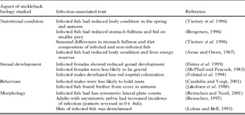
A number of studies have examined S. solidus infection prevalence and intensity in natural stickleback populations, and the phenotype (including the appearance, energetic condition, reproductive capacity and behaviour) of naturally infected sticklebacks has been well documented. Observations on the phenotype of infected fish from populations where S. solidus is endemic are summarised in Table 1. The proportion of fish harbouring infections can be extremely high, in some cases approaching 100% (Dick, Reference Dick1816; Smyth, Reference Smyth1947; Hopkins and Smyth, Reference Hopkins and Smyth1951), but this varies considerably between populations (MacColl, Reference MacColl2009), and temporally within them (Chappell, Reference Chappell1969). Typical features of classical ‘schistocephalosis’ include characteristic distension of the fish's abdomen, an altered swimming gait, increased risk-taking behaviour, reduced body condition and functional (if not physiological) castration. However, as more host populations are studied it is becoming clear that there is significant variation in infection phenotype, and there are a number of exceptions to these ‘typical’ infection phenotypes. Notably, intensive studies of some Alaskan populations suggest that the traditional view of S. solidus as an absolute castrator of female sticklebacks may need to be revised (Heins and Baker, Reference Heins and Baker2008), and in a small number of populations infection is additionally associated with almost complete demelanisation (Lobue and Bell, Reference Lobue and Bell1993; Ness and Foster, Reference Ness and Foster1999).
Table 1. Selected field studies documenting traits associated with Schistocephalus solidus infection in natural stickleback populations

In vitro culture of S. solidus
Pioneering work by the parasite physiologist J. D. Smyth in the 1940–50s developed protocols for the in vitro culture of adult helminths, including S. solidus (Smyth, Reference Smyth1946, Reference Smyth1990; Smyth and McManus, Reference Smyth and Mcmanus1989). In brief, plerocercoids recovered from infected sticklebacks are removed and placed into sterilised culture vessels containing a buffered medium that provides a suitable physico-chemical environment for parasite development (such as horse serum and/or a cell culture medium such as RPMI-1640). Antibiotics may be added to reduce contamination. The plerocercoid(s) are not placed directly into the medium but are instead constrained within narrow diameter semi-permeable dialysis tubing, which mimics the small intestine of the definitive host and provides the constriction needed for successful fertilisation (Smyth, Reference Smyth1990). Culture vessels are placed into a water bath set to 40°C and shaken gently to aid the dissolution of metabolic products from developing worms. Due to the progenetic development of pseudophyllidean plerocercoids, sexual maturation occurs rapidly and within 48 h the adult worms begin producing eggs. The fact that S. solidus attains its final size in the fish body cavity and matures so rapidly to the adult form made it possible for the first time to maintain adult cestodes under sterile experimental conditions and undertake detailed physiological studies. Such studies had been impossible with most other tapeworm species, which typically grow and mature over prolonged time periods in the host intestine, and S. solidus became an extremely valuable model in parasite physiology (Smyth and McManus, Reference Smyth and Mcmanus1989).
Using Smyth's techniques – or slightly modified versions of them – large numbers of eggs can be generated and hatched to yield coracidia, which in turn can be used to infect lab-bred copepods (Wedekind, Reference Wedekind1997). After a period of development to the infective cercomer-bearing stage, procercoids can be fed to sticklebacks inside infected copepods, either by gavage or by natural feeding, to generate experimentally infected fish hosts and allowing detailed experimental studies of fish-parasite interactions.
MECHANISMS OF RESISTING S. SOLIDUS INFECTION: HOST BEHAVIOUR, IMMUNE responseS and host manipulation
Animals have evolved three major types of mechanisms to avoid, or reduce the likelihood of developing, debilitating parasite infections; behavioural mechanisms that limit contact with infectious agents, physical barriers to invasive stages and immune systems. The ability to infect intermediate hosts experimentally in large numbers means that it is possible to examine the responses of hosts to controlled experimental challenge. In recent years, experimental infection studies have been used to examine both behavioural and immunological aspects of stickleback responses to infective stages of S. solidus.
Behavioural resistance
The strong selection pressure placed on host organisms to avoid debilitating parasite infections, together with the typically high costs of mounting immune responses against invading pathogens, has led to the evolution of a wide range of strategies of behavioural resistance in animals (Hart, Reference Hart1990, Reference Hart1992, Reference Hart, Clayton and Moore1997). For parasites that are transmitted trophically between hosts, avoiding infected prey intuitively reduces the level of exposure to infection, but this requires that infected prey are identifiable and that the benefits of avoiding parasitized prey outweigh the costs of ignoring them. In some cases, feeding on parasitized prey that are easy to catch may benefit potential hosts if the risks of becoming infected and/or the costs of infection, are low (Lafferty, Reference Lafferty1992).
Copepods infected with procercoids of S. solidus and other related pseudophyllidean cestodes behave differently from those that are non-infected, providing the potential for avoidance by discriminating fish. However, the behaviours that are altered by infection, which include activity patterns, swimming ability and responses to disturbance, make infected copepods more susceptible to human ‘predators’ armed with pipettes (Pasternak et al. Reference Pasternak, Huntingford and Crompton1995; Urdal et al. Reference Urdal, Tierney and Jakobsen1995; Wedekind and Milinski, Reference Wedekind and Milinski1996) and so potentially make them more visible to, and/or more easily caught by, fish predators. Infected copepods have also been reported to actively approach sticklebacks (Jakobsen and Wedekind, Reference Jakobsen and Wedekind1998). It is therefore likely that behaviour changes in infected copepods are adaptations of the parasite to facilitate transmission (Parker et al. Reference Parker, Ball, Chubb, Hammerschmidt and Milinski2009), and recent evidence examining temporal aspects of their behaviour change supports this (Hammerschmidt et al. Reference Hammerschmidt, Koch, Milinski, Chubb and Parker2009; Hammerschmidt and Kurtz, Reference Hammerschmidt and Kurtz2009). So do sticklebacks avoid eating infected copepods? The results of two critical experimental tests suggest that sticklebacks have no behavioural defence against S. solidus, and may even feed on infected copepods preferentially (Urdal et al. Reference Urdal, Tierney and Jakobsen1995; Wedekind and Milinski, Reference Wedekind and Milinski1996). However, there is still further work to be done in this area. One possibility for the apparent non-evolution of avoidance behaviour is that sticklebacks are simply unable to discriminate infected from non-infected copepods, so avoiding infection would mean excluding an important prey type, which is just too costly. Also, because few tests have been carried out, it is not known whether all populations are equally non-discriminating, or whether some populations have evolved to be more selective in their prey choice. Further, individual sticklebacks are known to vary in key personality traits (Bell and Stamps, Reference Bell and Stamps2004; Bell, Reference Bell2005); so, are all individuals within populations as likely as others to approach infected copepods, or do fish exhibiting particular personality types suffer increased exposure? Because the probability of acquiring infections after feeding appears to be relatively high (at least in laboratory studies, see below), and the consequences of infections are typically severe, it seems unlikely that feeding on more easily caught infected copepods could be beneficial to sticklebacks. However, it is possible that wild fish develop better immune responses than those reared under sterile laboratory conditions and face a lower risk of infection per infective stage ingested, reducing the pressure to evolve discrimination (see also Hammerschmidt and Kurtz, Reference Hammerschmidt and Kurtz2009).
The presence of debilitating parasites in the environment can also drive the evolution of mate preferences, either by the avoidance of mates harbouring directly transmissible parasites, or by the selection of individuals with genes that confer parasite resistance on offspring (Keymer and Read, Reference Keymer, Read, Toft, Aeschlimann and Bolis1991; Andersson, Reference Andersson1994). In sticklebacks, preferences for males with the brightest red nuptial colouration have evolved at least in part as a mechanism for avoiding parasitised males (Milinski and Bakker, Reference Milinski and Bakker1990; Bakker and Milinski, Reference Bakker and Milinski1991). To test the hypothesis that females selecting brightly ornamented males gain indirect genetic benefits by producing more resistant offspring, Barber et al. (Reference Barber, Arnott, Braithwaite, Andrew and Huntingford2001) produced clutches of maternal half-sibling fish that differed only the brightness of the male parent, and exposed them to infective S. solidus procercoids. The results showed that male brightness significantly affected the proportion of half-sibships that developed infections, with brighter males producing the most resistant offspring, suggesting indirect benefits of ornamentation based mate choice.
Dynamics of S. solidus transmission from copepods to sticklebacks
Interactions of S. solidus with its first intermediate copepod host were recently reviewed in detail by Hammerschmidt and Kurtz (Reference Hammerschmidt and Kurtz2009). With no strong evidence that sticklebacks are capable of adopting behaviours to avoid ingesting infected copepods, those in populations with endemic infection are likely to be exposed regularly to infective S. solidus procercoids. After the ingestion of a S. solidus-infected copepod the prey is digested in the stomach and procercoids are released from copepod tissues. Procercoids retain their outer layer, which is rich in PNA-binding sugars (GalNac, D-galactose), in the stickleback stomach, and presumably this protects the parasite from enzymatic digestion (Hammerschmidt and Kurtz, Reference Hammerschmidt and Kurtz2007). During passage through the stomach, the outer layer is shed (or digested) together with the cercomer and in the intestine the underlying tegument with microtriches is exposed. Typically for vertebrate cell surfaces this tegument is rich in sialic acid residues, which may help the parasite evade the stickleback's immune system (Hammerschmidt and Kurtz, Reference Hammerschmidt and Kurtz2005). From the intestine, the parasites penetrate the gut wall and enter the body cavity.
The period from ingestion to establishment in the stickleback body cavity is critically important in determining infection success, with 50–75% of ingested parasites failing to complete this phase (Hammerschmidt and Kurtz, Reference Hammerschmidt and Kurtz2007). In an experimental infection, the majority (>90%) of parasites recovered from sticklebacks were alive in the stomach 16 h post-exposure (p-e). After 22 h p-e, approximately 40% were still alive in the stomach, 40% had entered the body cavity and about 20% of parasites recovered (by dissection and histological analysis) were dead. By 24 h p-e, the majority (>90%) of detectable parasites were alive in the body cavity whereas dead or damaged parasites were no longer detectable, presumably due to progressive degradation in the digestive tract (Hammerschmidt and Kurtz, Reference Hammerschmidt and Kurtz2007). This indicates that parasites are vulnerable to the aggressive environment in the digestive tract after losing their outer layer, and progression to the intestine and passage through the gut wall must be achieved quickly to avoid significant losses in viability. In terms of preventing the establishment of S. solidus, the relative contribution of hostile conditions in the fish digestive tract and the host immune system is difficult to estimate; however, no attachment of phagocytic cells to, or encapsulation of, S. solidus stages during gut wall penetration or in the body cavity was observed (Hammerschmidt and Kurtz, Reference Hammerschmidt and Kurtz2007).
There is little evidence yet for a prominent reduction of the viability of S. solidus once the body cavity is reached (i.e. clearance by the immune system). In another infection experiment, relatively few dead parasites (n=4) were found in the body cavity of infected sticklebacks at 7d and 17d p-e, while 78 S. solidus plerocercoids were recovered alive after these times (Scharsack et al. Reference Scharsack, Koch and Hammerschmidt2007). During this experiment, infection rates changed from >60% at 7d and 17d p-e to approximately 50% after 27-67d-e. This suggests that while clearance of S. solidus plerocercoids in the body cavity is possible during early stages of infection it occurs less frequently later on.
Stimulation of immunity soon after S. solidus infection seems to reduce the infection success of S. solidus. Wedekind and Little (Reference Wedekind and Little2004) triggered activation of the host immune system by tissue injury through spine clipping at 7d p-e to S. solidus. At 90d p-e, the spine-clipped sticklebacks showed significantly lower infection rates compared to controls without spine clipping (Wedekind and Little, Reference Wedekind and Little2004). The time at which plerocercoids were cleared was not recorded in this study, but the results might indicate that immune stimulation was most efficient in the early (1-2w) stage of infection, when S. solidus is still vulnerable to immune attack.
Lymphatic organs and leukocytes during S. solidus infection
Responses from different immunologically active organs have been recorded in sticklebacks infected experimentally with S. solidus. The spleens of S. solidus-infected sticklebacks were enlarged compared to non-infected fish (Arnott et al. Reference Arnott, Barber and Huntingford2000). Enlargement of the spleen is often observed during parasite infections of fish, e.g. in common carp, Cyprinus carpio, infected with the blood flagellate Trypanoplasma borreli, due to proliferation of leukocytes and increased amounts of antigen-antibody immune complexes, which are removed from the blood stream by spleen macrophages (Bunnajirakul et al. Reference Bunnajirakul, Steinhagen, Hetzel, Korting and Drommer2000). In the stickleback-S. solidus system, specific reasons for the enlargement of spleens are to date unclear and await further investigation. In the blood of S. solidus-infected sticklebacks, distinct changes of leukocyte subsets have been observed. Early in the infection the proportion of granulocytes increased, while the proportion of lymphocytes decreased in the peripheral blood, with both trends levelling out after 60-96d p-e (Scharsack et al. Reference Scharsack, Kalbe, Derner, Kurtz and Milinski2004). The offspring of more brightly ornamented male sticklebacks showed elevated white blood cell counts and were less susceptible to S. solidus infection (Barber et al. Reference Barber, Arnott, Braithwaite, Andrew and Huntingford2001). These observations suggest that peripheral blood leukocytes may indeed play a role in the S. solidus infection, but the underlying mechanisms are not yet well understood. Most information about leukocyte responses to S. solidus has been generated from experiments with stickleback head kidney leukocytes (see later). To date, limited information is available on the interplay of lymphatic tissues during S. solidus infection, in particular the role of (cellular) immune defence at the site of infection, the body cavity. However, because the teleost head kidney is a site of antigen presentation, leukocyte activation, proliferation and maturation, and consequently interacts closely with immunological activity in the periphery (Manning, Reference Manning and Turner1994; Van Muiswinkel, Reference Vanmuiswinkel and Woo1995), information derived from head kidney leukocyte studies can be regarded as representative for immune activity in the periphery, even if specific interactions at the site of infection might remain concealed.
Cellular innate immunity
Respiratory burst and monocyte proliferation
The respiratory burst activity of head kidney leukocytes (HKL) is one of the most important mechanisms of cellular innate immunity, so it may be expected to be up-regulated at an early stage in exposed sticklebacks. The HKL respiratory burst from S. solidus exposed sticklebacks 7-37d after an experimental challenge did not differ from that of sham-exposed controls, suggesting the mechanism is not important in early defence against infection (Scharsack et al. Reference Scharsack, Koch and Hammerschmidt2007). Interestingly, the respiratory burst of HKLs was up-regulated from 47-67d p-e, but as neither the survival nor the growth rates of S. solidus plerocercoids were affected during this period this appears to be an ineffective defence mechanism.
Nevertheless, the proliferation of head kidney monocytes, a component of the cellular innate immune response, was up-regulated among exposed sticklebacks at 7d p-e, suggesting that the mobilisation of monocytes could play a role in early defence against S. solidus. Among fish that developed infections, monocyte proliferation dropped below sham-exposed controls at 17d p-e, recovered and dropped again, indicating possible immune-manipulation by S. solidus (Scharsack et al. Reference Scharsack, Koch and Hammerschmidt2007) (see below). Interestingly, the kinetics of monocyte proliferation in exposed fish that did not develop infections followed a similar pattern, suggesting an early priming of monocyte responses.
Monocyte manipulation?
The idea that S. solidus is capable of substantially manipulating stickleback monocyte responses is supported by in vitro experiments. Monocytic leukocytes (granulocytes and macrophages) isolated from the head kidney of experimentally infected sticklebacks at 45d p-e failed to respond to S. solidus antigens in vitro (Scharsack et al. Reference Scharsack, Kalbe, Derner, Kurtz and Milinski2004). This was not a general anergy, as monocytes from the same sticklebacks responded to stimulation with a non-specific antigen (poke weed mitogen, PWM) in a manner comparable to cells from sham-exposed controls. Thus S. solidus does not appear to immunologically compromise its stickleback host, but is apparently capable of manipulating (evading) immune traits that are specifically directed against parasite antigens.
Immune priming and susceptibility
Priming of the immune system by S. solidus does not induce resistance in G. aculeatus, as super infections are possible by sequential exposures (i.e. there is no ‘vaccination effect’). Experimentally infected nine-spined sticklebacks (Pungitius pungitius) reject S. solidus plerocercoids more rapidly after pre-exposure to the parasite (Orr et al. Reference Orr, Hopkins and Charles1969). However, these plerocercoids were unable to survive in P. pungitius longer than 14d. Detailed analysis of infections harboured by three-spined sticklebacks that had been sequentially exposed to S. solidus showed that plerocercoids from later exposures survived better and grew larger than ‘pioneering’ worms (Jäger and Schjørring, Reference Jäger and Schjørring2006). These results include exposures where only the secondary S. solidus survived and may be explained by the first invading worm paying higher costs of immune manipulation/priming (Jäger and Schjørring, Reference Jäger and Schjørring2006).
These findings, together with the observation that priming of monocytes is detectable in exposed sticklebacks that do not develop infections and the loss of responsiveness of monocytes to (secondary) in vitro exposure to S. solidus antigens (Scharsack et al. Reference Scharsack, Kalbe, Derner, Kurtz and Milinski2004, Reference Scharsack, Koch and Hammerschmidt2007), suggest that S. solidus has a strong impact on the stickleback immune system. Immune priming, initiated to protect the invading parasite from host immune attack, seems to be so efficient that it persists even if the first invader is cleared, facilitating the establishment of subsequent infections.
To what extent immune priming by S. solidus can influence susceptibility of sticklebacks to other parasites has not yet been experimentally investigated. In populations with endemic S. solidus infection, fish harbouring plerocercoids tend to be more heavily infected by Gyrodactylus sp. parasites than those free from S. solidus (M. Kalbe, personal communication). Experimental exposure of S. solidus infected sticklebacks to other parasites could reveal the extent to which S. solidus can influence susceptibility to additional parasites.
Adaptive immunity
Clearance of S. solidus infection seems to depend mainly on an early innate immune response, potentially facilitated by previous exposure of the parasite to the aggressive environment in the digestive tract. An adaptive immune response, including the presence of specific antibodies, would need about 2–3 weeks to be fully in place in fish maintained at 18°C (Rijkers et al. Reference Rijkers, Frederixwolters and Vanmuiswinkel1980). Thus substantial involvement of antibody-mediated immunity in early defence against invading S. solidus is unlikely, and since clearance of infection at later stages (beyond 17d p-e) was not observed, antibody-mediated responses to S. solidus infection are not expected to make a significant contribution. However, due to the lack of specific tools such responses have not yet been fully investigated.
Lymphocyte activation
Nevertheless, in a study of the kinetics of immune parameters following exposure to S. solidus, the proliferation of lymphocytes (B and T cells) was measured in head kidney isolates. The clonal expansion and proliferation of lymphocytes forms a significant component of the adaptive immune response and is expected 1–4 weeks after infection. In S. solidus-infected sticklebacks, significant changes in lymphocyte proliferation, compared to sham-exposed controls, were only observed among exposed fish that did not develop infections. Among these fish, lymphocyte proliferation was elevated at 7d p-e, dropped below controls at 17d p-e before returning to control values from 27 to 67d p-e (Scharsack et al. Reference Scharsack, Kalbe, Derner, Kurtz and Milinski2004). (A less prominent and statistically non-significant pattern was recorded among sticklebacks that developed infections). The pattern of lymphocyte proliferation among exposed sticklebacks that did not become infected suggests a possible role in defence against S. solidus. Since lymphocyte proliferation dipped below controls at 17d p-e, B cell proliferation and production of antibodies (T helper cell 2 [Th2]- mediated humoral immunity) is unlikely. Early lymphocyte proliferation might alternatively be explained by the proliferation of T cells, maintaining a Th1 response that activates cellular immunity. This corresponds to the observation that monocyte proliferation was regulated contemporarily (see Scharsack et al. Reference Scharsack, Koch and Hammerschmidt2007).
Potential role of the Th1-Th2 system?
In mammals, helminth parasites are considered classical inducers of Th2 responses which have the potential to damage parasites and clear infections (Maizels and Yazdanbakhsh, Reference Maizels and Yazdanbakhsh2003; Wang et al. Reference Wang, Cao and Shi2008). However, the nature of interactions between helminth parasites and the Th1/Th2 system remains controversial (Maizels and Yazdanbakhsh, Reference Maizels and Yazdanbakhsh2003); for example, schistosomes appear to have evolved immune evasion strategies in which the Th1/Th2 system is driven towards a Th1 response, thereby avoiding humoral responses (Herve et al. Reference Herve, Angeli, Pinzar, Wintjens, Faveeuw, Narumiya, Capron, Urade, Capron, Riveau and Trottein2003).
Information on Th1/Th2-mediated immune function in teleost fish is scarce, but molecular studies indicate that the Th1/Th2 system is at least present (Takizawa et al. Reference Takizawa, Araki, Kobayashi, Moritomo, Ototake and Nakanishi2008a, Reference Takizawa, Mizunaga, Araki, Moritomo, Ototake and Nakanishib). The available information does not point towards a typical Th2 response in S. solidus-infected stickleback, as lymphocyte proliferation after an initial weak increase remained unaffected (Scharsack et al. Reference Scharsack, Koch and Hammerschmidt2007) and degenerative changes at the surface of procercoids (as a result of a Th2-induced humoral response) were not detected by means of electron microscopy (Orr et al. Reference Orr, Hopkins and Charles1969). The extent to which protection against S. solidus infections in stickleback hosts might involve a Th1 response (or an abrogated Th2 response) therefore requires further investigation.
The MHC and influences on plerocercoid growth
Overall, adaptive immunity seems unlikely to protect three-spined sticklebacks from S. solidus infection, but there is evidence that it can restrict parasite growth during ongoing infection. Proteins of the major histocompatibility complex (MHC) play a central role in presenting antigens to the adaptive immune system. Using three-spined sticklebacks that varied in their individual MHC class IIB allelic diversity, Kurtz et al. (Reference Kurtz, Kalbe, Aeschlimann, Haberli, Wegner, Reusch and Milinski2004) observed that S. solidus grew larger in sticklebacks with low and high MHC diversity compared with those having an intermediate number of MHC alleles. The underlying molecular mechanism is unknown, but these results support observations that sticklebacks with intermediate (optimal) MHC IIB diversity suffered less from parasite infections compared to fish with high and low (suboptimal) MHC IIB diversity (Wegner et al. Reference Wegner, Kalbe, Kurtz, Reusch and Milinski2003).
Summary: innate and adaptive immunity
Clearance of S. solidus by the immune system of its specific second intermediate host, the three-spined stickleback, appears to be the exception rather than the rule. Damage to the parasite by the aggressive gut environment might reduce the infection success at least as prominently as attack by the immune system. S. solidus does not appear to be very vulnerable to immune attack, but rather appears to be capable of substantial immune evasion and manipulation. The typically rapid death of plerocercoids following experimental transfer to fish species other than three-spined sticklebacks (Bråten, Reference Bråten1966; Orr et al. Reference Orr, Hopkins and Charles1969) strongly suggests that fish immune systems can, in principle, clear S. solidus infections. It therefore seems most likely that specific adaptation of S. solidus to the immune system of the three-spined stickleback permits its invulnerability to host immune responses. From an evolutionary perspective, adaptation to a host immune system is costly and balancing selection on S. solidus has resulted in an extremely high degree of specialisation towards the three-spined stickleback.
The data on immune responses of stickleback against S. solidus described here are mainly derived from laboratory experiments. In the wild, S. solidus infection success and development in sticklebacks might be constrained by factors acting on the host immune system, such as activation of the immune system by pre-exposure to other parasites, and by other environmental stressors, both natural and anthropogenic.
IMPACTS OF EXPERIMENTAL S. SOLIDUS INFECTIONS ON HOST ENERGETICS AND BEHAVIOUR
Schistocephalus solidus infections are expected to impact host energetics and behaviour of host sticklebacks for two main reasons. First, plerocercoids grow to a large size and, because the nutrients to fuel this growth are entirely host-derived, this incurs a considerable energetic burden on host fish (Walkey and Meakins, Reference Walkey and Meakins1970; Lester, Reference Lester1971). Second, the parasite relies on the ingestion of the stickleback host to complete its life cycle, facilitating the evolution of parasite adaptations that increase the predation risk of host sticklebacks. A number of studies have quantified infection-associated variation in stickleback energetics and behaviour among fish from naturally infected populations. Experimental infection studies allow a number of fitness correlates to be measured under standardised conditions.
Effects of experimental infections on host energetics, growth and sexual development
Laboratory investigations of the impact of S. solidus on the growth and development of stickleback hosts have been the subject of a recent review (Barber et al. Reference Barber, Wright, Arnott and Wootton2008), so here we provide an overview of the major findings of these studies and outline future approaches and potential research questions.
In naturally infected populations, fish, harbouring S. solidus typically exhibit low growth and poor body condition, and as a result, in most studied populations at least, they suffer reduced sexual development and are unlikely to participate successfully in spawning (Arme and Owen, Reference Arme and Owen1967; Pennycuick, Reference Pennycuick1971; Tierney et al. Reference Tierney, Huntingford and Crompton1996; Bagamian et al. Reference Bagamian, Heins and Baker2004; Heins and Baker, Reference Heins and Baker2008). When naturally or experimentally-infected fish are maintained under laboratory conditions such effects are less frequently observed, often because ethical guidance on animal husbandry requires fish to experience benign environments, with access to abundant, high quality food. The growth and energetic condition of infected fish can even exceed that of non-infected individuals under certain types of laboratory housing.
Experimental studies of fish held under benign conditions
Barber and Svensson (Reference Barber and Svensson2003) experimentally exposed laboratory-bred juvenile sticklebacks to single infective procercoids and held them under a constant host ration of 8% body mass per day. The length of infected sticklebacks followed approximately the same trajectory as non-exposed, control fish over the 16-week p-e period. The mass of infected fish (including plerocercoid mass) also followed a similar trajectory to controls, with infected fish actually showing elevated growth rates during weeks 5–7 p-e. However, when the mass contributed by developing plerocercoids was removed, the trajectory of mass increase of infected fish clearly differed from that of controls, and infected fish weighed significantly less at the end of the experiment. On dissection, infected females had equivalent liver masses, but lower perivisceral fat reserves and, surprisingly, larger ovaries than non-exposed control fish. One explanation for the counterintuitive investment in gonad development is that it may reflect a life history change that could compensate for the likely reduction in survival associated with infection (Minchella, Reference Minchella1985).
Other studies under similarly benign conditions have recorded a similar lack of detectable impact of the parasite on host growth. In a recent infection experiment, sticklebacks were fed ad libitum with frozen chironomids 3 times a week. Here, the mass of infected stickleback including parasite mass was significantly higher at 57 and 67d p-e compared to controls, but equally high with parasite mass subtracted (Koch, Scharsack, Hammerschmidt, unpublished data). In a study by Arnott et al. (Reference Arnott, Barber and Huntingford2000), experimentally infected fish were held individually and fed ad libitum to excess each day. Under these conditions, infected sticklebacks outgrew non-infected fish, weighing significantly more than the latter at the end of the study even when correcting for plerocercoid mass. Infected female fish (though not males) held under these conditions also developed significantly larger livers relative to their body size, and they had an equivalent amount of perivisceral fat to fish that did not develop infections after exposure.
Experimental studies of fish held under more natural conditions
The results described above suggest that the feeding regime experienced by hosts has considerable influence on the energetic costs of infection in the host fish, and hence the phenotype exhibited by S. solidus infection. Synthesising the results from a number of lab studies, Barber et al. (Reference Barber, Wright, Arnott and Wootton2008) showed that infection phenotypes, more closely reflecting those found in natural populations, were more commonly found when experimentally infected fish are reared under less benign conditions in the laboratory. For example, when housing exposed and non-exposed sticklebacks together in groups, effectively forcing competition between infection classes, Barber (Reference Barber2005) found the relative liver mass (hepatosomatic index, HSI) to be significantly reduced among experimentally-infected, compared to sham-exposed, females. Wright et al. (Reference Wright, Wootton and Barber2007) examined the effect of temporary food restriction on the growth and energetics of sticklebacks experimentally infected with S. solidus. In contrast to sham-exposed sticklebacks, which undertook rapid compensatory growth on commencement of ad libitum feeding to catch up to the mass of continually-fed fish after only three weeks of re-feeding, experimentally infected sticklebacks showed only partial compensation, reaching just 80% of the mass of continually-fed infected fish after six weeks of re-feeding. Infected fish reared under the compensatory regime also developed smaller livers than sham-exposed ‘compensatory’ fish, whereas infection status did not affect liver size among fish held under a continual feeding regime. Analysis of the food intake of individual fish revealed that the likely cause of the inability of infected fish to compensate was their failure to mount significant hyperphagic responses post-deprivation (Wright et al. Reference Wright, Wootton and Barber2007). A subsequent study confirmed that the maximum voluntary meal size of infected fish was reduced in infected sticklebacks (Wright et al. Reference Wright, Wootton and Barber2006). Because fish in natural environments, with temporally unpredictable food availability, are expected to rely heavily on compensatory growth responses, the inability to undertake such responses may exacerbate the growth effects of S. solidus and represent a hitherto ‘hidden cost’ of infection.
The goal of laboratory investigations of the stickleback-S. solidus system is generally to better understand the selective role that parasites play in nature, so it is becoming increasingly clear that investigating the growth and development effects of S. solidus in laboratory studies presents certain challenges. At the same time there is an urgent need to better understand how parasites and hosts interact under altered environments. A possible way in which laboratory studies of the stickleback-S. solidus system could contribute considerably to our understanding of host-parasite interactions in nature is to investigate the role of variation in the rearing environment experienced. To date few studies have systematically investigated such effects, but the effects of factors such as temperature, food availability and other environmental stressors (including pollutants) could readily be examined in an experimental framework.
Behavioural effects of infection
Infection-associated behavioural variation among wild-caught fish
A number of authors have compared the behaviour of wild caught sticklebacks naturally infected with S. solidus with non-infected fish from the same population. These studies have identified a wide range of behaviours in which individual variation is associated with infection status, including shoaling behaviour (Barber and Huntingford, Reference Barber and Huntingford1995; Barber et al. Reference Barber, Huntingford and Crompton1995, Reference Barber, Downey and Braithwaite1998) antipredator and risk-taking behaviour (Giles, Reference Giles1983; Milinski, Reference Milinski1985; Giles, Reference Giles1987a, Reference Gilesb; Godin and Sproul, Reference Godin and Sproul1988; Tierney et al. Reference Tierney, Huntingford and Crompton1993; Ness and Foster, Reference Ness and Foster1999), prey choice (Milinski, Reference Milinski1984; Ranta, Reference Ranta1995) and competitive ability (Barber and Ruxton, Reference Barber and Ruxton1998). In many cases behaviour studies are carried out to investigate hypotheses about the basis of altered behaviour, and specifically whether they may constitute examples of host ‘manipulation’ by parasites (Poulin, Reference Poulin1994). Although the results of these studies often suggest adaptive manipulation by parasites, such an approach can only ever produce correlational data, as infection status is not imposed experimentally. Alternative explanations, including the possibility that pre-existing behavioural variation influences exposure to infections, or underlying ‘quality’ factors that impact both susceptibility to infection and behaviour, mean that experimental infection studies are needed to unambiguously assign causality.
Behaviour change in experimentally infected fish
In contrast to studies of wild-caught, naturally infected sticklebacks, relatively few have examined the behaviour in experimentally infected sticklebacks. Aeschlimann et al. (Reference Aeschlimann, Häberli and Milinski2000) tested the risk taking behaviour of experimentally infected sticklebacks under threat of predation by pike Esox lucius during the early phase of infection before the parasite was infective to the definitive host. The aim of the study was to examine whether experimentally infected fish increased their risk-taking behaviour in order to maximise food intake, to reach sexual maturity early in an attempt to reduce the fitness impacts of infection. The results showed that during these early stages of infection, when host behaviour was predicted to reflect host responses to infection rather than being influenced by ‘manipulative’ parasites, there was no effect of infection status on the propensity to taking risks whilst foraging. This suggests that infected fish do not respond to infection by exploiting risky yet available prey, and is consistent with the finding that increased food intake actually appears to benefit parasites as well as hosts (Barber, Reference Barber2005).
Studies of naturally and experimentally infected sticklebacks suggest that reduced predator avoidance behaviour is typically shown when fish harbour either a high burden (parasite index >25%) of S. solidus (Milinski, Reference Milinski1984, Reference Milinski1985) or when parasite mass exceeds 50 mg (Tierney et al. Reference Tierney, Huntingford and Crompton1993), but not during early stages of infection (Aeschlimann et al. Reference Aeschlimann, Häberli and Milinski2000). Given the fact that 50 mg appears to be the threshold mass for successful production of fertile eggs in the avian host (Tierney and Crompton, Reference Tierney and Crompton1992), these observations are consistent with adaptive manipulation of behaviour. To examine this more closely, Barber et al. (Reference Barber, Walker and Svensson2004) experimentally infected juvenile sticklebacks and used image analysis to track parasite growth alongside behavioural analysis of host escape responses over a 16 week p-e period. Reduced antipredator behaviour responses to a heron model were only observed in experimentally infected fish once plerocercoids had reached an estimated mass of 50 mg, corroborating observational studies of the behaviour of naturally infected stickleback. This was the first study to use experimentally infected sticklebacks to demonstrate that S. solidus was responsible for the observed changes in behaviour.
Potential for laboratory artefacts in behavioural studies
Extrapolating results from experimental laboratory studies to the field situation may be difficult, as benign laboratory conditions can also affect the behaviours exhibited by S. solidus-infected sticklebacks. Candolin and Voigt (Reference Candolin and Voigt2001) captured nest-holding males from a population in which 26% of males harboured S. solidus and showed that nest holders were almost exclusively (33/35) non-infected. They then transferred naturally infected fish to the laboratory, and found that after a 7d period of ad libitum feeding, with access to nesting territory and materials, most infected fish readily built nests and courted females. Schistocephalus solidus therefore appears to have influenced reproductive performance of males in this population indirectly, by reducing the ability of host sticklebacks to gain access to resources (food, territory, nesting material) essential for successful reproduction. There also appears to be population variation in the capacity to reverse the effects of infection under lab housing, with naturally infected males from different populations being differentially capable of reproductive behaviour following a period of benign housing (Rushbrook and Barber, Reference Rushbrook and Barber2006; MacNab et al. Reference MacNab, Katsiadaki and Barberin press).
Manipulation of host behaviour: potential role of the immune system
Reduced predator avoidance behaviour in the stickleback-S. solidus system is thought to be caused by increased concentrations of monoamine neurotransmitters in neuronal tissues of the brain in S. solidus-infected sticklebacks (Øverli et al. Reference Øverli, Páll, Borg, Jobling and Winberg2001). Whether the neuronal changes are a consequence of changes in energy or endocrine status of the fish or are induced directly by the parasite, for instance by the release of a neuroactive substance, is unclear. Changes in neuroendocrine status are consistent with a chronic stress response in infected fish, which could be, among other stressors, the result of an immune response (Øverli et al. Reference Øverli, Páll, Borg, Jobling and Winberg2001).
Investigations of immune kinetics in S. solidus-infected sticklebacks, detailed above, reveal distinct changes in immune parameters during the period of parasite growth that corresponds with host behaviour change, when the parasites had passed the 50 mg threshold weight between 40-60d p-e. Nonetheless, effects on parasite survival and fitness were not observed and plerocercoids kept on growing to attain 150 mg at 67d p-e (Scharsack et al. Reference Scharsack, Koch and Hammerschmidt2007) and about 200 mg by 98d p-e (Scharsack et al. Reference Scharsack, Kalbe, Derner, Kurtz and Milinski2004). As immune responses are presumably very costly for the host, but do not appear to have any effect on parasite survival at this late stage of infection, one possible explanation for the observed pattern is that S. solidus, on attaining an infective size, triggers the stickleback immune system (in a ‘controlled’ manner that is not harmful for the parasite) to interfere with the crosstalk between neuro-endocrine system and immune system to induce reduced predation avoidance behaviour of its stickleback host (Scharsack et al. Reference Scharsack, Koch and Hammerschmidt2007). A second explanation could be that S. solidus interacts directly with the neuro-endocrine system and that the observed changes in immune parameters are side-effects of a host stress response. Stress responses generally result in a broad (ubiquitous) activation of immunity, to which S. solidus is apparently vulnerable (Wedekind and Little, Reference Wedekind and Little2004; see above). The survival and growth of S. solidus plerocercoids relies on their ability to control the stickleback immune system, which would be more costly to maintain in a stress-induced activation of several immune traits, instead of single immune traits manipulated specifically by the parasite.
Due to the complex interactions between immunity and the host nervous system, it is difficult to distinguish whether a parasite directly or indirectly manipulates its host behaviour (Milinski, Reference Milinski, Barnard and Behnke1990; Adamo, Reference Adamo2002; Thomas et al. Reference Thomas, Adamo and Moore2005). In mammals, the crosstalk between the immune response and brain is known as acute sickness behaviour, where behavioural changes that are associated with acute infections are typically immunologically mediated (Vollmer-Conna, Reference Vollmer-Conna2001). For S. solidus infections of sticklebacks, the exact mechanism responsible for translating the immune signal into a neural signal is still unclear, but it is well established in teleosts and mammals that activation of innate immunity interferes with the neuro-endocrine system (Engelsma et al. Reference Engelsma, Huising, Van Muiswinkel, Flik, Kwang, Savelkoul and Verburg-Van Kemenade2002; Dantzer, Reference Dantzer2004). The observed changes in leukocyte responses during late infection with S. solidus could thus lead to the neuronal changes, which induce behavioural modifications of the stickleback and so elegantly enhance parasite transmission to the final host (Scharsack et al. Reference Scharsack, Koch and Hammerschmidt2007).
STUDIES OF PARASITE GROWTH AND FITNESS IN VIVO
The large size of S. solidus plerocercoids relative to their stickleback hosts, and the subsequent distension of the host body cavity that is associated with infection, provides a useful tool to examine the growth of plerocercoids in vivo. The degree of distension caused by such parasites can be accurately measured using digital photography and image analysis software and converted into an estimate of plerocercoid mass (Barber, Reference Barber1997; Loot et al. Reference Loot, Giraudel and Lek2002; Barber and Svensson, Reference Barber and Svensson2003), enabling plerocercoid growth to be examined non-invasively over the post-infection period. If fish are fed singly-infected copepods then the mass of individual plerocercoids can be tracked. This approach enabled Barber and Svensson (Reference Barber and Svensson2003) to construct the growth curve for plerocercoids infecting stickleback hosts fed on a fixed ration of 8% body weight per diem and could be of considerable value in future studies designed to establish the impacts of host environmental factors on plerocercoid performance.
Furthermore, the ability to sexually mature the worms recovered from sticklebacks and collect data on adult fecundity makes it possible to examine the effects of host rearing environments and plerocercoid growth history on the fecundity of adult parasites (Dörücü et al. Reference Dörücü, Wilson and Barber2007) as well as providing useful models for investigating egg production strategies, mate choice and the ‘hermaphrodite's dilemma’ (Lüscher and Wedekind, Reference Lüscher and Wedekind2002).
CONCLUSIONS
In many ways S. solidus plerocercoids are unusual parasites; their typically extreme body size relative to the stickleback host is a dominant feature of infection that is not often observed in other host-parasite systems. It is likely that some of the effects of the parasites on stickleback hosts will be rather specific to this and a limited number of other systems that involve large bodied parasites, such as Ligula intestinalis infections of cyprinid fish, Spirometra mansonoides infections of mammals and some invertebrate-parasitoid systems. On the other hand, S. solidus exhibits features common to many ecologically important parasites, such as an indirect life cycle with trophic transmission and the potential to affect host growth, reproduction and survival. The great utility of the system lies in three key attributes; the typically important consequences that infections have for host performance, the ready availability of experimental infection techniques and the fact that the host fish is an extremely well characterized model organism. These attributes combine to facilitate experimental investigations into the role of parasites as agents of selection in host populations.
ACKNOWLEDGEMENTS
We are grateful to the editor and two anonymous referees for constructive, insightful comments on the manuscript. We also thank D. W. T. Crompton and Felicity Huntingford (IB) and Manfred Milinski and his research group (JPS) for introducing us to this remarkable model system.