


Introduction
Cortical mastoidectomy, along with repair of the tympanic membrane perforation and the ossicular chain, has for long been considered the surgical procedure of choice for tubotympanic, chronic, suppurative otitis media (CSOM). As a precautionary measure, many surgeons perform both procedures routinely irrespective of the stage of the disease, fearing recurrence and graft failure. This has made the surgical procedure very elaborate and time-consuming. It has become difficult to increase the number of such procedures performed within one day, in response to the continuing high incidence of tubotympanic CSOM and the increasing demand for corrective surgery in developing countries. If a mastoidectomy could be avoided in at least some of these cases, where it was unnecessary, much time and effort could be saved. This begs the question of whether mastoidectomy is always routinely necessary in every case of tubotympanic CSOM.
The present study was devised against this background. To the best of our knowledge, no such randomised, controlled study has been previously reported which attempted to address this clinical question.
Materials and methods
This was a randomised, controlled study undertaken in a tertiary referral, teaching hospital. It comprised a single-blinded, efficacy study of two surgical procedures. The total study period was three years, during which patients were recruited only in the first 2.5 years, from July 2003 to December 2005. Sixty-eight patients who fulfilled the eligibility criteria were recruited into the study and randomly allocated into two groups (Figure 1). In group one, 35 ears underwent cortical mastoidectomy together with type one tympanoplasty. In group two, 33 ears underwent type one tympanoplasty alone.
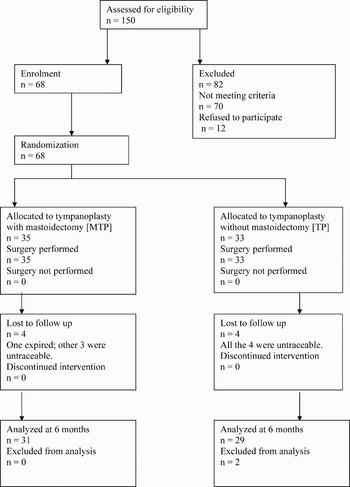
Fig. 1 CONSORT flow chart showing study details.
The inclusion criteria comprised tubotympanic CSOM in the quiescent stage together with the following: (1) more than one month but less than six months elapsed since last ear discharge; (2) wet middle-ear mucosa; and (3) central perforation with congested margins.
The intactness of the ossicular chain was confirmed by otoendoscopy, pure tone audiometry (PTA) with patch test and presence of a round window reflex during surgery.
The exclusion criteria comprised the presence of the following: (1) mucopurulent ear discharge or completely dry ear; (2) granulation tissue, cholesteatoma or polyp in the ear; (3) ossicular pathology; (4) multiple tympanic membrane perforations; (5) subtotal or total perforation of the pars tensa; (6) clinically significant predisposing focus of infection in the nose or throat; (7) complications of otitis media; (8) age below 12 years; (9) mixed hearing loss; and (10) systemic co-morbidities (e.g. diabetes mellitus, hypertension or immunosuppression).
Prospective study candidates were examined and investigated in the ENT out-patient clinic to confirm eligibility. All eligible patients had a normally functioning eustachian tube as tested by impedance audiometry. Pre-operative radiography (X-ray) of the mastoids was performed in all cases. Any predisposing focus in the nose or throat was evaluated by diagnostic nasal endoscopy. Pure tone audiometry with masking was performed with a digital audiometer calibrated to American National Standard Institute (ANSI) standards.
Consent was obtained from the patient for both surgical procedures (i.e. cortical mastoidectomy with type one tympanoplasty, and type one tympanoplasty alone).
Randomisation
Sixty-eight ears were randomly assigned to one of the two surgical groups. Group one (cortical mastoidectomy with type one tympanoplasty) included 35 ears and group two (type one tympanoplasty alone) included 33 ears. Permuted block randomisation of two, four and six block sizes was used. Blocks were divided in three strata – S1, S2 and S3 – in each group, corresponding to the three operating surgeons. All three surgeons were sufficiently experienced and familiar with both procedures. A random sequence of the numbers one and two was generated using Ralloc computer software. Number one corresponded to cortical mastoidectomy with type one tympanoplasty (i.e. group one) and number two to type one tympanoplasty alone (i.e. group two). Patients underwent one of the two surgical treatments as per their computer-generated, random allocation number.
If necessary, randomisation was delayed until patients fulfilled all the eligibility criteria. Eligibility was finally confirmed after a tympanotomy was performed in the operating theatre and the middle-ear findings were established.
Allocation concealment
The computer-generated random numbers were carefully marked onto a paper slip, separately for each of the three surgeons (S1, S2 and S3). This paper slip was then sealed in an opaque, dark envelope which was numbered separately for each surgeon. The numbered envelopes were collected together and tagged separately for each surgeon. These steps were performed by a neutral observer who was not involved in the study.
Surgical procedure and intervention
Most of the surgical procedures were performed under local anaesthesia (cortical mastoidectomy with type one tympanoplasty, n = 29; type one tympanoplasty alone, n = 29). However, general anaesthesia was used in a few patients under 18 years of age (cortical mastoidectomy with type one tympanoplasty, n = 6; type one tympanoplasty alone, n =4). Tympanomeatal flap elevation via a postauricular approach was performed in all cases. The underlay technique with temporalis fascia graft was employed in all cases. The graft was placed under the skeletonised handle of the malleus and tucked anteriorly under the rim of the perforation. The graft was supported by a few pieces of dry Gelfoam (absorbable gelatin sponge; Virchow Biotech Private Limited, Ranga Reddy District , Andhra Pradesh, India) in the middle ear.
In group one (cortical mastoidectomy with type one tympanoplasty) the patency of the aditus was checked and established, and an external tube drain from the mastoid cavity was kept in situ for two days post-operatively.
In group two (type one tympanoplasty alone), the patency of the aditus was not checked, the mastoid antrum was not opened, and no post-operative drains were used.
Post-operative management
All the patients in both groups were treated with the same medications. In group one, the drain was removed on the second post-operative day. Dressing and skin sutures were removed on the seventh post-operative day and the patient was discharged from hospital.
Follow up
Patients were followed up in the ENT out-patient clinic three and six months after surgery. During each follow-up appointment, patients underwent otoendoscopy, PTA and impedance audiometry. Cases of surgical failure (i.e. failure of perforation healing or recurrence of ear discharge, or both) additionally underwent diagnostic nasal endoscopy and culture and sensitivity analysis of an ear swab.
Primary outcomes
These were: (1) eradication of disease, evaluated by presence or absence of recurrent, mucopurulent ear discharge and confirmed by otoendoscopy; (2) closure of tympanic membrane perforation, evaluated by otoendoscopy; and (3) improvement in hearing status, evaluated by PTA.
Secondary outcomes
These were: (1) change in compliance of the tympanic membrane, evaluated by impedance audiometry; (2) external ear canal stenosis, evaluated by measuring the horizontal and vertical dimensions of the ear canal using callipers; and (3) complications of surgery.
This study was completed and reported in accordance with the Revised CONSORT Statement guidelines for evaluating the structure and analysis of randomised, controlled trials.Reference Altman, Schulz, Moher, Egger, Davidoff and Elbourne1
Results
Demographic characteristics
Group one (cortical mastoidectomy with type one tympanoplasty) comprised 19 male and 16 female patients. Patients' ages ranged from 12 to 35 years (mean, 22.91 years).
Group two (type one tympanoplasty alone) comprised 17 male and 16 female patients. Patients' ages ranged from 13 to 52 years (mean, 24.09 years).
Follow-up results
The post-operative results at three and six months for both groups are given in Tables I and II, respectively. Cases were analysed with an intention to treat.
Table I Ear surgery results: third post-operative month

n represents number of ears. Perfn = perforation; MTP = mastoidotympanoplasty; TP = tympanoplasty alone; S = surgeon
Table II Ear surgery results: 6th post-operative month

n represents number of ears. Perfn = perforation; MTP = mastoidotympanoplasty; TP = tympanoplasty alone; S = surgeon
Statistical analysis
The data for both groups were compared statistically using the unpaired t-test, Fisher's exact test and analysis of variance (ANOVA) where appropriate. Statistical significance was assigned to p < 0.05. Outcomes were analysed using the statistical software packages Graphpad Instat version 3 and Minitab version 14.
Outcomes and estimation
The time taken for post-operative healing was the same in both groups.
Primary outcomes
The first primary outcome was perforation closure and disease eradication. Findings at three and six months post-operatively are given in Tables I and II, respectively. Contingency tables (2×2) were prepared to compare the results of the two surgical procedures with respect to each surgeon separately (S1,S2,S3) and combined (S1+S2+S3) at the third and sixth postoperative months (Table III). However, no statistically significant difference was found (p > 0.05) between the results of the two groups for any comparison.
Table III Comparison of the results of two surgical procedures at the 3rd and 6th post operative month*

* Assessed by achievement of primary outcomes, i.e. perforation closure and disease eradication, used, calculated from contingency tables. Mth = month; S = surgeon
The second primary outcome was improvement in hearing status. Patients' pure tone average was calculated by adding the hearing thresholds (air conduction) at 500 Hz and at 1, 2 and 4 kHz and then dividing by four. The improvement in hearing at the third and sixth post-operative month was calculated by subtracting the post-operative pure tone average from the pre-operative pure tone average. No post-operative sensorineural hearing loss was noted in any case.
The effect of stratification on patients' post-operative hearing results was studied using one-way ANOVA. The mean of the pure tone averages was calculated for S1, S2 and S3. The mean difference in the pure tone averages between the three strata, the standard deviation (SD) and the standard error of mean were also calculated for group one (cortical mastoidectomy with type one tympanoplasty) at the third post-operative month. The Tukey–Kramer multiple comparisons test was used to calculate the value of ‘q’. It was found that the variation among the means of the various strata was not significantly greater than that expected by chance (p = 0.8075). Hence, it was concluded that stratification had no effect on the hearing results of cortical mastoidectomy with type one tympanoplasty at three month follow up. Therefore, the means for surgeons one, two and three could be combined and analysed together. The same calculations were repeated for type one tympanoplasty alone at three month follow up (p = 0.7756), for cortical mastoidectomy with type one tympanoplasty at six month follow up (p = 0.6728), and for type one tympanoplasty alone at six month follow up (p = 0.9111). Again, in all these cases stratification was found to have no effect on hearing results, and it was therefore concluded that combined analysis of the hearing results of the three surgeons could be performed.
The mean pure tone averages for S1, S2 and S3 and for S1 + S2 + S3 were assessed at the third post-operative month, comparing cortical mastoidectomy with type one tympanoplasty versus type one tympanoplasty alone (Table IV). The unpaired t-test was used for each comparison, with significance limits set at 95 per cent confidence interval (CI). We found no statistically significant difference between group one and group two, either considering S1, S2 and S3 separately or together (i.e. S1 + S2 + S3). Thus, at the third post-operative month, patients' hearing results did not differ significantly, comparing the two surgical treatments.
Table IV Pure tone averages for MTP and TP patients: 3rd post-operative month

MTP = mastoidotympanoplasty; TP = tympanoplasty alone; PTA = pure tone average; S = surgeon; SD = standard deviation
Table V compares the mean pure tone averages for S1, S2 and S3 and for S1 + S2 + S3 at the sixth post-operative month, comparing cortical mastoidectomy with type one tympanoplasty and type one tympanoplasty alone. The same statistical tests were used. Again, no statistically significant difference between groups one and two was found, either considering S1, S2 and S3 separately or together (i.e. S1 + S2 + S3). Thus, at the sixth post-operative month, patients' hearing results did not differ significantly, comparing the two surgical treatments.
Table V Pure tone averages for MTP and TP patients: 6th post-operative month

MTP = mastoidotympanoplasty; TP = tympanoplasty alone; PTA = pure tone average; S = surgeon; SD = standard deviation
Secondary outcomes
Regarding the first secondary outcome, change in tympanic membrane compliance, a type A curve was observed in 22.87 per cent of group one patients and in 25.80 per cent of group two patients at the third post-operative month. However, at the sixth post-operative month these percentages had risen to 40 and 44.82 per cent, respectively (Figure 2).

Fig. 2 Status of the tympanogram curve at the sixth month follow up.
The second secondary outcome, external ear canal stenosis, was not noted to any significant degree in any patient from either group.
Regarding the third secondary outcome, post-operative complications, two group one patients were noted to have tympanosclerosis post-operatively. No other post-operative complications were noted in any patient of either group.
Discussion
Cortical mastoidectomy is widely performed, along with tympanoplasty, to treat active tubotympanic disease, in order to address mastoid disease when present. While the mastoid reservoir of infection theory holds good in active tubotympanic CSOM, the same may not be true in the quiescent or dry phase when active ear discharge is absent. The question thus arises whether to routinely address the mastoid surgically or not. Even though it is desirable to expose the mastoid antrum in order to confirm the absence of disease there, the procedure itself is not without disadvantages. The addition of mastoidectomy to tympanoplasty carries several disadvantages, such as: increased risk of damage to the incus, dura, sigmoid sinus and facial nerve; prolongation of surgery; and higher morbidity due to bone drilling, especially in the hands of an inexperienced surgeon. The advantages and disadvantages of adding mastoidectomy to tympanoplasty in noncholesteatomatous chronic otitis media have been the focus of much controversy and debate. Previous research findings have provided evidence both for and against the use of mastoidectomy in noncholesteatomatous otitis media (Tables VI and VII). Most of these studies used retrospective case series; very few were prospective, controlled studies. Some authors performed cortical mastoidectomy together with tympanoplasty, especially for discharging ears, while others preferred tympanoplasty alone. Many authors considered mastoidectomy to be an unnecessary and avoidable procedure in many cases of tubotympanic CSOM (Table VII).
Table VI Studies supporting mastoidectomy in noncholesteatomatous otitis media

Post-op = post-operative; mth = months; yr = years; MTP = mastoidotympanoplasty; TP = tympanoplasty; ABG = air–bone gap; CSOM = chronic, suppurative otitis media
Table VII Studies not supporting mastoidectomy in noncholesteatomatous chronic otitis media

Post-op = post-operative; MTP = mastoidotympanoplasty; TP = tympanoplasty; yr = years; mth = months; ABG = air–bone gap; CSOM = chronic, suppurative otitis media; MRSA = methicillin-resistant Staphylococcus aureus; MSSA = methicillin-sensitive S aureus
In our study, follow up of the recruited cases was technically feasible only up to six months post-operatively, due to ethical constraints. This was because, as per the study protocol, no surgical revision of failed cases was permitted in the follow-up period, in order to maintain uniform post-operative outcomes which could be compared among all cases. Any failed cases requiring revision surgery underwent the same after six months' follow up. Medical treatment, however, was not denied to any of these cases over the same period.
Most of the surgical failures in both groups had a reactivated, predisposing focus in the nose or throat. Such predisposing foci included persistent adenoids, sinusitis, nasal polyp, allergic rhinitis and atrophic rhinitis. In failed cases, dormant foci in the nose and throat could have become clinically significant after surgery and could have been responsible for the recurrence of ear disease. Viral upper respiratory tract infections (highly prevalent in developing countries) probably activate these dormant foci. Prophylactic surgery to eliminate such clinically dormant foci prior to study recruitment could not be undertaken in many cases for ethical reasons. Further studies are required to explore and substantiate the contribution of these foci to the recurrence of tubotympanic CSOM. The co-variation between ear infection and tonsillitis, sinusitis and atopic diseases was studied in 1996 by Kverner et al. Reference Kverner, Tambs, Harris, Mair and Magnus14 They found a clustering tendency among the upper respiratory tract infections. A few authors have advocated delay in ear surgery in children because tubal function has been shown to improve with age.Reference Bylander and Tjernstorm15 However, we observed that poor tubal function can persist into adolescence and even young adulthood in many people, especially in low socioeconomic groups, in which the prevalence of upper respiratory tract infections is high. Smith-Vaughan et al. found a significantly higher nasal bacterial load among Australian Aboriginal children, which explained their increased risk of suppurative otitis media.Reference Smith-Vaughan, Byun, Nadakarni, Jacques, Hunter and Halpin16 This study found that the nasal bacterial load of respiratory pathogens was a highly sensitive measure of suppurative otitis media, but had a low specificity. The study demonstrated a significant association between nasal bacterial load and ear disease.Reference Smith-Vaughan, Byun, Nadakarni, Jacques, Hunter and Halpin16 The authors suggested that nasal bacterial load should be reduced in order to control the incidence of suppurative otitis media.Reference Smith-Vaughan, Byun, Nadakarni, Jacques, Hunter and Halpin16
In the present study, we attempted to make the two treatment groups as homogeneous as possible. Paediatric patients were excluded from the study, as predisposing foci in the nose and throat are more active at that age and hence more likely to interfere with the results of surgery.
• The incidence of chronic, suppurative otitis media (CSOM) continues to be high in developing countries, and the demand for corrective surgery is ever-increasing
• It has yet to be confirmed, by a randomised, controlled study, whether cortical mastoidectomy is routinely required in the quiescent stage of this disease
• This randomised, controlled study compared the outcomes of mastoidotympanoplasty and tympanoplasty alone in cases of quiescent, tubotympanic CSOM
• No statistically significant difference was found between the two procedures with respect to hearing improvement, tympanic perforation closure, graft uptake or disease eradication
In the present study, the relatively high failure rates in both treatment groups can probably be attributed to the uniform, homogeneous surgical techniques performed on all the ears, as dictated by the strict study protocol, rather than to the demands of the individual operations. Some authors have reported higher surgical failure rates in cases involving large perforations.Reference Sade, Berco, Brown, Weinberg and Avraham17 This may have been yet another reason for the observed surgical failures. Since there was a large variation in the pre-operative hearing levels of the ears recruited into the study, the same was also found in post-operative hearing levels. This post-operative heterogeneity in hearing levels might have contributed to the overall poor degree of hearing improvement noted after surgery.
Booth et al., in an analysis of failed myringoplasty cases, stated that the pre-operative presence of a ‘dry’ ear did not affect surgical success rates.Reference Booth18 Glasscock et al. reviewed 1556 tympanic membrane graft cases, and they too opined that an ear did not have to be dry to achieve a good result.Reference Glasscock, Jackson, Nissen and Schwaber19 They also stated that the majority of surgical failures occurring six to 12 months after surgery were associated with infection, in contrast to later failures which were not necessarily associated with infection.Reference Glasscock, Jackson, Nissen and Schwaber19
Blakley et al. studied the relationship between pre- and post-operative hearing in 124 patients undergoing tympanoplasty.Reference Blakley, Kim and VanCamp20 They found that poor hearing before surgery was associated with poor healing after surgery, regardless of anatomy. They concluded that, in ears with persistent infection, the hearing outcome after tympanomastoidectomy surgery depended more on pre-operative hearing levels than on the type of tympanoplasty performed.Reference Blakley, Kim and VanCamp20 We too observed that ears with a wider pre-operative air–bone gap fared more poorly after surgery, compared with those with a narrower air–bone gap.
Conclusion
This study found that the addition of cortical mastoidectomy to tympanoplasty for the surgical treatment of quiescent-stage, tubotympanic CSOM was not beneficial in terms of hearing improvement or disease eradication, over a short term follow-up period. Hence, it may not be necessary to explore the mastoids routinely at this stage of the disease. These results could be useful when deciding on the need for mastoid exploration in cases of quiescent, tubotympanic CSOM, especially for the inexperienced surgeon. Analysis of surgical failures revealed the influence of predisposing disease foci in the nose and throat on graft failure and disease recurrence, especially in young adults. Such analysis also revealed the possible activation of dormant, predisposing foci in the nose and throat in adolescence and early adulthood. The degree of post-operative hearing improvement in both our treatment groups was dependent on pre-operative hearing status; the larger the pre-operative air–bone gap, the poorer the reduction in post-operative air–bone gap.


