


Case report
A term male baby was born to a mother of African-American ethnicity who was a carrier of cystic fibrosis mutation via spontaneous vaginal delivery. A fetal echocardiogram obtained during pregnancy demonstrated a right-sided aortic arch. Maternal prenatal labs which included testing for hepatitis B, human immunodeficiency virus, syphilis, and rubella were all negative. The baby did well after birth and had Apgar scores of 8 and 9 at 1 and 5 minutes, respectively. A transthoracic echocardiogram was obtained to clarify the findings on the prenatal echocardiogram and showed retroaortic left innominate vein (Figs 1–3) and a right aortic arch with mirror image branching. The intra-cardiac anatomy was otherwise normal.

Figure 1. The retroaortic innominate vein is seen posterior to the aorta and the pulmonary artery in this suprasternal frontal two-dimensional echocardiographic image.
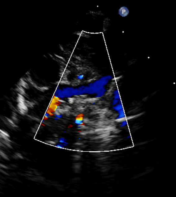
Figure 2. Colour image of the retroaortic innominate vein in the same view.
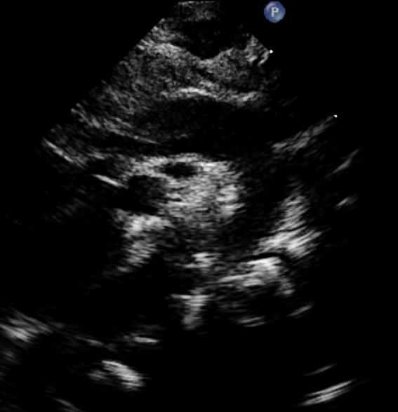
Figure 3. The vein is seen posterior to the aortic arch (along with the right pulmonary artery) in this suprasternal sagittal two-dimensional echocardiographic image.
Discussion
Innominate vein, also known as brachiocephalic vein, is formed by the union of left internal jugular and left subclavian veins, typically courses superoanterior to the aortic arch to join the right innominate vein to form the superior caval vein. A retroaortic innominate vein, courses underneath the aortic arch, posterior to the ascending aorta. It is uncommon with a reported prevalence of 0.55–0.57% in patients with congenital heart disease (CHD). However, a slightly higher prevalence (1.9%) has been reported in a smaller study from the Middle East.Reference Kulkarni, Jain, Kasar, Garekar and Joshi 1 – Reference Corno, Alahdal and Das 3 Associated CHDs include tetralogy of Fallot with or without pulmonary atresia, total anomalous pulmonary venous return, coarctation of aorta, truncus arteriosus, interrupted aortic arch, right atrial isomerism, atrial septal defects, ventricular septal defects (VSDs), pulmonary atresia with VSD, double outlet right ventricle with mitral atresia, and hypoplastic left ventricle.Reference Kulkarni, Jain, Kasar, Garekar and Joshi 1 , Reference Nagashima, Shikata and Okamura 2 , Reference Morhy Borges Leal, Andrade and de Souza 4 – Reference Sukulal, Bijulal and Thakran 8 Aortic arch variants or anomalies such as right-sided aortic arch, high cervical arch, or double aortic arch are frequently associated with this venous anomaly.Reference Kulkarni, Jain, Kasar, Garekar and Joshi 1 , Reference Nagashima, Shikata and Okamura 2 , Reference Chen, Liu, Chen, Chiu, Lee and Wu 6 , Reference Curtil, Tronc and Champsaur 9 , Reference Bartoli, Chagnaud, Moulin, Di Stefano-Louineau, Bory and Kasbarian 10
Isolated retroaortic innominate vein without any associated CHD as seen in our patient is exceedingly rare with a reported prevalence of 0.02%.Reference Nagashima, Shikata and Okamura 2 Consequently, only a handful of patients have been reported so far.Reference Nagashima, Shikata and Okamura 2 , Reference Gülsün, Gökoğlu, Ariyürek, Demirkazik and Hazirolan 11 – Reference Semionov and Kosiuk 14 All of the previously reported patients with isolated retroaortic innominate vein had a left-sided aortic arch. To the best of our knowledge, this is the first patient with isolated retroaortic innominate vein with a right-sided aortic arch.
The exact embryogenesis of this anomaly is not known. Several hypotheses have been postulated including double pre-cardinal anastomoses, with regression of the upper resulting in an anomalous innominate veinReference Adachi 15 and formation of an alternative channel when the normal course of the left innominate vein is obstructed.Reference Minami, Noda and Kawauchi 16 Kim et al postulated that a pre-cardinal anastomosis can be formed in any pathway where space is available after development of the aortic arch.Reference Chen, Liu, Chen, Chiu, Lee and Wu 6 , Reference Kim, Chung and Im 17
Isolated retroaortic innominate vein is usually not of clinical significance. However, it has been mistaken for persistent left superior caval vein,Reference Gerlis and Ho 18 the ascending vertical vein in total anomalous pulmonary venous connection,Reference Minami, Noda and Kawauchi 16 and right pulmonary artery in patients with hypoplastic or atretic pulmonary arteries on echocardiography.Reference Kulkarni, Jain, Kasar, Garekar and Joshi 1 , Reference Chen, Liu, Chen, Chiu, Lee and Wu 6 , Reference Curtil, Tronc and Champsaur 9 , Reference Choi, Jung, Kim, Noh and Yun 19 It is important for the interventional cardiologist or surgeon to be aware of this anomaly as it can present with technical difficulties during cannulation of the superior caval vein and left-sided venous access during procedures such as central venous catheterisation and pacemaker and defibrillator lead placement.Reference Kulkarni, Jain, Kasar, Garekar and Joshi 1 , Reference Chen, Liu, Chen, Chiu, Lee and Wu 6 Problems related to adequate surgical exposure of cardiovascular structures have been reported in patients with CHD and this venous anomaly during Glenn anastomosis, systemic-to-pulmonary artery shunt or patent ductus arteriosus ligation.Reference Kulkarni, Jain, Kasar, Garekar and Joshi 1 , Reference Gerlis and Ho 18 , Reference Mill, Wilcox, Detterbeck and Anderson 20 Retroaortic innominate vein has been used for pulmonary artery reconstruction during cavopulmonary anastomosis and right atrial pulmonary anastomosis in a patient with complex cardiac anomalies such as tricuspid atresia/pulmonary atresia, superior–inferior ventricles, and pulmonary atresia with a single ventricle and atrium; therefore, it is important for the surgeon to be aware of its presence.Reference Agrawal, Krishnan, Kulkarni and Cherian 21 – Reference Nakayama, Itou, Abe and Yoshizumi 23
To conclude, isolated retroaortic innominate vein is an exceedingly rare venous anomaly which has previously not been reported in association with right-sided aortic arch in the absence of cardiac defects. Though usually not of any clinical significance, its presence can present technical difficulties during some cardiac procedures.
Author ORCIDs
Utkarsh Kohli 0000-0003-3410-840X
Financial Support
This research received no specific grant from any funding agency, commercial or not-for-profit sectors.
Conflict of Interest
None.
Ethical Standards
This report does not include any human or animal experimentation.


