Introduction
The nasal septum is one of the main contributors to the structural integrity of the nose. It is often necessary to excise certain parts of the cartilaginous septum in order to obtain grafts or to improve the nasal obstruction caused by septal deviation; however, an adequate amount of the quadrangular cartilage in the dorsocaudal L-strut should be preserved to maintain stability.Reference Killian and Foster1 The L-strut must be strong enough to withstand the forces of gravity, the nasal soft tissue envelope and minor trauma.Reference Paul, Messenger, Yuan, Kwon, Kim and Inman2, Reference Fattahi and Quereshy3
The posterior septal angle is an important landmark during septal surgery. It is the point at which the caudal septum articulates with the anterior nasal spine. In certain surgical techniques (e.g. swinging door technique or extracorporeal septoplasty), the septum is disarticulated from the anterior nasal spine and it needs to be re-attached as part of the surgical procedure.Reference Wu and Hamilton4–Reference Akduman, Haksever and Yanilmaz6 Also, during the septal cartilage harvest for grafts, an adequate amount of the caudal (and dorsal) cartilaginous septum should be left behind for structural support, as the less contact the caudal L-strut has with the maxillary crest, the higher the risk of instability and collapse.Reference Lee, Lee, Ha, Kim and Cho5 The relationship of the anterior nasal spine and posterior septal angle should be observed by the operating surgeon in these situations, as inaccurate repositioning or septal cartilage harvest can result in changes to the nasal profile view, particularly the nasolabial angle and the supratip area, and can lead to reduced structural support.
The senior author (SA) has noted in his surgical experience that there is great variability in the distance between the posterior septal angle and the anterior nasal spine. Knowledge of the average distance between these two points is surgically valuable when performing nasal surgery, especially when measurement of this distance is not possible at the time of surgery (e.g. when repositioning a dislocated septum or reconstructing a new septum). We thus reviewed the literature to see if such information was already available.
A literature search conducted in July 2017 using the Medline search engine with the keyword ‘posterior septal angle’ did not produce any results. Articles with keywords ‘septal anatomy’ and ‘nasal spine’ were reviewed, and there were no studies primarily investigating the anatomical relationship between the posterior septal angle and anterior nasal spine. We therefore undertook a study to measure the distance between the anterior nasal spine and the posterior septal angle.
Materials and methods
We planned to measure the distance between the most anterior part of the anterior nasal spine and the posterior septal angle on sinus computed tomography (CT) scans performed on Caucasian patients being investigated for their sinus symptoms.
A retrospective cross-sectional study was conducted in an urban (inner-city), university affiliated teaching hospital. The hospital served an ethnically diverse population. Sinus CT scans conducted over a three-month period (from February to April 2016) were reviewed.
Data were obtained from the radiology department database, and scans were reviewed on the hospital imaging platform Agfa Impax. All Caucasian patients aged above 18 years were included in the study. Patients who had radiological evidence or documented evidence of previous septal surgery were excluded from the study. Any scans in which the authors were unable to clearly identify the posterior septal angle or anterior nasal spine, or were unable to undertake the measurements, were excluded from the study. Information on ethnicity was collected from the hospital ethnicity data set. Demographic data were also collected for these patients.
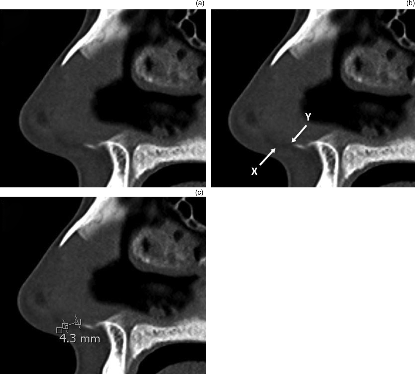
The sagittal CT scan images were reviewed by two authors (DK and SG) independently. The posterior septal angle and the anterior nasal spine were marked out, and the distance between them was then measured digitally. These measurements were made using a digital calliper available on radiology imaging viewer software (Figure 1). The mean measurement of our two raters for each case was used for further analysis.

Fig. 1. (a–c) Sagittal computed tomography images of the nasal septum and the relevant anatomical landmarks. In part (b), ‘X’ indicates the location of the posterior septal angle and ‘Y’ indicates the location of the anterior nasal spine. Part (c) shows the measurement of the anterior nasal spine and posterior septal angle distance (in this case, 4.3 mm) with the digital calliper.
Results
In total, 122 scans were reviewed. Of these scans, 15 (12.3 per cent) were deemed to be inadequate, as either the CT slices were too large (slices were more than 1.00 mm apart) or the image was not clear enough to measure the distance accurately. Seven scans (5.73 per cent) had evidence of previous septal surgery and were thus excluded from the study. As a result, 100 eligible scans were reviewed and their data used for analysis.
Our sample of 100 eligible scans were from 49 male and 51 female patients. Their mean age was 52.5 years (standard deviation (SD) = 16.5 years; range = 22–93 years).
The Shapiro–Wilk test for normality was performed on the distance between the posterior septal angle and the anterior nasal spine (W = 0.990, p = 0.656, 95 per cent confidence interval (CI)= 0.975 to 1.000), which allowed us to assume normality for our data. A paired t-test analysis of the distance between the posterior septal angle and the anterior nasal spine from our two raters showed no significant difference in measurements (t = −0.201, p = 0.841, 95 per cent CI = -0.097 to 0.119). Therefore, the distance between the posterior septal angle and the anterior nasal spine for each scan was obtained from the averaged measurement from our two raters.
The mean distance between the posterior septal angle and the anterior nasal spine for our sample was 5.13 mm (SD = 1.24 mm; range = 1.85–8.00 mm). The mean distance between the posterior septal angle and the anterior nasal spine for males was 5.18 mm (SD = 1.36 mm; range = 1.85–8.00 mm) and for females the mean was 5.07 mm (SD = 1.13 mm; range = 2.80–7.30 mm). However, a t-test analysis showed that this was not significantly different (t =0.450, p = 0.654, 95 per cent CI = -0.382 to 0.606) (Table 1).
Table 1. Distance between posterior septal angle and anterior nasal spine

*n = 49; †n = 51; ‡n = 100. SD = standard deviation
Discussion
The relation of the anterior nasal spine to the posterior septal angle is an important factor to consider when undertaking septal surgery. There are a variety of techniques described in the literature regarding caudal septal surgery. However, it is generally accepted that to maintain the structural integrity of the nose, it is important to leave an adequate dorsocaudal L–strut. It has traditionally been suggested that at least approximately 10 mm of the caudal septum should be left behind to maintain stability.Reference Lee, Lee, Ha, Kim and Cho5
The two surgically relevant key points of septum fixation are located at the confluence with the nasal bones, at the keystone region, and at the interface of the anterior nasal spine and the posterior septal angle. Traditional teaching states that the posterior septal angle rests on the anterior nasal spine. However, our study has shown that the two points often do not articulate at one point; the posterior septal angle sits proud of the anterior nasal spine by a few millimetres (i.e. on average by 5.13 mm, with a range of 1.85–8.00 mm). This finding has significant clinical implications when the surgeon reattaches the caudal septum to the nasal spine or harvests the graft and leaves a caudal strut behind. Incorrect repositioning of the caudal septum will result in changes to the profile view, especially in extracorporeal septoplasty. Preserving the relationship of these landmarks will prevent unwanted changes to the nasolabial angle and supratip area, and prevent columellar retraction.
During septal cartilage harvest, the distance between the posterior septal angle and the anterior nasal spine should be taken into account, so that enough contact is preserved between the caudal septal strut and the maxillary crest (including the anterior nasal spine). The traditional view that 10 mm of the caudal septum is left behind was expanded on by Lee et al., who suggested that the 10 mm caudal strut needs at least 45 per cent contact with the anterior nasal spine to be able to stabilise the septum.Reference Lee, Lee, Ha, Kim and Cho5 As shown in our study, often a large portion of the caudal septum, up to 8 mm, may not be resting on the anterior nasal spine. In these cases, leaving a 10 mm caudal strut may not be adequate, as the contact between the septal cartilage caudal strut and the nasal spine could be only a few millimetres.
• The posterior septal angle is part of the caudal septum that articulates with the anterior nasal spine
• This study demonstrates that the posterior septal angle sits proud of the anterior nasal spine
• The mean distance between the posterior septal angle and anterior nasal spine was 5.13 mm (standard deviation = 1.24 mm; range = 1.85–8.00 mm)
• The caudal septum amount unsupported by the anterior nasal spine should be considered when judging the caudal L-strut width to be preserved
• Inaccurate caudal septum to anterior nasal spine reattachment (e.g. in extracorporeal septoplasty) will result in unwanted profile view changes (e.g. columella retraction)
Our study used CT imaging to assess the distance of the anterior nasal spine to the posterior septal angle in patients who underwent a sinus CT for other indications, as it is not routine practice in the UK to obtain pre-operative imaging for septal surgery. The authors of this study recommend that rhinoplastic surgeons should observe the relationship and the distance between the anterior nasal spine and the posterior septal angle in each individual case intra-operatively. These anatomical landmarks become clear, especially once flaps are raised and the septum is exposed. Surgeons may wish to measure this distance if they need to detach and re-attach the septum from the spine (e.g. during extracorporeal septoplasty) so that reinsertion can be conducted accurately, without resulting in unintended changes in the patient's profile view (e.g. columella retraction or supratip depression). In addition, surgeons should be aware that some portion of the caudal septum may not be resting on the nasal spine; they should harvest the graft in such a way that an adequate segment of the caudal septum remains resting on the anterior nasal spine, to provide support and avoid instability.
Conclusion
As seen in our study, there is great variability in the distance between the anterior nasal spine and the posterior septal angle. Being aware of this variability in anatomy is vital to maintain stability and to avoid unwanted aesthetic changes when performing septal cartilage harvest and caudal septal reattachment and repositioning.
Competing interests
None declared


