


Introduction
Cystic echinococcosis (CE) is a cyclo-zoonotic disease of global importance that is caused by the larval stage of the dog tapeworm, Echinococcus granulosus sensu lato (s.l.). CE occurs as an infection in intermediate hosts, the herbivores and omnivores, and man as a ‘dead-end host’. Dogs, other canids and felids are the definitive hosts that harbour the adult cestode in the small intestines (Thompson, Reference Thompson2017). CE occurs worldwide and is of great public health and socioeconomic importance in pastoral communities (Deplazes et al., Reference Deplazes, Rinaldi and Alvarez Rojas2017). The burden of CE in humans is estimated to be between one and three million disability-adjusted life years, and, according to the World Health Organization, up to three billion US dollars is spent annually on the treatment of human CE cases and due to losses incurred in livestock industry (WHO, 2015).
Molecular phylogeny studies have classified E. granulosus s.l. into five species – namely, Echinococcus granulosus sensu stricto (s.s.) (G1, G3), Echinococcus equinus (G4), Echinococcus ortleppi (G5), Echinococcus canadensis (G6–8, 10) and Echinococcus felidis (Nakao et al., Reference Nakao, Lavikainen, Yanagida and Ito2013a, Reference Nakao, Yanagida, Konyaev, Lavikainen, Odnokurtsev, Zaikov and Itob). All these CE agents besides E. canadensis (G8 and G10) have been detected in Kenya (Romig et al., Reference Romig, Deplazes, Jenkins, Giraudoux, Massolo, Craig, Wassermann, Takahashi and de la Rue2017). The epidemiology of CE is well established in the traditionally known endemic areas of Turkana and Maasailand in Kenya (Dinkel et al., Reference Dinkel, Njoroge, Zimmermann, Walz, Zeyhle, Elmahdi, Mackenstedt and Romig2004; Addy et al., Reference Addy, Alakonya and Wamae2012; Mulinge et al., Reference Mulinge, Magambo and Odongo2018; Odongo et al., Reference Odongo, Tiampati and Mulinge2018; Nungari et al., Reference Nungari, Mbae, Gikunju, Mulinge, Kaburu, Zeyhle and Magambo2019). Recent studies outside the known endemic areas have highlighted the differences in prevalence of CE, fertile cysts vs. calcified cysts, the most important intermediate host and the diversity of Echinococcus spp. (Mbaya et al., Reference Mbaya, Magambo, Njenga, Zeyhle, Mbae, Mulinge, Wassermann, Kern and Romig2014; Gachengo et al., Reference Gachengo, Kikuvi, Mulinge, Zeyhle and Mbae2017; Kere et al., Reference Kere, Joseph, Jessika and Maina2019) – for example, the abundance of E. ortleppi in livestock from Meru and Isiolo (Mbaya et al., Reference Mbaya, Magambo, Njenga, Zeyhle, Mbae, Mulinge, Wassermann, Kern and Romig2014) and E. canadensis (G6/7) in dogs from Turkana (Mulinge et al., Reference Mulinge, Magambo and Odongo2018), despite being rare in characterized humans cases (Dinkel et al., Reference Dinkel, Njoroge, Zimmermann, Walz, Zeyhle, Elmahdi, Mackenstedt and Romig2004; Casulli et al., Reference Casulli, Zeyhle, Brunetti, Pozio, Meroni, Genco and Filice2010; Mutwiri et al., Reference Mutwiri, Magambo, Zeyhle, Mkoji, Wamae, Mulinge, Wassermann, Kern and Romig2013). The differences could be due to sample size, cultural, slaughtering and livestock husbandry practices among the respective communities. Therefore, there is a need for further studies on CE in other regions outside the known endemic areas to understand the epidemiology of CE in those areas.
The epidemiology of CE in Garissa and Wajir counties is poorly understood due to lack of data, while some baseline data on livestock and dog infection do exist from Isiolo county (Mbaya et al., Reference Mbaya, Magambo, Njenga, Zeyhle, Mbae, Mulinge, Wassermann, Kern and Romig2014; Mulinge et al., Reference Mulinge, Magambo and Odongo2018). Garissa, Isiolo and Wajir counties are arid and semi-arid areas in north-eastern Kenya, whose residents largely depend on livestock keeping for their livelihood. These regions are characterized by the presence of many stray dogs, informal and home slaughtering of livestock and cultural conditions that facilitate CE transmission. The present study provides baseline data on the presence of species causing CE in livestock in Isiolo, Garissa and Wajir counties of Kenya.
Materials and methods
Study area
The study was undertaken in Isiolo, Garissa and Wajir counties, which are located in the north-eastern part of Kenya (fig. 1). The counties have a predominantly semi-arid and hot desert climate, with ample sunshine all through the year and a large land area with sparse vegetation. Rain falls infrequently and quite sporadically. Most regions are dry and receive less than 150 mm of rainfall annually, while the mean annual temperature is 28°C (KNBS, 2009). The study counties are predominantly rural and characterized by a large number of pastoralists, with the main economic activity being livestock production.

Fig. 1. Map of Kenya showing the study counties.
Collection and identification of hydatid cysts
During the months of October and November 2018, daily abattoir visits were made for one week in the main abattoirs per county in Isiolo, Garissa and Wajir, where carcasses and organs of cattle, sheep, goats and camels were inspected visually and physically through palpation and subsequent incision to establish presence of cysts. Data on total number of slaughtered animals, species, number infected with hydatid cysts and location of the cysts were recorded. Each cyst was labelled and stored separately in 70% ethanol in a plastic container while awaiting transportation to the Kenya Medical Research Institute (KEMRI), parasitology laboratory in Nairobi, for examination and further analysis.
Microscopic examination
The contents of all cysts were examined microscopically at 40× magnification for presence of protoscoleces. Fertile cysts were those with protoscoleces, while non-fertile cysts lacked protoscoleces or were calcified.
Deoxyribonucleic acid extraction
Crude DNA (lysates) were obtained by lysing of single protoscolex or cyst material in 0.02 M sodium hydroxide at 99°C for 10 min. In instances where the aforementioned process failed to yield sufficient DNA, extraction was performed on cyst material using the DNeasy Blood and Tissue kit (Qiagen, Hilden, Germany) following the manufacturer's guidelines.
Polymerase chain reaction (PCR) and restriction fragment length polymorphism
A nested PCR was undertaken to amplify the NADH dehydrogenase subunit 1 (nad1) as previously described by Hüttner et al. (Reference Hüttner, Nakao and Wassermann2008). In both PCR assays, the reaction mixture contained 2 μl of DNA, 1× DreamTaq™ Green Buffer (Thermo Fisher Scientific, Waltham, MA, USA) (20 mm Tris (hydroxymethyl) aminomethane hydrochloride (pH 8.0), 1 mm dithiothreitol, 0.1 mm ethylenediaminetetraacetic acid, 100 mm Potassium chloride, 0.5% (v/v) Nonidet P40 (Thermo Fisher Scientific, Waltham, MA, USA), 0.5% (v/v) Tween 20 (Thermo Fisher Scientific, Waltham, MA, USA), 0.2 mm deoxyribonucleotide triphosphates, 0.25 μM of forward and reverse primers, 2 mm magnesium chloride and 0.625 units of DreamTaq™ Green DNA Polymerase (Thermo Fisher Scientific) in 25 μl final volume. The conditions for amplification included prior denaturation at 94oC for 5 min, followed by 40 cycles of denaturation at 94 oC for 30 s, annealing at 55°C for 30 s and elongation at 72oC for 1 min, followed by final elongation at 72°C lasting 5 min. The nad1 amplicons were digested, as earlier reported by Hüttner et al. (Reference Hüttner, Siefert, Mackenstedt and Romig2009), using the restriction enzyme, HphI (New England Biolabs, Ipswich, MA, USA). The total reaction mixture was 20 μl, including 7.5 μl distilled water, 2.0 μl of 10× CutSmart Buffer (New England Biolabs’, Ipswich, MA, USA), 0.5 μl HphI (five units) and 10 μl PCR product. The reaction mixture was incubated overnight at 37 oC and then separated on 3% agarose gel. From the samples, the Echinococcus species were detected by contrasting their banding patterns to those of already established patterns according to Hüttner et al. (Reference Hüttner, Siefert, Mackenstedt and Romig2009). DNA sequencing was carried out on the nad1 gene for samples whose restriction banding patterns were not identified. The secondary PCR product was first purified in accordance with the QIAquick PCR purification kit (Qiagen, Hilden, Germany) manual following the manufacturer's guidelines. The purified samples were carefully labelled and sent to Inqaba Biotec Laboratory, South Africa, for sequencing using the nested reverse primer.
Statistical data analysis
Data were first entered into MS Excel (Microsoft Inc., Sacramento, California, USA) and then imported to STATA 15.1 (StataCorp LLC, College station, Texas, USA) for analysis. Initially, the data were checked for accuracy, coded and analysed using descriptive statistics. Proportions were determined for categorical variables and presented as a percentage of the overall number, along with a 95% confidence interval, where applicable. Univariable analysis using simple logistic regression was performed to determine unconditional associations between animal county and species with the presence of E. granulosus. A multivariable model with county of origin and animal species was built to allow estimation of coefficient. The area under the curve of the receiver operating characteristic was used to evaluate the overall performance of the model. Echinococcus and Taenia species sequences were carefully edited using GENtle version 1.9.4 (http://gentle.magnusmanske.de). The species were identified by comparing sequences with those available in the National Centre for Biotechnology Information database (NCBI) using the basic local alignment search tool (BLAST) (http://www.ncbi.nlm.nih.gov/BLAST/; Altschul et al., Reference Altschul, Madden, Schaffer, Zhang, Zhang, Miller and Lipman1997).
Results
Prevalence of CE in livestock in Isiolo, Garissa and Wajir counties
A total of 1368 carcasses were inspected in the three counties. The overall CE proportions per inspected livestock species were camels 29.1% (68/234), goats 9.9% (68/687), sheep 8.2% (27/329) and cattle 14.4% (17/118) (table 1). In Isiolo county, the proportions were 58% in camels (36/62), 15.3% in goats (39/255), 16.9% in sheep (10/59) and 17.2% in cattle (16/93). Garissa county recorded a species proportion of 10.4% in camels (13/125), 8.4% in goats (26/310), 3.6% in sheep (8/222) and 4.7% in cattle (1/21). In Wajir county, the prevalence rates were 40.4% in camels (19/47), 2.5% in goats (3/122), 18.8% in sheep (9/48) and 0% in cattle (0/4).
Table 1. Proportion of CE and predilection site in the livestock species in Isiolo, Garissa and Wajir counties.
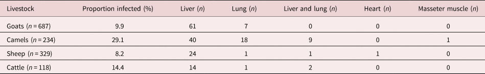
n, number of animals.
Location, number and viability of the cysts
A total of 295 cysts were recovered from 180 infected animals, and the liver was the major predilection site for the cysts with 72.9% (215/295), followed by the lungs at 26.4% (78/295), with the heart and masseter muscle each having a single cyst (table 1). The number of cysts per animal largely varied among the sampled livestock, ranging between one and 11 cysts (table 2). Fertile cysts 14/295 (4.7%) were found only in the lungs of camels from Isiolo county. The majority of the cysts were calcified 244/295 (82.7%) and these included all the cysts from sheep (table 3). The cysts identified were generally ovoid in shape with varying diameters, ranging between 2.5 mm and 10 cm.
Table 2. Number of recovered cysts per species in Isiolo, Garissa and Wajir counties.

Table 3. Conditions of cyst and Echinococcus/Taenia species from livestock isolated in Isiolo, Garissa and Wajir counties.

Genotyping of cyst materials/protoscoleces
Genomic DNA or cyst lysate was obtained from 250 of 295 cysts, while the remaining 45 cysts were excluded for not being appropriate for DNA extraction. Crude DNA was obtained from protoscoleces originating from 22 samples and from cyst wall material of the remaining 228 samples, and included DNA extraction using the DNeasy Blood and Tissue kit (Qiagen, Hilden, Germany) being undertaken on 195 cyst samples. Of the cysts tested by nested PCR, only 139 (55.6%) were PCR positive and, hence, genotyped up to the species level. Two species each of E. granulosus s.l. and Taenia were found. A total of 79.9% (111/139) cysts were identified as E. canadensis (G6/7) and 14.4% (20/139) as E. granulosus s.s. Echinococcus granulosus s.s. was found mainly in goats and cattle from Isiolo county, but was rare in all the species from Garissa and Wajir counties. Echinococcus canadensis (G6/7) was the most common species in livestock in all three counties, the only species in sheep and predominantly in camels (table 3). A total of 11 isolates were sequenced and included eight isolates with unknown restriction patterns whose representative sequences were deposited in GenBank. The E. granulosus s.s. isolate with accession number MT525963 showed 99.78% identity to reference sequence EF367333, cattle isolate from Morocco (Azlaf et al., unpublished data). Echinococcus canadensis (G6/7) sequences with accession numbers MT525964–MT525967 were 99.89–100% identical to reference sequences KX010873, KX010875 and KX010879 previously derived from cattle, goats and camels in Kenya (Addy et al., Reference Addy, Wassermann and Kagendo2017). Three cysts (3/139) were identified as Taenia spp. One was Taenia saginata from a calcified liver cyst of cattle from Isiolo, with accession number MT535753, which showed 99.55% identity to the reference sequence AY684274 from Korea (Jeon et al., Reference Jeon, Kim and Eom2007). The other two Taenia spp., with accession numbers MT535754 and MT535755, were 99.66% identical to the reference sequence AB905200 from hyena in Ethiopia (Terefe et al., Reference Terefe, Hailemariam, Menkir, Nakao, Lavikainen, Haukisalmi, Iwaki, Okamoto and Ito2014). Both Taenia spp. isolates were calcified and located in camel liver from Garissa and Wajir, one of which occurred as co-infection with E. canadensis (G6/7). PCR products from 5/139 cysts could not be genotyped due to low yield of amplicon.
Risk factors for CE in livestock
The variables that had significant association between positivity to E. granulosus s.l. infection in the final multivariable analysis were county of animal origin and animal species (table 4). The odds of an animal being positive for E. granulosus s.l. were lower for Garissa and Wajir counties as compared with Isiolo county. The odds of being positive for E. granulosus s.l. were higher for camels as compared to cattle, sheep and goats.
Table 4. Multivariable logistic regression association of cystic echinococcosis with animal species and county of origin in Isiolo, Garissa and Wajir counties.

a Overall P-value. CI, confidence interval.
Discussion
The results of this study confirmed the presence of CE for the first time in the counties of Garissa and Wajir, and the current situation in Isiolo county. The proportions of CE from this study – 29.1% in camels, 9.9% in goats, 8.2% in sheep and 14.4% in cattle – were significantly higher than those of Mbaya et al. (Reference Mbaya, Magambo, Njenga, Zeyhle, Mbae, Mulinge, Wassermann, Kern and Romig2014) in their survey of Isiolo and Meru counties, where the prevalence were 6.9% in camels, 0.4% in goats, 4.6% in sheep and 1.9% in cattle. However, this study is comparable to that of Addy et al. (Reference Addy, Alakonya and Wamae2012) and Nungari et al. (Reference Nungari, Mbae, Gikunju, Mulinge, Kaburu, Zeyhle and Magambo2019) from Maasailand, where infection rates were 10.8% goats, 16.5% in sheep and 25.8% in cattle, and 15.2% in goats, 14.9% in sheep and 14.2% in cattle, respectively. The findings that camels were the most infected species in this study confirms previous reports in Sudan (Elmahdi et al., Reference Elmahdi, Ali, Magzoub, Ibrahim, Saad and Romig2004; Omer et al., Reference Omer, Dinkel, Romig, Mackenstedt, Elnahas, Aradaib, Ahmed, Elmalik and Adam2010; Ibrahim et al., Reference Ibrahim, Thomas, Peter and Omer2011) and Algeria (Bardonnet et al., Reference Bardonnet, Benchikh-Elfegoun, Bart, Harraga, Hannache, Haddad, Dumon, Vuitton and Piarroux2003). Livestock in Isiolo county were more likely to be infected than those in Garissa and Wajir counties. This could be because of the arid climatic conditions that characterize Garissa and Wajir counties coupled with high environmental temperatures and low vegetation cover as compared to Isiolo county. Under similar environmental conditions, camels were more likely to be parasitized compared to the other livestock species. Camels had the highest CE prevalence rate, with the greatest number of fertile cysts and an odds ratio of about 12 for infection compared to sheep and goats, which had odds ratios of 0.89 and 0.51, respectively. The findings of this study simulate those of another study in Isiolo and Meru counties (Mbaya et al., Reference Mbaya, Magambo, Njenga, Zeyhle, Mbae, Mulinge, Wassermann, Kern and Romig2014) and elsewhere, as camels are usually slaughtered at an advanced age and, therefore, acquire and develop CE over a period of time (Elmahdi et al., Reference Elmahdi, Ali, Magzoub, Ibrahim, Saad and Romig2004). The analysis of comparable sample sizes from these counties is necessary to confirm with certainty the differences in the three counties.
Hydatid cysts have been reported in various internal organs, including the lungs, liver, kidney and heart (Varcasia et al., Reference Varcasia, Canu, Kogkos, Pipia, Scala, Garippa and Seimenis2007; Addy et al., Reference Addy, Alakonya and Wamae2012). The majority of the cysts in this study were isolated from the liver, which is common since the liver is the first organ encountered by the migrating oncosphere after leaving the intestines. This finding agrees with observations in Maasailand, Kenya (Addy et al., Reference Addy, Alakonya and Wamae2012; Odongo et al., Reference Odongo, Tiampati and Mulinge2018; Nungari et al., Reference Nungari, Mbae, Gikunju, Mulinge, Kaburu, Zeyhle and Magambo2019) and in Sudan (Elmahdi et al., Reference Elmahdi, Ali, Magzoub, Ibrahim, Saad and Romig2004). These results, however, differed with reports from other regions in Kenya that found the lung as the most parasitized organ (Njoroge et al., Reference Njoroge, Mbithi, Gathuma, Wachira, Gathura, Magambo and Zeyhle2002; Kere et al., Reference Kere, Joseph, Jessika and Maina2019). There was multiple organ infection of mostly the liver and lungs, with one cyst recovered from the heart of a sheep and another from the masseter muscle of a camel. The multiple organ infection signifies the huge economic losses accrued from the condemnation of the organs at slaughter. The multiple organ infection also details the need of meat inspectors to be keen in conducting a thorough inspection of all internal organs and the carcass in general.
Based on the fertility rates of cysts, it can be postulated that camels are potential intermediate hosts that facilitate the transmission cycle of CE in the north-eastern region of Kenya. All cysts from the other livestock species were non-fertile and mostly calcified. The role of camels in the transmission of CE is difficult to explain as they are rarely slaughtered at home compared to small ruminants. However, transmission could have been due to the improper disposal of offal at abattoirs/slaughter slabs that was observed in this region and has been reported in Turkana and Maasailand (Mulinge et al., Reference Mulinge, Magambo and Odongo2018). All cysts recovered from sheep were calcified possibly due to the rare occurrence of E. granulosus s.s, a trend contrary to other studies in Kenya (Mbaya et al., Reference Mbaya, Magambo, Njenga, Zeyhle, Mbae, Mulinge, Wassermann, Kern and Romig2014; Gachengo et al., Reference Gachengo, Kikuvi, Mulinge, Zeyhle and Mbae2017; Odongo et al., Reference Odongo, Tiampati and Mulinge2018; Nungari et al., Reference Nungari, Mbae, Gikunju, Mulinge, Kaburu, Zeyhle and Magambo2019) that reported high cyst fertility in sheep. Indeed, calcified cysts accounted for 82.7% of all samples in this study – a common trend in recent studies in Kenya – 32.9% in Maasailand (Nungari et al., Reference Nungari, Mbae, Gikunju, Mulinge, Kaburu, Zeyhle and Magambo2019) and 80% in Turkana (Zeyhle, unpublished data). The sampling procedure of cysts employed in recent studies in Kenya is responsible for the characterization of many calcified cysts; however, the cause of calcification remains unknown (Nungari et al., Reference Nungari, Mbae, Gikunju, Mulinge, Kaburu, Zeyhle and Magambo2019).
Echinococcus canadensis (G6/7) was the most frequent species isolated, which is contrary to previous studies in Kenya (Dinkel et al., Reference Dinkel, Njoroge, Zimmermann, Walz, Zeyhle, Elmahdi, Mackenstedt and Romig2004; Addy et al., Reference Addy, Alakonya and Wamae2012; Mbaya et al., Reference Mbaya, Magambo, Njenga, Zeyhle, Mbae, Mulinge, Wassermann, Kern and Romig2014; Odongo et al., Reference Odongo, Tiampati and Mulinge2018; Nungari et al., Reference Nungari, Mbae, Gikunju, Mulinge, Kaburu, Zeyhle and Magambo2019). However, it should be noted that 75% of the genotyped cysts originated from camels, the primary intermediate host for E. canadensis (G6/7), and confirms the findings of Mbaya et al. (Reference Mbaya, Magambo, Njenga, Zeyhle, Mbae, Mulinge, Wassermann, Kern and Romig2014) where 36 out of 39 camel's cysts belonged to E. canadensis (G6/7). In this study, the proportion of camel cysts that belonged to E. canadensis was 94.5%. Although goats are considered important in the maintenance of the life cycle of E. canadensis (G6/7), especially in the absence of camels (Varcasia et al., Reference Varcasia, Canu, Kogkos, Pipia, Scala, Garippa and Seimenis2007; Addy et al., Reference Addy, Alakonya and Wamae2012), in this study goats infected with E. canadensis (G6/7) were almost equal in number to those infected with the E. granulosus s.s. The calcification of cysts and low prevalence of CE in goats in the current and a previous study in Isiolo (Mbaya et al., Reference Mbaya, Magambo, Njenga, Zeyhle, Mbae, Mulinge, Wassermann, Kern and Romig2014) indicate that goats may be playing a minor role in the maintenance of CE in these areas. Moreover, all the cysts from sheep in this study were E. canadensis (G6/7) and calcified, despite examining more sheep (329) compared to the previous study (65) (Mbaya et al., Reference Mbaya, Magambo, Njenga, Zeyhle, Mbae, Mulinge, Wassermann, Kern and Romig2014). These findings confirm the suggestion that sheep are not suitable hosts for E. canadensis (G6/7) (Dinkel et al., Reference Dinkel, Njoroge, Zimmermann, Walz, Zeyhle, Elmahdi, Mackenstedt and Romig2004; Varcasia et al., Reference Varcasia, Canu, Kogkos, Pipia, Scala, Garippa and Seimenis2007; Omer et al., Reference Omer, Dinkel, Romig, Mackenstedt, Elnahas, Aradaib, Ahmed, Elmalik and Adam2010).
The most notable difference between this study and those of high-prevalence regions is the rare occurrence of E. granulosus s.s. Although previous studies in Kenya have confirmed sheep as the main intermediate host of E. granulosus s.s. where cysts attained high fertility rates (Dinkel et al., Reference Dinkel, Njoroge, Zimmermann, Walz, Zeyhle, Elmahdi, Mackenstedt and Romig2004; Addy et al., Reference Addy, Alakonya and Wamae2012; Odongo et al., Reference Odongo, Tiampati and Mulinge2018; Nungari et al., Reference Nungari, Mbae, Gikunju, Mulinge, Kaburu, Zeyhle and Magambo2019), in this study, no cyst from sheep presented this species. The analysis of more cysts from sheep in this region is required. In cattle, the majority of the cysts recovered were of E. granulosus s.s. and all were non-fertile, showing that cattle are poor hosts for this species. Echinococcus ortleppi is known to occur in cattle and has been reported in Kenya in cattle, goats and sheep (Addy et al., Reference Addy, Alakonya and Wamae2012; Mbaya et al., Reference Mbaya, Magambo, Njenga, Zeyhle, Mbae, Mulinge, Wassermann, Kern and Romig2014; Nungari et al., Reference Nungari, Mbae, Gikunju, Mulinge, Kaburu, Zeyhle and Magambo2019). None of the samples from this study presented this species, although the low number of cattle sampled does not permit further conclusions.
The Taenia species found in this study were calcified and could not be differentiated morphologically from hydatid cysts. Bovine cysticercosis is rare in Kenya, even in areas with high CE prevalence (Zeyhle, pers. comm.). In Isiolo, a prevalence of 4.95%, 30.69% and 6.93% was reported in cattle by meat inspection, antigen and antibody enzyme-linked immunosorbent assay (ELISA), respectively (Onyango-Abuje et al., Reference Onyango-Abuje, Nginyi, Rugutt, Wright, Lumumba, Hughes and Harrison1996). Similarly, a high prevalence of cysticercosis by antigen ELISA was observed in western Kenya, possibly due to cross-reaction with other common Taenia species such as Taenia hydatigena (Fèvre et al., Reference Fèvre, de Glanville, Thomas, Cook, Kariuki and Wamae2017). An unknown Taenia spp. was identified in two camels and was 99.66% identical to a Taenia spp. isolated from hyena in Ethiopia (Terefe et al., Reference Terefe, Hailemariam, Menkir, Nakao, Lavikainen, Haukisalmi, Iwaki, Okamoto and Ito2014). Although this Taenia spp. was first isolated from hyena, it was found to be phylogenetically close to Taenia solium. The host range of this Taenia spp. and its pathogenicity to humans remains unknown. Domesticated animals were suggested as possible prey for hyena in Ethiopia (Terefe et al., Reference Terefe, Hailemariam, Menkir, Nakao, Lavikainen, Haukisalmi, Iwaki, Okamoto and Ito2014); however, the involvement of camels in the transmission of this species is not clear as both cysts were calcified. Further studies to understand the epidemiology of this Taenia spp. are required.
In conclusion, this study reports, for the first time, the characterization of Echinococcus species in livestock from Garissa and Wajir counties, and the current situation in Isiolo county, Kenya. The fertility of cysts in camels and dominance of E. canadensis (G6/7) highlights the importance of camels in the maintenance of CE in livestock in the north-eastern counties of Kenya. This study provides an opportunity for further studies in humans and domestic dogs to understand fully the epidemiology of CE in these regions.
Acknowledgements
We are grateful to Michael Mugo and Lucy Nungari of KEMRI for their technical assistance during the study. We acknowledge the respective County Directors of Veterinary Services and meat inspectors for their help in selecting slaughterhouses and collecting cysts. This work is published with permission from the Director General, KEMRI.
Financial support
This work was implemented under the One Health Research, Education and Outreach Centre funded by the Federal Ministry of Economic Cooperation and Development (BMZ) (grant number BMZ002). Part of the funding was provided by the CGIAR Research Program on Agriculture for Nutrition and Health led by the International Food Policy Research Institute.
Conflicts of interest
None.
Ethical approval
The study complies with the University of Nairobi regulations. This study was approved by the Department of Veterinary Services, Kenya, and permission was obtained from the Directors of Veterinary Services of Isiolo, Garissa and Wajir counties, Kenya.


