


Introduction
Robert Koch discovered the tubercular bacilli on 24 March 1888.Reference Shimao1 Today, tuberculosis is still a major, world-wide public health problem, resulting in over 8.8 million new cases and nearly two million deaths per year.Reference Okada and Kobayashi2
Tuberculosis has been known since antiquity. During the Middle Ages in England and France, the touch of the sovereign was thought to be curative. Today, the problem is more prevalent in developing countries, although there has been a steady increase of cases in the developed world, due to increased immigration from endemic countries, increased levels of human immunodeficiency virus (HIV) infection, worsening urban and social conditions, and the abandonment of tight control programmes.Reference Barnett and Medzon3 In the United States, there was a decline in the disease until 1980 but a resurgence thereafter, seeming to coincide with an increase in HIV infections. Since this time, tuberculosis has become a new clinical entity due to its changing patterns of clinical presentation and dissemination, with increasing prevalence of extrapulmonary forms.Reference Fanlo and Tiberio4, Reference Golden and Vikram5
Extrapulmonary tuberculosis can affect almost any organ of the body, with cervical lymphadenitis, or scrofula, being the commonest form.
The present study included a large series of patients with cervical mycobacterial lymphadenopathy. It aimed to determine the relative contribution of tubercular lymphadenitis as a cause of persistent cervical adenopathy, and also to document the disease's clinical presentation and the appropriate diagnostic investigation and treatment modalities.
Patients and methods
A total of 1827 cases of cervical lymphadenopathy were screened, and 893 patients with tubercular cervical lymphadenitis were evaluated. All patients had visited our surgery, ENT or paediatrics out-patients departments over a three-year period from June 2004 to May 2007.
After a detailed clinical history and examination, all patients underwent fine needle aspiration cytology (FNAC) of involved nodes. Excisional biopsy was performed in 35 patients in whom FNAC was negative on two occasions despite clinical suspicion of tuberculosis. All patients underwent routine investigation, including haemogram (total leucocyte count (TLC) and differential leucocyte count (DLC)) with erythrocyte sedimentation rate (ESR), Mantoux test and chest radiography. Sputum analysis for acid-fast bacilli was performed in 402 cases. Ziehl–Neelson staining of aspirated material or excised nodes was performed in 394 cases.
Polymerase chain reaction analysis of either excised nodes or aspirated material was performed in 136 cases. Ultrasonography of the neck was performed in 102 cases. Computed tomography (CT) of the neck was performed in 18 patients suspected of scrofula.
Results
Out of a total of 1827 patients with cervical lymphadenopathy, 893 (48.87 per cent) cases were of tubercular origin. Other causes of cervical lymphadenopathy were non-specific inflammation (in 571; 31.20 per cent), metastases (in 202; 11.05 per cent) and various other causes (in 161; 8.81 per cent).
The ages of these 893 patients ranged from 10 months to 74 years, with a mean age of 20.2 years. The commonest age group affected was 11–20 years (406 patients; 45.46 per cent), followed by 21–30 years (282 patients; 31.57 per cent). There were 371 males and 522 females, with a male to female ratio of 1:1.4.
The main symptomatology was presentation with cervical lymphadenopathy, without any constitutional or other symptoms of tuberculosis (Table I).
Table I Symptomatology of patients with tubercular cervical lymphadenopathy

*With or without expectoration.
The commonest presentation was posterior triangle, unilateral, matted nodes with an average size of 3 × 3 cm. An accompanying neck abscess was also evident in 4 per cent of cases (Figure 1).
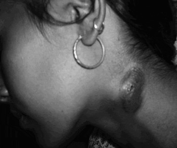
Fig. 1 Clinical photograph of a girl with tubercular cervical abscess and sinus, with underlying lymphadenopathy.
On cytological analysis, granulomas, Langerhan's giant cells, epithelioid cells, plasma cells, lymphocytes, macrophages, neutrophils and necrosis were noted in 804 (90 per cent) of our patients.
An excisional biopsy was undertaken in 35 patients, resulting in a definitive diagnosis in 32, whereas three patients required polymerase chain reaction analysis for diagnostic confirmation. The ESR was found to be raised in 92 per cent of patients, with the increase being more than 30 mm in the first hour. The Mantoux test was positive in 84 per cent of patients, mostly in children and young adults.
Pulmonary tuberculosis was seen in 18 per cent of patients.
Sputum testing for acid-fast bacilli was performed in 402 cases, with a positive yield in 22. Ziehl–Neelson staining of aspirated material was performed in 394 cases, with a positive yield in 102 (25.8 per cent of patients).
In 126 cases, a portion of the needle aspirate was sent for Mycobacterium tuberculosis polymerase chain reaction analysis (testing for the mycobacterial deoxyribonucleic acid (DNA) sequence); 63 per cent were positive for tuberculosis.
The combination of FNAC and polymerase chain reaction had a diagnostic yield of 96 per cent. Excisional node biopsy combined with polymerase chain reaction, performed in three cases, had 100 per cent positivity.
Ninety-six of the 102 ultrasound-guided cervical node aspiration procedures (Figure 2) yielded positive results for tubercular adenitis. Computed tomography (Figure 3) was also used as a diagnostic tool, in 18 patients, with a diagnostic accuracy of 77.7 per cent.

Fig. 2 High resolution ultrasonogram of the neck, showing matted cervical nodes in the posterior triangle.

Fig. 3 Axial contract enhanced computerized tomography (CECT), showing matted cervical nodes with necrotic centre and rim enhancement.
None of our patients tested positive for HIV.
Rifampicin, isoniazid, pyrazinamide and ethambutol were the main drugs used in the initial two months of treatment; rifampicin and isoniazid were used for a further four months. In cases in which treatment response was delayed, a longer treatment of nine months was initiated. All but eight patients were successfully treated with the six-month regimen.
Three of our patients needed surgical excision. In these patients, the main surgical treatment was excision of ulcers and/or sinuses along with affected underlying lymph nodes. All of these patients were nonresponsive to anti-tubercular treatment for the first four months of treatment.
Discussion
Tuberculosis still remains a major, world-wide public health challenge. The disease is common in developing countries such as India, where the incidence of all cases of tuberculosis is in the order of 1.5 per cent.Reference Mukerjee6 In addition, there has been a resurgence of the disease in developed countries, due to immunosuppressive conditions such as HIV acquired immunodeficiency syndrome and also to immigration. In a study by Choudhury et al.,Reference Choudhury, Bruch, Kothari, Rao and Simo7 most of the 33 cases of tuberculous adenitis diagnosed in a south London hospital were of non-British origin.
Peripheral cervical lymphadenopathy is the most common form of extrapulmonary tuberculosis, and cervical tubercular adenopathy, or scrofula, is its commonest variant. Jha et al. Reference Jha, Dass, Nagarkar, Gupta and Singhal8 noted a much higher incidence of lymph node tuberculosis (63.8 per cent), compared with the present study (48.87 per cent).
In our study, the prolonged time from initiation of symptoms to presentation (mean 2.5 months) could be attributed to the fact that most of our patients were illiterate, lived in rural villages and were of very low socioeconomic status. Tubercular lymphadenopathy has been found to be more common in underprivileged patients living in thatched, improvised houses.Reference Narang, Narang, Narang, Mendiratta, Sharma and Tyagi9
Hussain and RizviReference Hussain and Rizvi10 reported an 8 per cent incidence of ulcers and sinuses in patients with tubercular cervical lymphadenitis, whereas we found a much lower incidence of 1.67 per cent.
Jha et al. Reference Jha, Dass, Nagarkar, Gupta and Singhal8 found tubercular lung involvement along with tubercular cervical adenopathy in 16 per cent of their patients, a slightly lower proportion compared with our findings; however, Davies' Clinical Tuberculosis textbook cites a much higher incidence, of 40–50 per cent.Reference Humphries, Lam, Teoh and Davies11
Fine needle aspiration cytology can readily be used to diagnose tuberculosis of the lymph nodes. The technique is simple, cost-effective and can be performed in the out-patients' clinic, with a high diagnostic accuracy both in adults and children. This technique also averts the need for more invasive procedures undertaken for the diagnosis of tubercular adenopathy. Diagnostic cytological findings comprise epithelioid cell granulomas and multinucleated giant cells with or without caseous necrosis. The presence of granulomatous features on FNAC is highly suggestive of tuberculosis, especially in developing countries where other granulomatous diseases are rare and tuberculosis is common.Reference Lau, Wei, Hsu and Engzell12
We were able to diagnose tubercular adenitis on the basis of FNAC in 90 per cent of our patients; however, Nemish et al. Reference Nemish, Mah, Mahmood, Bannatyne and Khan13 found the sensitivity of FNAC to be as low as 46 per cent. Chao et al. Reference Chao, Loh, Tan and Chong14 and Hussain and RizviReference Hussain and Rizvi10 have noted higher FNAC sensitivities, of 88 and 83 per cent, respectively.
Ziehl–Neelson staining of aspirated material gave a positivity of 25.8 per cent in the present study. Osores et al. Reference Osores, Nolasco, Verdonck, Arévalo, Ferrufino and Agapito15 noted a corresponding positivity of 18.8 per cent, while Ueda et al. Reference Ueda, Murayama, Hasegawa and Bando16 found an almost identical proportion, 17 per cent.
• The commonest form of extrapulmonary tuberculosis is tubercular cervical lymphadenitis, or scrofula
• This study investigated the clinical characteristics of 893 patients with cervical lymphadenopathy of tubercular origin
• The commonest clinical picture was unilateral, matted adenopathy, predominantly in females aged 11–20 years, without constitutional symptoms of tuberculosis
• Medical treatment with anti-tubercular drugs for six months formed the mainstay of treatment and cure; surgical treatment was reserved for selected refractory patients
Microbiological culture studies of aspirated or biopsied material are time-consuming, although the diagnosis may be enhanced. Other studies have reported positive microbiological cultures in only 28.6 and 19 per cent of patients.Reference Osores, Nolasco, Verdonck, Arévalo, Ferrufino and Agapito15, Reference Kim, Chung, Kim, Lee and Ha17
After FNAC has been performed, polymerase chain reaction analysis of the remaining aspirate for M tuberculosis DNA sequence can enhance the diagnostic positivity of the procedure. The latter technique is capable of detecting the presence of as few as 10–50 tubercular bacilli under quality controlled conditions.Reference Singh and Seth18
Excisional biopsy and polymerase chain reaction analysis should be recommended only for patients in whom polymerase chain reaction analysis of fine needle aspirate is negative, or when there is discrepancy with the overall clinical impression.
In our study, CT scanning generally showed lesions to have a central area of low density. Two lesions showed large, confluent areas of low density. These findings have been substantiated by other studies.Reference Sharafeldin, Khalil, El Hag, Elsiddig, Elsafi and Aijafari19
A short, six-month course of anti-tubercular chemotherapy formed the mainstay of medical treatment. Campbell et al. Reference Campbell, Ormerod, Friend, Jenkins and Prescott20 have also confirmed the success of a six-month regime of daily treatment for tubercular lymphadenitis. Most of our patients were successfully treated by anti-tubercular chemotherapy alone; however, Sun et al. Reference Sun, Liu and Liang21 reported the use of surgical treatment, in the form of either total mass resection or regional cervical lymph node dissection, in more than 80 per cent of their patients.
Except for the eight patients who were lost to follow up, all our patients were reviewed monthly for a period ranging from nine months to one year.
Conclusion
Tubercular cervical lymphadenitis, or scrofula, can be readily diagnosed by FNAC, a simple and freely available test. Contrary to the assumption that a large number of patients require surgical treatment, a short, six-month course of anti-tubercular therapy usually cures the disease completely. Extended treatment and surgical options are reserved for more refractory cases.


