Introduction
Ménière's disease is a condition characterised by attacks of vertigo, which are accompanied by tinnitus, low-frequency hearing loss and the perception of aural fullness.1 It is thought that Ménière's disease symptoms can be attributed to endolymphatic hydrops – a pathological excess of endolymph in the scala media of the inner ear, resulting in distension of membranous structures such as Reissner's membrane and the semicircular canals.Reference Naganawa and Nakashima2,Reference Salt and Plontke3
Compared to other symptoms, aural fullness is considered a lesser complaint of Ménière's disease, as it is usually less debilitating than the vertigo, tinnitus and hearing loss.Reference Levo, Kentala, Rasku and Pyykko4 Although the exact mechanism of how aural fullness occurs is unclear, it could be due to associated degeneration in the trigeminal ganglion.Reference Vass, Shore, Nuttall and Miller5 The recent identification of nociceptive fibres in the mammalian cochlea may indicate an alternative mechanism.Reference Flores, Duggan, Madathany, Hogan, Marquez and Kumar6,Reference Liu, Glowatzki and Fuchs7 Levo et al.Reference Levo, Kentala, Rasku and Pyykko4 has shown that although several conservative methods such as salt restriction may be attempted to alleviate aural fullness, only relaxation had statistically significant results. This suggests a possible psychological element to the symptom.
Studies investigating aural fullness in Ménière's disease have been lacking, potentially because of the perceived mildness of this complaint compared to the other disease symptoms. During active Ménière's disease and even in remission, the symptom of aural fullness can be extremely troublesome in older patients. This complaint is not uncommon in the ENT clinic; it was the chief complaint in almost 1.5 per cent of patients seen in one clinic, of which 23 per cent had an inner-ear cause.Reference Park, Lee, Kang, Ryu, Lee and Yeo8
In the treatment of Ménière's disease, the insertion of ventilation tubes, or ‘grommets’, has been a popular intervention amongst UK otolaryngologists for decades. Smith et al.Reference Smith, Sankar and Pfleiderer9 reported a national survey of UK otolaryngologists and found that 8 per cent of responders chose to insert a ventilation tube as initial surgical management. Harcourt et al.Reference Harcourt, Barraclough and Bronstein10 also acknowledged the intervention's popularity in their review of Ménière's disease in 2014. This practice is changing with the concept of evidence-based medicine; however, it is still used to treat Ménière's disease. Historically, studies looking at treatments for Ménière's disease have included ventilation tube insertion.Reference Thomsen, Bonding, Becker, Stage and Tos11,Reference Montandon, Guillemin and Hausler12 Whether this is to treat the symptom of aural fullness or the disease itself is unclear. The use of ventilation tubes is attractive as it potentially carries far fewer risks than alternatives such as intratympanic gentamicin therapy and endolymphatic sac surgery.Reference Ogawa, Otsuka, Hagiwara, Inagaki, Shimizu and Nagai13
The reasons why ventilation tubes may help alleviate endolymphatic hydrops are not well understood.Reference Park, Chen and Westhofen14 Proposed mechanisms include: decreasing middle-ear pressure, which consequently reduces endolymphatic pressures; and the alleviation of hypoxic inner-ear environments that lead to hydrops by the introduction of oxygenated air from the external ear.Reference Sugawara, Kitamura, Ishida and Sejima15,Reference Kimura and Hutta16 In the presence of a functioning Eustachian tube, it is difficult to understand these hypotheses fully.Reference Syed and Aldren17
Despite being a relatively popular surgical treatment option for the condition,Reference Harcourt, Barraclough and Bronstein10 there are no existing systematic or scoping reviews targeting the effects of ventilation tube insertion, nor the symptom of aural fullness, on Ménière's disease.
Objectives
This review aimed to catalogue the data on the use of ventilation tubes on aural fullness in Ménière's disease. The primary objective of this paper is not to determine whether ventilation tube insertion is effective in treating this symptom, but to give an overview of data available in this field, regardless of the quality of evidence, and to summarise key findings.
Materials and methods
This review follows the five-stage methodological framework outlined by Arksey and O'Malley for conducting a scoping review.Reference Arksey and O'Malley18
Inclusion criteria
Studies of all types, published in English-language journals and grey literature, were included, as long as they described how ventilation tube insertion affected the degree of perceived aural fullness in participants with Ménière's disease.
Exclusion criteria
The exclusion criteria were the following: (1) studies showing the effect of ventilation tubes exclusively on other Ménière's disease symptoms; (2) studies showing the effect of ventilation tube insertion on aural fullness exclusively on conditions other than Ménière's disease; (3) studies that only showed how aural fullness changed after the administration of a secondary therapy to ventilation tube insertion (e.g. pressure therapy, gentamicin treatment); (4) studies not specifying aural fullness specifically as the symptom being affected by ventilation tube insertion (e.g. only mentioning how ventilation tube insertion improved general patient functioning, without specifying aural fullness as the improved symptom); and (5) review articles synthesising existing data.
Electronic searches
The following databases were searched: PubMed, Embase, Medline, Scopus, Web of Science, Central, OpenGrey, DART-Europe (Digital Access to Research Theses), ProQuest and Google Scholar. The search terms used were: (‘menier* disease’ or ‘menier* syndrome’ or ‘endolymphatic hydrops’) and (‘grommet’ or ‘ventilation tube’ or ‘transtympanic tube’ or ‘transtympanic ventilation tube’) and (‘fullness’ or ‘pressure’ or ‘otalgia’). The search was conducted on 14 January 2018. Search results were limited to include only English-language and human studies.
Searching other resources
The reference lists of identified studies were also screened for papers that were not found by the electronic search. Any additional studies meeting the criteria for this review were added to the results.
Selection process
Two authors independently scanned the search hits based on titles, keywords and available abstracts. The titles and abstracts of the search results were screened for relevant articles to be included in the review, based on the inclusion and exclusion criteria. The full texts of the remaining articles were then acquired and screened. In cases where there was uncertainty regarding the relevance of a record based on its abstract, the full text was screened. Discrepancies about which articles to include were discussed and subsequently resolved, by third-party involvement if necessary.
Data analysis
The following data, if available, were extracted from studies meeting the criteria for this review: (1) aims and methods of the study; (2) details of patients involved (e.g. age, gender, number); (3) the type of disease being treated (i.e. whether Ménière's disease or syndrome, laterality, and so on); (4) method of aural fullness assessment; (5) time elapsed after ventilation tube insertion until aural fullness was measured; (6) changes in aural fullness, both qualitative and quantitative; and (7) discussion on the safety of the intervention.
Results
Search results
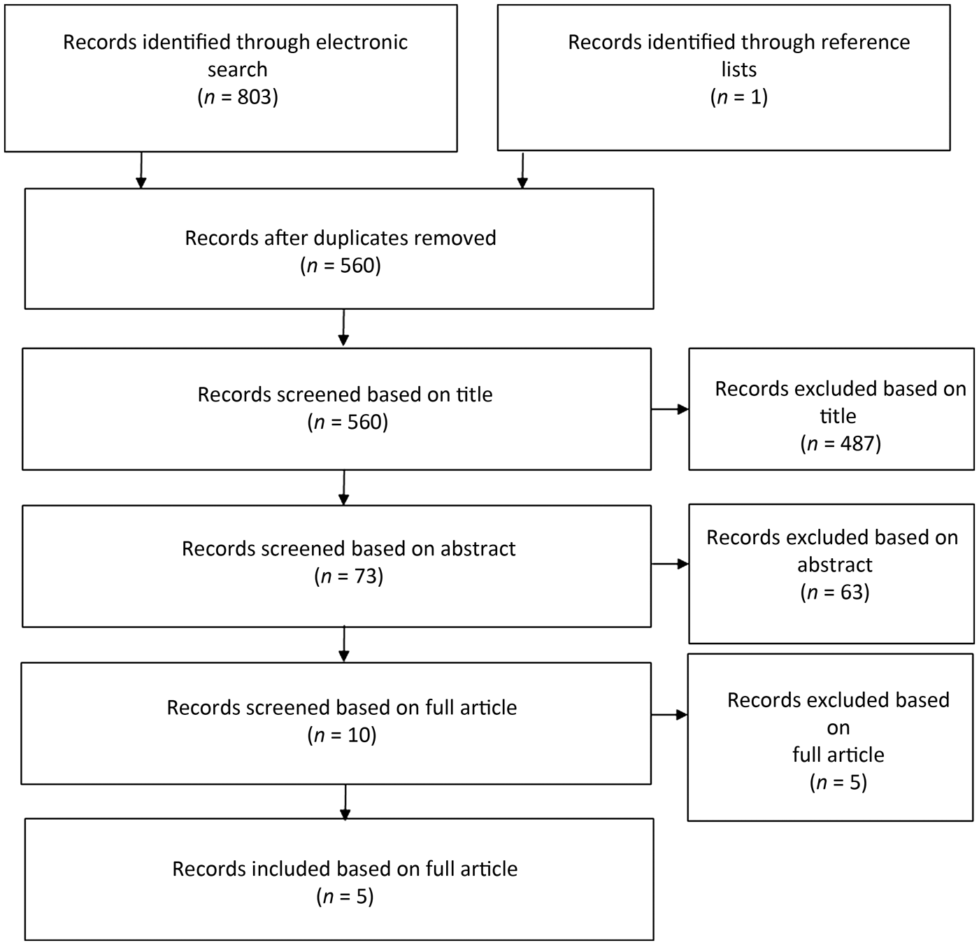
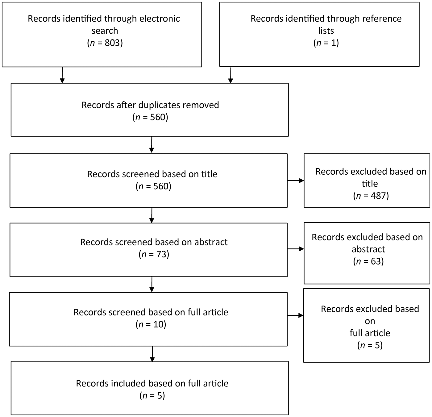
A total of 803 search hits were acquired through the electronic search strategy previously described. After duplicate study removal, 559 results remained for scrutiny by 2 authors under the set criteria (Figure 1). Another study was found through searching the references of relevant papers.

Fig. 1. Flowchart showing literature searching process for review.
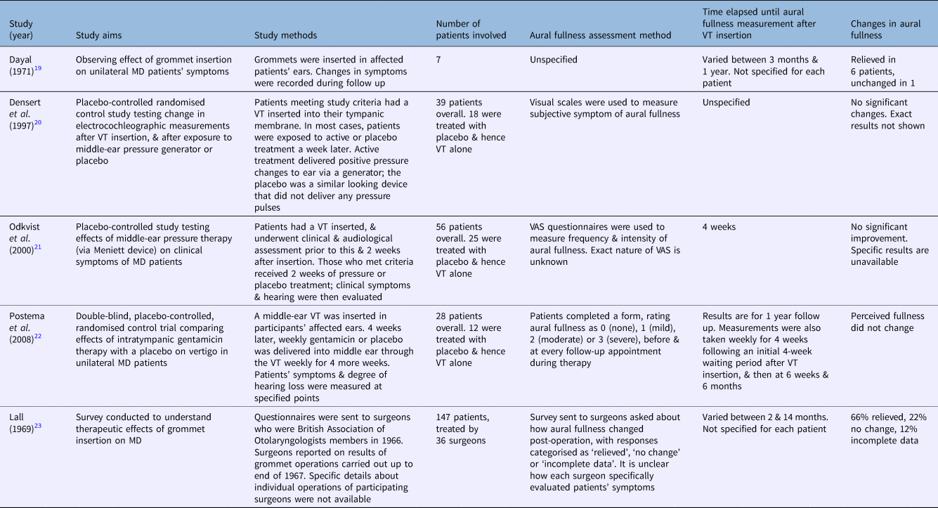
Ten records remained after the exclusion of records based on title and abstract. Four studies meeting the criteria based on their full articles were found via the electronic search. These were studies by Dayal,Reference Dayal19 Densert et al.,Reference Densert, Densert, Arlinger, Sass and Ödkvist20 Odkvist et al.Reference Odkvist, Arlinger, Billermark, Densert, Lindholm and Wallqvist21 and Postema et al.Reference Postema, Kingma, Wit, Albers and Van Der Laan22 An additional study by Lall,Reference Lall23 which was identified from the references of papers, also met the criteria and was included. Table 1 summarises the details of these studies.Reference Dayal19–Reference Lall23
Table 1. Summary of studies included in reviewReference Dayal19–Reference Lall23

VT = ventilation tube; MD = Ménière's disease; VAS = visual analogue scales
Study aims and characteristics
A detailed analysis of the quality of the studies included is beyond the scope of this review, as its aim is not to determine the efficacy of this treatment. However, it is notable that only two of the included studies directly measured the effect of ventilation tube insertion on aural fullness;Reference Dayal19,Reference Lall23 the other studies included ventilation tube insertion as a placebo to assess another therapy.Reference Densert, Densert, Arlinger, Sass and Ödkvist20–Reference Postema, Kingma, Wit, Albers and Van Der Laan22
For example, in the study by Odkvist et al.,Reference Odkvist, Arlinger, Billermark, Densert, Lindholm and Wallqvist21 the effect of ventilation tube insertion on aural fullness could be inferred from the placebo treatment, as a ventilation tube was inserted, but the placebo device did not deliver any pressure pulses. Therefore, any changes in aural fullness could be attributed to ventilation tube insertion alone. LallReference Lall23 compiled data received from questionnaires sent to members of the British Association of Otolaryngologists at the time about the effect of ventilation tube insertion on the symptoms of their patients with Ménière's disease. The case series by DayalReference Dayal19 directly observed the effects of ventilation tube insertion on the symptoms of patients with Ménière's disease. The studies by Odkvist et al.,Reference Odkvist, Arlinger, Billermark, Densert, Lindholm and Wallqvist21 Densert et al.Reference Densert, Densert, Arlinger, Sass and Ödkvist20 and Postema et al.Reference Postema, Kingma, Wit, Albers and Van Der Laan22 were randomised placebo-controlled trials, although none directly measured the effects of ventilation tube insertion on Ménière's disease symptoms.
Patient demographics
In studies where patient ages were specified, all were adults (20–65 years old).Reference Dayal19–Reference Postema, Kingma, Wit, Albers and Van Der Laan22 Only DayalReference Dayal19 specified the gender of participants; all seven patients in that study were male. Table 2 shows the number of participants in each study.Reference Dayal19–24
Table 2. Details of patients in included studies

MD = Ménière's disease; VT = ventilation tube; AAOHNS = American Academy of Otolaryngology – Head and Neck Surgery
Although Lall'sReference Lall23 study had the highest number of patients (147 patients), this was a conglomerate of results that the author acquired from a questionnaire sent out to multiple surgeons, and the specific age, gender and disease type of these patients are unknown. DayalReference Dayal19 and Postema et al.Reference Postema, Kingma, Wit, Albers and Van Der Laan22 were the only ones to measure aural fullness in all patients receiving ventilation tubes, although data were lost for two patients in the latter study. Additionally, it is only clear in Dayal'sReference Dayal19 study that all of his patients had aural fullness prior to intervention. In the others, it is unknown how many of the patients receiving intervention had complained of aural fullness prior to this and how severe the symptom was.
Ménière's disease diagnostic criteria
The vast majority of patients were defined as having ‘Ménière's disease’, with only 15 patients in the study by LallReference Lall23 being defined with ‘Ménière's syndrome’. Two studies diagnosed Ménière's disease based on the criteria set out by the 1995 American Academy of Otolaryngology – Head and Neck Surgery.Reference Densert, Densert, Arlinger, Sass and Ödkvist20,Reference Postema, Kingma, Wit, Albers and Van Der Laan22 This is also likely the case for the study by Odkvist et al.,Reference Odkvist, Arlinger, Billermark, Densert, Lindholm and Wallqvist21 as they described their patients as all having ‘definite Ménière's disease’. Specific diagnostic criteria for patient recruitment are not stated in the other two papers. The patients in the studies by Densert et al.Reference Densert, Densert, Arlinger, Sass and Ödkvist20 and Postema et al.Reference Postema, Kingma, Wit, Albers and Van Der Laan22 all had unilateral Ménière's disease, while disease laterality was unspecified in the other three studies.
Aural fullness measurement
Visual analogue scales were used in the studies by Densert et al.Reference Densert, Densert, Arlinger, Sass and Ödkvist20 and Odkvist et al.Reference Odkvist, Arlinger, Billermark, Densert, Lindholm and Wallqvist21 The exact nature of both scales is unknown, although Odkvist et al.Reference Odkvist, Arlinger, Billermark, Densert, Lindholm and Wallqvist21 stated that their scale measured symptom frequency and intensity. Postema et al.Reference Postema, Kingma, Wit, Albers and Van Der Laan22 asked participants to use a 0–3 scale to rate their symptom severity, with 0 equating to no symptoms and 3 reflecting severe symptoms.
LallReference Lall23 acquired data from several surgeons, using a questionnaire, on how aural fullness changed after ventilation tube insertion. Although the exact nature of this questionnaire is unknown, the changes in symptoms were categorised as either ‘relieved’, ‘no change’ or ‘incomplete data’. Changes in the other Ménière's disease symptoms were also assessed in this paper, and options for these included ‘worsened’, ‘complete lasting relief’, ‘slight lasting relief’ and ‘temporary relief’. It is unknown if these options were also present to describe the change in aural fullness. Additionally, it is not stated how any of the surgeons responding to the questionnaire measured aural fullness.
DayalReference Dayal19 did not specify how aural fullness was assessed after the operation, although their results were presented in the paper.
Time elapsed until aural fullness measurement post-treatment
Odkvist et al.Reference Odkvist, Arlinger, Billermark, Densert, Lindholm and Wallqvist21 made a clinical and audiological assessment two weeks after ventilation tube insertion. At this point, patients still fulfilling the entry criteria for having Ménière's disease continued in the study. It is unstated if aural fullness measurement was part of this clinical assessment. After a further two weeks, aural fullness was measured in both the active pressure treatment and placebo group, and it is the result of this placebo group that is considered by this review.
Postema et al.Reference Postema, Kingma, Wit, Albers and Van Der Laan22 left a four-week waiting period after ventilation tube insertion, and then administered gentamicin treatment or placebo weekly for four weeks. Aural fullness was measured at each visit, and then at six weeks, six months and one year post-treatment initiation. Aural fullness data are only available for one year after placebo therapy.
The ranges in follow-up periods for participants in Lall'sReference Lall23 and Dayal'sReference Dayal19 studies were 2–14 months and 3–12 months respectively, although specific patient follow-up periods are unstated. Densert et al.Reference Densert, Densert, Arlinger, Sass and Ödkvist20 did not specify when they measured aural fullness.
Changes in aural fullness post-treatment
Densert et al.Reference Densert, Densert, Arlinger, Sass and Ödkvist20 and Odkvist et al.Reference Odkvist, Arlinger, Billermark, Densert, Lindholm and Wallqvist21 both stated that their placebo, and hence ventilation tube only, groups showed no significant changes in aural fullness post-treatment. Specific quantitative results from their visual scales are not presented.
Postema et al.Reference Postema, Kingma, Wit, Albers and Van Der Laan22 also showed that their placebo group's perceived aural fullness did not change. They presented their results as histograms showing the distribution of perceived aural fullness as stated by patients according to their numerical scale before and after therapy. However, it is unknown how the aural fullness of each individual patient changed, only that the overall distribution of symptom severity did not change. Two of these patients were lost to follow up, and so data were only available for 10 patients at 12 months.
DayalReference Dayal19 stated that six out of seven patients experienced relief of aural fullness after ventilation tube insertion, while one patient experienced no change in symptoms. However, the degree of relief or initial symptom severity was not stated.
Finally, Lall'sReference Lall23 survey showed that, of all the patients treated by the responding surgeons, 66 per cent showed relief of aural fullness after ventilation tube insertion and 22 per cent showed no change. Data were incomplete for the remaining 12 per cent. Again, the data reflecting the degree of relief and change from initial symptoms were not available for individual patients. Additionally, it is not stated how many of these patients experiencing symptomatic relief were classified as having either ‘Ménière's disease’ or ‘Ménière's syndrome’.
Safety of intervention
DayalReference Dayal19 and LallReference Lall23 both commented on the general ease and safety of ventilation tube insertion compared to more destructive surgical interventions; however, this is not discussed in detail. DayalReference Dayal19 also highlighted the limitation that the patient must be careful not to allow water to enter the ear.
LallReference Lall23 reported that 28 patients experienced relapse of symptoms following ventilation tube extrusion, though it is unclear what these symptoms were. Some of these patients underwent another procedure for re-insertion, although the safety implications of these were not discussed. In the same study, it is noted that 3 per cent, 3 per cent and 6 per cent of patients experienced worsened symptoms of vertigo, deafness and tinnitus, respectively, although it is not stated whether these could be attributed at all to the intervention.
The other three studies did not comment on the safety of ventilation tube insertion. None of the studies commented on whether the ventilation tube was inserted under local or general anaesthetic.
Levels of evidence
The studies by DayalReference Dayal19 and Lall,Reference Lall23 which directly addressed the effect of ventilation tube insertion on aural fullness, were case series (level 4 evidence). The other three studies, which used this treatment as a placebo,Reference Densert, Densert, Arlinger, Sass and Ödkvist20–Reference Postema, Kingma, Wit, Albers and Van Der Laan22 were randomised controlled trials (level 2 evidence).24 However, as these trials did not measure the effect of ventilation tube insertion on aural fullness as their primary outcome, this level of evidence cannot be correctly applied to these studies.Reference Densert, Densert, Arlinger, Sass and Ödkvist20–Reference Postema, Kingma, Wit, Albers and Van Der Laan22 The case series are a lower form of evidence given the risk of bias by the author's opinions, as well as the lack of confounding factor control.Reference Burns, Rohrich and Chung25
Discussion
Importance of research question
This review has identified studies that attempted to determine the effects of ventilation tube insertion on aural fullness in Ménière's disease patients. However, despite a wide search strategy that encompassed all study types and all patient demographics, and that accepted ventilation tube insertion and measurement of aural fullness as secondary aspects to the investigation, only five studies met the criteria. From the outset, this indicates a gap in the literature.
Ventilation tube insertion is a safe, simple procedure, and it would be beneficial to the patient if their symptoms could be controlled with this approach, prior to more destructive interventions.Reference Syed and Aldren17,Reference Lall23 It is notable that only two studies measured the changes in aural fullness as a result of ventilation tube insertion used as the primary intervention, and in neither of those studies was that symptom the primary outcome.Reference Dayal19,Reference Lall23 It is also noteworthy that the two studies directly measuring the effects of ventilation tube on aural fullness were published around fifty years ago. Although considered as part of the diagnostic criteria of Ménière's disease,Reference Goebel1 aural fullness may not be regarded as a main feature of the condition by some clinicians, being overshadowed by the classic triad of vertigo, hearing loss and tinnitus.Reference Yardley, Dibb and Osborne26,Reference Belinchon, Perez-Garrigues, Tenias and Lopez27 However, aural fullness is still an important feature of the condition, as severe manifestations can significantly impact a patient's quality of life, leading to social isolation.Reference Levo, Kentala, Rasku and Pyykko4 The lack of recent studies directly measuring the effect of ventilation tube insertion on aural fullness shows scope for an update to research on this topic.
Measuring aural fullness
Three out of the five studies measured aural fullness using a patient-reported, subjective, visual analogue scale, indicating that this is the preferred way of assessing changes in the symptom.Reference Densert, Densert, Arlinger, Sass and Ödkvist20–Reference Postema, Kingma, Wit, Albers and Van Der Laan22 However, as two of these studies did not specify what was included in their questionnaires, it cannot be ascertained what should be included in such a scale to best measure aural fullness.Reference Densert, Densert, Arlinger, Sass and Ödkvist20,Reference Odkvist, Arlinger, Billermark, Densert, Lindholm and Wallqvist21 LallReference Lall23 employed a different approach, and simply asked patients whether the symptom was relieved or not; this does not allow measurement of the degree of symptom change. No objective measure of aural fullness was mentioned in any of the studies, most likely because the mechanism of development of this symptom is not understood.
Additionally, no consensus exists regarding the optimal time to measure changes in aural fullness after ventilation tube insertion, with follow-up measurements ranging from 4 weeks to 14 months. Given the lack of understanding concerning the pathophysiology behind aural fullness development in Ménière's disease, it is unknown when best to measure the effects of ventilation tube insertion on the symptom. However, the minimum timeframe until measurement of symptoms was four weeks after ventilation tube insertion in the included studies; it is therefore unknown whether there were any changes in aural fullness prior to this. Knowing how quickly aural fullness is relieved, if at all, may help direct understanding of why this symptom occurs in Ménière's disease.
Efficacy of intervention
The studies included in this review have shown mixed results as to the effects of grommet insertion on aural fullness in Ménière's disease. While this review cannot determine the effectiveness of ventilation tube insertion in Ménière's disease aural fullness, it has shown that no randomised controlled trial exists to answer this question.
Finally, the possibility of a placebo effect being the cause of symptomatic improvement after grommet insertion must be considered. A placebo-controlled trial would be needed to distinguish the true effect of ventilation tube insertion in Ménière's disease aural fullness from placebo effects,Reference Probst, Grummich, Harnoss, Hüttner, Jensen and Braun28 a practice supported by the Royal College of Surgeons.29 We recognise that this may not be feasible or required, given the lack of evidence to support this treatment in Ménière's disease.
Conclusion
Although the quality of evidence was not formally appraised, this scoping review reveals a severe lack of literature detailing the effects of grommet insertion on aural fullness in patients with Ménière's disease, with the latest direct evaluation published in 1971.Reference Dayal19 Although there is a theoretical need for future research to fill this gap in knowledge, in order to definitively confirm or disprove the efficacy of this intervention on this often debilitating symptom, the evidence presented does not lend much weight to its efficacy.
Acknowledgements
We would like to thank Dr Derek Hoare for his advice when writing this review. Author David Baguley's involvement was funded by the National Institute of Health Service Research. (The opinions expressed herein are his own, not those of the National Institute of Health Service Research nor the UK Department of Health and Social Care.)
Competing interests
None declared


