


Introduction
Maternal obesity during gestation and lactation has been shown to affect offspring development and thereby affect health in later life.Reference Barker 1 Currently, the majority of women of childbearing age are overweight or obese in relation to western lifestyle,Reference Seidell 2 especially with a high dietary intake of fat.Reference Dubuisson, Lioret and Touvier 3 Excess fat intake has deleterious effects on maternal health, and lipids transferred from the maternal compartment to the fetus can have a significant impact on pregnancy complications and fetal growth.Reference Lawlor, Relton, Sattar and Nelson 4 Recent data in humans indicate that these effects may be self-perpetuating to the next generation,Reference Shankar, Harrell and Liu 5 – Reference Frias and Grove 8 confirming that the effects may be mediated through the germline.
Nutritional environment in utero may also affect reproductive function in offspring, as suggested by data collected from animal models and human cohorts.Reference Gardner, Ozanne and Sinclair 9 – Reference Dupont, Cordier and Junien 11 In mice, maternal nutritional restriction was shown to reduce the reproductive success of daughters.Reference Meikle and Westberg 12 In a rat model of maternal undernutrition, food restriction reduced the number of primordial, secondary and antral follicles in the offspring ovary, in association with decreased expression of genes involved in follicular maturation, ovulation (GDF9, ERB and leptin receptor) and antioxidant mechanisms (Prx3).Reference Bernal, Vickers, Hampton, Poynton and Sloboda 13 Nutrient reduction in pregnant cows was shown to increase maternal plasma testosterone concentrations and to decrease the ovarian reserve [reduction in the number of antral follicles in the ovary, increased follicle-stimulating hormone (FSH) concentrations] in their heifer offspring.Reference Mossa, Carter and Walsh 14 In sheep, maternal undernutrition during gestation increased DNA oxidative lesions in mid-gestational fetal oogonia compared with controls,Reference Murdoch, Van Kirk, Vonnahme and Ford 15 but germ cell numbers were not modified.Reference Rae, Palassio and Kyle 16 Reduced plasma progesterone and ovarian expression of the steroidogenic enzymes StAR and P450scc were also observed in a limited number (n=4) of 6-year-old offspring born to ewes undernourished during pregnancy.Reference Long, Ford and Nathanielsz 17
In humans, the relationship between prenatal famine exposure and subsequent reproductive performance has been studied, with inconsistent results. In one study, women who were exposed to the Dutch famine in utero were more reproductively successful than women who were not exposed to famine during their fetal development.Reference Painter, Westendorp and de Rooij 18 In contrast, two other studies using the same cohort, but at a different time point, demonstrated either a no effect or a negative impact of famine exposure on female fertility.Reference Yarde, Broekmans and van der Pal-de Bruin 19 , Reference Lumey and Stein 20 Natural menopause also occurred earlier in women who were exposed to Dutch famine during their fetal development.Reference Yarde, Broekmans and van der Pal-de Bruin 19 Recently, early pubertal development was also reported in intrauterine growth restriction girls.Reference Hernandez, Martinez-Aguayo and Cavada 21
In obese women, systemic alterations associated with obesity extend directly into the ovarian follicular microenvironment and the expression of lipoprotein receptors CD36 and SR-BI genes is increased in the granulosa cells.Reference Robker, Akison and Bennett 22 Molecular changes were also observed in the ovary in obese animal models. In lethal yellow mice, which are affected by adult-onset obesity, altered metabolic regulation and early reproductive senescence, obesity increases the expression of genes involved in cholesterol biosynthesis in the ovary (but this study was conducted only on three animals per group).Reference Brannian, Eyster and Greenway 23 In obese hyperinsulinemic (fa/fa) rats carrying the fa allele of the leptin receptor gene (Lepr), there are more atretic follicles in the ovaries compared with controls, with a positive association between follicular atresia and the expression of the proapoptotic transcription factor FOXO1.Reference Kajihara, Uchino and Suzuki 24 Another study on the mouse ob/ob model has shown that excess lipid storage induces ovarian function disorders with advanced follicular atresia, apoptosis, defective steroidogenesis and significantly higher cleaved caspase-3 protein expression.Reference Serke, Nowicki and Kosacka 25 In mice fed a high-fat diet for 7 months, the expression of genes involved in inflammatory response (Il1b, Il6 or Tnfα), apoptosis (Foxo3a) and xenobiotic biotransformation (Ephx1, Cyp2e1, Glutathione S-transferase) is higher in the ovaries from obese mouse, potentially affecting ovarian function.Reference Nteeba, Ortinau, Perfield and Keating 26 , Reference Nteeba, Ross, Perfield Ii and Keating 27
Unfortunately, the role of maternal obesity on offspring reproductive function is not yet very well documented, although early puberty was reported by some studies on daughters from overweight or obese mothers,Reference Keim, Branum, Klebanoff and Zemel 28 , Reference Boynton-Jarrett, Rich-Edwards and Fredman 29 and despite a rising concern on this issue.Reference Sauerbrun-Cutler and Segars 30 In rats, an early onset of puberty was reported in females born to dams fed a high-fat diet,Reference Hilakivi-Clarke, Clarke and Onojafe 31 , Reference Sloboda, Howie, Pleasants, Gluckman and Vickers 32 with irregular estrus cycles and prolonged estrus periods;Reference Connor, Vickers, Beltrand, Meaney and Sloboda 33 however, general effects of a maternal high-fat diet during gestation and lactation appear transient compared with effects because of the postnatal diet.Reference Mitra, Alvers, Crump and Rowland 34 In sheep, the number of ovarian follicles was significantly reduced in female offspring born to adolescent obese ewes compared with controls.Reference Da Silva, Aitken, Rhind, Racey and Wallace 35 , Reference Da Silva, Aitken, Rhind, Racey and Wallace 36 There was no effect on the expression of LH-β nor FSH-β mRNA in the fetal pituitary.Reference Da Silva, Aitken, Rhind, Racey and Wallace 36 In sows of Iberian breed, which are obese because of leptin resistance, fertility is reduced. Follicular estrogen secretion and ovulation rates, however, are not reduced.Reference Gonzalez-Anover, Encinas and Sanz 37
A rabbit model of dietary-induced dyslipidemia has been previously established in our laboratory. The rabbit was chosen as a model because of its decisive advantages over the rodent models for longitudinal studies.Reference Fischer, Chavatte-Palmer, Viebahn, Navarrete Santos and Duranthon 38 Moreover, in contrast to rodents, rabbits develop high plasma concentrations of low-density lipoproteins in response to high-fat diets and are thus particularly relevant for the study of lipid abnormalities such as hypercholesterolemia.Reference Montoudis, Simoneau and Brissette 39 – Reference Napoli, Witztum, Calara, de Nigris and Palinski 41 It was previously demonstrated that a high-fat high cholesterol diet (H diet) administered to dams from 10 weeks of age decreased the number of tertiary follicles and increased the number of atretic follicles in the ovary of female rabbits.Reference Cordier, Leveille and Dupont 42 Fertility was conserved, but offspring were growth retarded in utero and developed hypertension and excess fat mass as adults.Reference Picone, Laigre and Fortun-Lamothe 43 The objective of the present study was to explore the effects of a maternal H diet administered from the prepubertal period on offspring’s selected reproductive hormones, ovarian folliculogenesis and gene expression in the ovary using the rabbit as a model.
Methods
Animal and experimental design (Fig. 1)

Figure 1 Schematic representation of the experimental protocol.
Fourteen F0 New Zealand female rabbits (INRA 1077 or PS 19 line) were housed individually with free access to water, under an 8-h light/16-h dark photoperiod, unless stated below. At 10 weeks of age, they were allocated to one of two groups and fed ad libitum with either a hyperlipidic hypercholesterolemic diet (H group; n=7) or a control diet (C group; n=7) containing, respectively, 7.71% or 1.83% fat (from soybean oil) and 0.2% or 0% cholesterol, as previously described.Reference Picone, Laigre and Fortun-Lamothe 43
At 27 weeks of age and after 1 week of synchronization with a photoperiod of 16 h of light/8 h of dark, F0 rabbit does were mated with three different control males. At birth, litters were equilibrated in number (five to seven pups) and the dams continued to receive the same diet as given previously throughout the gestation and lactation periods. At 5 weeks of age, a limited number of F1 female offspring were placed on either C or H diet, resulting in a total of four groups. Female rabbits were weighed every 15 days from 6 to 22 weeks of age. Between 18 and 22 weeks of age, they were mated as previously described and euthanized at 28 days of gestation. Animals were euthanized by exsanguination after electronarcosis in the local experimental slaughterhouse, according to the protocol approved by the local ethics committee and the veterinary services. The male rabbits were used for another experiment.
A few days before mating and just before euthanasia, F1 female rabbits were fasted overnight, weighed and blood was collected from the auricular vein into EDTA-coated vacutainers for biochemical assays (glucose, total cholesterol, high-density lipoprotein (HDL), triglycerides, progesterone, estradiol and testosterone). Immediately after euthanasia, the liver, kidney, interscapular and perirenal fat were weighed. The ovaries were flash frozen in liquid nitrogen and stored at −80°C for molecular analysis (one ovary per animal) or fixed in 10% formalin and processed for histological analysis (one ovary per animal).
Metabolic and hormonal parameters
Glucose was determined with Freestyle Optium Test Strips (Abbott, Rungis, France).
Total cholesterol, HDL and triglycerides were measured with a colorimetric enzymatic method, and hormones were measured with an electrochemiluminescence (ECLIA) method using a biochemical autoanalyzer (Cobas® 3000).
Ovarian histology
Freshly dissected ovaries were fixed in 10% formalin at room temperature for 24 h. After washing in PBS, the tissues were processed in an automated Shandon Citadel 2000 (ThermoFisher Scientific, Illkirch, France) before being embedded in paraffin wax for histological analysis.
For histological studies, coronal ovarian sections of 6 µm were stained with Hemalun Eosin (H&E) using standard protocols to examine basic tissue morphology and count follicles. Periodic acid–Schiff (PAS) staining method was used to detect polysaccharides such as glycogen.
Morphological observations (length, width and area) were realized on ovarian sections after scanning using a Nanozoomer Digital Pathology software (Hamamatsu, Japan) by the same researcher. The microscopic observation was performed blindly in two steps: five randomly selected fields per ovary were observed with ×10 magnification and six randomly selected fields with ×5 magnification. Follicles were classified as previously reported.Reference Cordier, Leveille and Dupont 42 Repeatability was tested by the same researcher measuring six times the same sample. The intra-assay variations coefficients were lower than 5%.
RNA extraction and quantitative RT-PCR
The expression of, respectively, eight and seven genes involved in ovarian development and inflammation/oxidative stress was studied by qRT-PCR. Five ovaries from five dams of each condition (C/C; C/H; H/C; and H/H) were randomly chosen for this analysis. Total rabbit RNAs were extracted as previously described.Reference Cordier, Leveille and Dupont 42 Real-time PCR analysis of the different genes was performed using the Step One Plus PCR system (Applied Biosystems). Cycle conditions were as follows: one cycle at 50°C for 2 min, followed by one cycle at 95°C for 10 min, followed by 45 cycles at 95°C for 15 s and 60°C for 1 min. Briefly, PCR was performed in triplicate with the iTaq™ Universal SYBR® Green Supermix (BioRad, Marnes-la-Coquette, France), using 5 ng of cDNA from the RT. The primers used are presented in Supplementary Table S1. Control experiments were conducted to ensure that the primers could not amplify any genomic products. All PCR products were purified by the Wizard® SV Gel and PCR Clean-Up System (Promega, Madison, USA) and confirmed by sequencing (Beckman Coulter Genomics, Essex, UK). All expression data were normalized using the mean expression level for each sample of two different genes (EIF4A and H2AFX). Results were analyzed using Qbase Software (Ghent University, Ghent, Belgium).
Statistical analysis
All statistical evaluations were performed using R Commander software (R foundation, http://www.R-project.org). Normality data and homogeneity of variance were tested using a Shapiro and a Levene’s test, respectively. Non-normally distributed data were transformed to achieve data normality. Effects of maternal and postnatal offspring diet on weight, metabolic, hormonal, histological and gene expression parameters were analyzed by ANOVA followed by post hoc Dunn’s test or using a Kruskall–Wallis test when a Levene’s test showed non-homogeneous variables. False Discovery Rate (FDR) procedures were used to control the expected proportion of incorrectly rejected null hypotheses. The Spearman test was used to analyze correlations. Weights are presented as median with interquartile range. Data on follicles, weights of organs, biochemical/endocrine parameters and gene expression are presented as box plots representing median with interquartile range. Data on prolificacy are presented as mean±s.e.m. Data were considered significantly different when P<0.05.
Results
Body weight from 6 weeks and until euthanasia (Fig. 2)

Figure 2 Weight of F1 female rabbits (g) according to group and age. Each point represents the median with error bars indicating the interquartile range (Q1; Q3). At 23 weeks of age, C/H were significantly heavier than H/C (P=0.04), but there was no statistical difference between other groups. *Indicates P<0.05 (Post hoc Dunn’s test).
Body weight increased significantly from 6 weeks to euthanasia at 23 weeks of age (P<0.001) with a group effect (P<0.001) and a tendency for an interaction between group and time (P=0.07) characterized by the progressive catch-up of animals born to H dams and fed the H diet. At 23 weeks of age, C/H offspring were significantly heavier than H/C offspring (P=0.04), but there was no statistical difference between any other group.
Fertility and prolificacy
Fertility was calculated as the ratio of the number of pregnant on non-pregnant female rabbits. Prolificacy is defined by the number of offspring per doe that delivered. There was no significant difference in fertility between the groups (fertility of 86%, 100%, 83% and 83% in C/C, C/H, H/C and H/H does, respectively). Prolificacy in pregnant does was not significantly different between the groups (10.8±0.7, 9.2±1.1, 8.0±2.0, 6.6±1.4 fetuses per doe in C/C, C/H, H/C and H/H does, respectively).
Organ weights
The perirenal, interscapular and the sum of the two fat masses were significantly heavier in the C/H group compared with C/C controls (perirenal fat: P<0.01; interscapular fat: P<0.01; sum: P<0.01, respectively), but there was no statistical difference with any other group. The same trends were observed when the fat weight was related to body mass (Fig. 3 for the sum of perirenal and interscapular fat). No difference was observed for liver and kidney weight (data not shown).

Figure 3 Median (Q1; Q3) weight of the total measured fat (perirenal and interscapular fat masses)/total body weight ratio according to group. **Indicates P<0.01 (Post hoc Dunn’s test).
Metabolic profiles before mating (18 weeks) and at euthanasia in late gestation
The metabolic profile and steroid hormone plasma concentrations were used to evaluate metabolic and endocrine perturbations. Fasting plasma/serum glucose, cholesterol, HDL, triacylglycerol, progesterone, estradiol and testosterone concentrations were determined in the four groups of does before mating and in late gestation.
At 18 weeks of age, before mating, HDL and total cholesterol concentrations were significantly different between the groups (P<0.01, Fig. 4). Plasma HDL and total cholesterol concentrations were significantly higher in C/H does compared with C/C (P<0.01) and H/C (P<0.05) does (Figs 4a and 4b). Total cholesterol concentrations were also significantly higher in H/H compared with C/C does (P<0.05; Fig. 4b). There was no significant effect of the prenatal diet for triglycerides (triacylglycerol), total cholesterol and HDL concentrations (comparison C/C v. H/C and C/H v. H/H).

Figure 4 Median (Q1; Q3) of serum HDL (a and c) and total cholesterol (b and d) concentrations at 18 weeks of age (before mating) and at euthanasia (21 weeks of age, late gestation) according to group. * and ** indicate P<0.05 and P<0.01, respectively (Post hoc Dunn’s test).
At 28 days of gestation, higher cholesterol concentrations were observed during gestation compared with before mating, reflecting the mobilization of fatty acids during pregnancy. Does from the C/H and H/H groups had significantly higher HDL and total cholesterol concentrations compared with controls (C/C; P<0.05 and P<0.01, respectively; Figs 4c and 4d), reflecting the effect of the postnatal diet; however, there was no significant difference between C/H and H/H between any other group. No difference was observed for triacylglycerol and glucose plasma concentrations (data not shown).
Plasma concentrations of progesterone and estradiol were not significantly different between treatment groups at 18 weeks and in late gestation. In contrast, although the plasma concentration of testosterone did not differ between the groups at 18 weeks of age, the H/C group had significantly lower testosterone compared with the C/C group at the time of euthanasia (P<0.01; see Supplementary Table S2).
Histological analysis of adult ovaries
To gain some insight into whether fat mass and related metabolic changes had a direct effect on ovarian function, follicles were counted and the expression of genes involved in ovarian development and inflammation/oxidative stress were measured.
Ovarian width differed significantly between the groups (P<0.05), with a significantly reduced ovarian width in female rabbits fed the H diet during the postnatal period (P<0.05). Moreover, the ovarian surface area was significantly lower in H/H compared with C/C does (P<0.05; Figs 5a and 5b, Table 1). The other groups did not differ significantly.

Figure 5 Hemalun Eosin staining of whole ovarian sections of pregnant rabbits (×0.43 magnification) collected in the C/C (a) and H/H (b) groups. The ovary in the H/H group is larger and the surface area is increased compared with that of the C/C group. The apparent difference in length is not significant.
Table 1 Morphological analysis of the ovaries (area, length and width) according to group
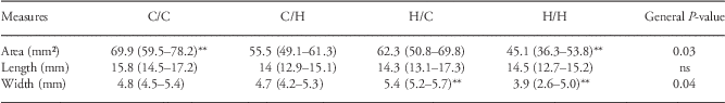
The data are shown as median (Q1; Q3). The general P-value indicates the P-value of the ANOVA for all groups together.
**Indicate P<0.001 between groups (post hoc Dunn’s test).
The number of primordial, primary and secondary follicles were not significantly different between the groups; however, a significantly higher number of atretic follicles (Fig. 6) was observed in the C/H ovaries compared with control C/C ovaries (P<0.001; Fig. 7). This postnatal diet effect was expected as previously reported.Reference Cordier, Leveille and Dupont 42 Prenatal nutrition also had a significant effect with more atretic follicles in the H/C v. C/C ovaries (P<0.001; Fig. 7). There was no significant difference between H/H and C/H, but the number of atretic follicles was significantly higher in H/H v. C/C ovaries (P<0.01; Fig. 7). Finally, numerous empty vacuoles were observed in the stroma of HH ovaries. PAS staining indicated that these vacuoles did not contain carbohydrates or glycogen, but it was not possible to assess whether these were lipid droplets because the samples had been paraffin embedded.

Figure 6 Hemalun Eosin staining of ovarian sections of pregnant rabbits from the C/C (a) and HH (b) groups at ×10 magnification. Compared with the control sample (a), numerous atretic follicle remnants (open arrowheads) are scattered in the ovarian parenchyma and empty vacuoles are observed (indicated by *) in the ovaries from the H/H group (b). Primordial (I), primary (II), secondary (III), tertiary follicles (IV) and atretic follicles (V) are observed in the control samples, whereas numerous fields in H/H group are composed of atretic follicle remnants. Scale bars=250 µm.

Figure 7 Median (Q1; Q3) of atretic follicle counts according to group. Each box plot represents the distribution of value in each group. ** and *** indicate P<0.01 and P<0.001, respectively (post hoc Dunn’s test).
Spearson correlation tests demonstrated a positive correlation between follicular atresia and total plasma cholesterol (r=0.51, P=0.03), plasma triacylglycerol (r=0.58, P=0.01) and a negative correlation with plasma testosterone (r=−0.54, P=0.02; Supplementary Figure S3).
Gene expression
The expression of, respectively, eight and seven genes involved in ovarian development and inflammation/oxidative stress was studied by qRT-PCR. These transcripts fell within functional categories that included: (i) ovary differentiation (FOXL2, WNT4, RSPO1), (ii) folliculogenesis (BMP15, FST),Reference Daniel-Carlier, Harscoet and Thepot 44 (iii) steroidogenesis and receptors (CYP19A1, ESR1), (iv) germ cell differentiation (VASA), (v) inflammation/oxidative stress (TNF-α, Adiponectin-R,) and (vi) oxidative stress (MnSOD, CuZnSOD, CAT, GPx1, eNOS). Of these quantified transcripts, none showed any significant difference in expression between the four groups. TNF-α gene expression analysis tended to be higher in the H/H compared with the C/C group (P=0.08; Supplementary Figure S4).
Discussion
This study evaluated the effect of prenatal and postnatal diet supplemented in soybean oil and cholesterol (H) on metabolic function and on ovarian function of offspring at adulthood. It was shown that, at adulthood, C/H F1 offspring had higher cholesterol and HDL plasma concentrations together with a higher fat mass weight compared with all other groups, indicating that the effect of the postnatal H diet may be reduced when the mother was also fed with an H diet. In contrast, the maternal diet did not affect plasma cholesterol concentrations and fat mass when offspring were fed a control diet after weaning.
In terms of ovarian function, the H diet administered to the F1 generation from 10 weeks of age, that is, before the onset of puberty, led to a high incidence of follicular atresia in the C/H compared with the C/C groups as previously demonstrated.Reference Cordier, Leveille and Dupont 42 Furthermore, adult offspring fed the H diet and the ones born to dams fed the H diet had a significantly decreased ovarian area and increased follicular atresia compared with C/C controls. These data suggest that ovarian function is sensitive to prenatal and postnatal H diet, although the number of healthy follicles and the expression of genes involved in ovarian development and oxidative stress were not modified. Interestingly, plasma concentrations of progesterone and estradiol were not different between the groups; however, plasma testosterone was significantly higher in the C/C group v. H/C group. Placental testosterone production has been reported,Reference Marchut 45 but the question requires further investigation.
In this rabbit model of nutrition, the present data clearly indicate that feeding the H diet in the prenatal and/or in postnatal period disturbs ovarian function through an increase in the atretic follicles. These data are strengthened by previous studies related to the administration of a high-fat diet. Indeed, a high-fat diet was shown to reduce the ovarian surface area in mice and rabbitsReference Nteeba, Ross, Perfield Ii and Keating 27 and to reduce the number of healthy follicles,Reference Sagae, Menezes and Bonfleur 46 increase ovarian atresia,Reference Cordier, Leveille and Dupont 42 , Reference Wu, Dunning and Yang 47 and impair ovarian function through the aberrant expression of genes involved in ovarian development and oxidative stress.Reference Nteeba, Ross, Perfield Ii and Keating 27 Data on the transgenerational effects of a maternal high-fat diet on reproductive fitness of offspring remain limited and unclear.Reference Hilakivi-Clarke, Clarke and Onojafe 31 , Reference Sloboda, Howie, Pleasants, Gluckman and Vickers 32 Here, the increase in the number of atretic follicles in the H/C group demonstrated that in utero nutritional history and/or lactation could affect reproductive function of offspring. Both ovaries of does born to mother fed the H diet and ovaries of does fed the H diet during postnatal period had more atretic follicles and remnants of atretic follicles, suggesting apoptotic mechanisms during folliculogenesis, compared with those whose dam was fed the control diet. Nevertheless, no difference was found for the primordial, primary, secondary and tertiary follicle counts, and fertility and prolificity were not affected. Finally, the large number (not quantified) of apparently empty vacuoles in the stroma of HH ovaries also suggests an accumulation of lipid droplets directly in the ovary, although it was not possible to conclude on their initial contents.
The molecular analysis of the ovarian expression of genes involved in ovarian development and oxidative stress was not significantly different between the groups. The gene expression analysis on whole ovarian tissue is an overall representation. We cannot exclude the hypothesis that high-fat diet may have altered cumulus–oocyte complex gene expression as previously described in obese women.Reference Wu, Dunning and Yang 47 TNF-α gene expression tended to be high in H does. TNF-α plays a critical role in ovarian atresia, as reflected by its expression in apoptotic granulosa cells of healthy and apopotic antral follicles.Reference Driancourt, Fair and Reynaud 48 These data, together with the positive correlation between cholesterol and triglycerides and the number of apoptotic follicles, are in agreement with previous data highlighting that cholesterol abundance promote reactive oxygen species and exacerbate apoptotic cell death in the ovarian cell.Reference Lee, Xu and Li 49 More work is needed to elucidate this question.
The correlations observed here between the concentration of circulating steroids and the number of atretic follicles are more difficult to explain. Estradiol has been reported to act as a survival factor against follicle atresia, and studies have reported estrogen-induced inhibition of granulosa cell apoptosis in antral follicles of rats.Reference Billig, Furuta and Hsueh 50 In humans, one study showed that testosterone suppressed ovarian tissue apoptosis in vitro,Reference Otala, Makinen and Tuuri 51 but the role of this mechanism is not clear and this finding remains controversial.Reference Billig, Furuta and Hsueh 50
In the rabbit ovary, the first germ cells are detected from the 9th day post coitum (dpc) Reference Chretien 52 and most germ cells have already entered the genital crests at 16 dpc. The first signs of meiosis have been reported in the postnatal life around 2 weeks after birthReference Gondos 53 and not during fetal life, as is the case in numerous other mammalian species. The majority of oocytes degenerate during their initial meiotic activities. The ovaries of 4- and 12-week-old rabbits already contain already mature follicles.Reference Lee, Britt and Dunbar 54 , Reference Lee and Dunbar 55 Previous data using this model have shown that the H diet administered from weaning (5 weeks) leads to impaired folliculogenesis with a reduced number of tertiary follicles.Reference Cordier, Leveille and Dupont 42 In the present study, the maternal diet may affect the key period of gonadal organogenesis before birth and during the postnatal, preweaning period.
In conclusion, this study highlights that offspring born to H-fed dams have same characteristics of impaired folliculogenesis as their dam. This is consistent with the data that suggest that nutritional environment in utero and in the postnatal period may be responsible for ovarian damage, without decreased fertility. Given the importance of maternal obesity prevalence and the growing evidence that reproductive health is regulated by early life events, it is necessary to evaluate these effects in children and adolescent girls.
Acknowledgments
The authors thank the staff of INRA Unité Commune d'Experimentation Animale for the care of the animals and Stephan Bouet of INRA GABI for his help with histological staining.
Financial Support
This research received no specific grant from any funding agency, commercial or not-for-profit sectors. It was financed by INRA through internal support from the “Animal Physiology and Breeding Systems” and “Human Nutrition” Departments.
Conflicts of Interest
None.
Ethical standards
The authors assert that all the procedures contributing to this work comply with the ethical standards of the International Guiding Principles for Biomedical Research involving Animals as promulgated by the Society for the Study of Reproduction and in accordance with the European Convention on Animal experimentation. The animal studies were approved by the local animal care and use committee (CSU UCEA Jouy en Josas) and received ethical approval from the local ethics committee (COMETHEA, N°15 on the National registry), under protocol number 12/029. Researchers involved in the work with the animals possessed an animal experimentation license (level 1 or 2) delivered by the French veterinary authorities.


