


Introduction
Historically, investigations into mood disorders have targeted the symptoms associated with the disorder, and many studies have concentrated entirely on behavioural aspects. However, less research has been undertaken examining the underlying cognitive impairments that manifest in these patients or the neurobiological substrates that underpin these deficits. While cognitive impairments are well recognised in idiopathic depression, fewer studies have investigated bipolar disorder (BD). The studies have reported subtle cognitive deficits that persist even while the patient is considered ‘well’ during the euthymic phase Reference Olley, Malhi and Bachelor(1).
Recent pathological findings in structures such as the subgenual prefrontal cortex Reference Drevets, Price and Simpson(2) in patients with mood disorders have precipitated the exploration of possible disturbances in information processing. The cognitive deficits explored have in many instances been likened to deficits commonly reported in patients with frontal lobe damage, such as impairments in executive functioning Reference Murphy and Sahakian(3). As a result of these findings, there has been a significant impetus to better understand the underlying neurobiology of BD, and hence, there have been a growing number of studies that are utilising modalities such as electroencephalogram (EEG) and event-related potentials (ERPs) to study these networks.
The EEG
The electrical activity of the brain can be measured directly off the scalp in the form of EEG, while the subject is resting or performing an experimental task. From the raw EEG, it is possible to extract brain potentials otherwise known as ERPs that are time-locked electrophysiological processes that occur in response to discrete stimuli. In unison, EEG/ERP measures provide a robust method for studying sensory pathways and cognitive processing in patients with BD.
EEG studies of BD have typically recorded neural responses from subjects in both eyes open and closed (Reference Kano, Nakamura, Matsuoka, Iida and Nakajima4–Reference Ikeda, Kato and Kato7) modalities, and some studies even utilised hyperventilation Reference Dewan, Haldipur, Boucher, Ramachandran and Major(8), drowsiness Reference Small, Milstein, Kellams, Miller, Boyko and Small(9) and sleep Reference Rao, Dahl and Ryan(10). Advances in computer technology have provided unlimited potential with respect to the presentation of visual stimuli, including affective images, through the use of video monitors Reference Flaisch, Junghofer, Bradley, Schupp and Lang(11) as well as a large range of auditory stimuli. However, the most common stimuli uncovered in this review were that of auditory tone stimuli used in the context of an auditory oddball paradigm to evoke an ERP waveform known as the P300.
Acquiring the EEG requires the subject to be fitted with a specialised cap that has electrodes incorporated into it that sit on the surface of the scalp. The electrodes have predefined spatial locations on the cap based on the international 10–20 system. The electrode cap along with additional reference electrodes (that may be located on the earlobe, nose or other mastoid areas) is connected to a biological amplifier for amplification, and the resultant signals are then transferred to a computer for acquisition and post hoc analysis.
The EEG is often acquired during resting ‘eyes closed’ and ‘eyes open’ conditions. Following this, there is often an activation-type condition where EEG is recorded while the subject is completing an experimental task, which may include the presentation of auditory and/or visual stimuli. This section of the experiment evokes specific waveforms known as ERPs. Visual-based experimental paradigms, such as facial affect recognition tasks, are presented through a high-resolution monitor synchronised to a stimulus presentation computer. Auditory stimuli because of the precise nature of the tone characteristics are produced through dedicated sound generation hardware and are often presented through earphones.
ERP data are viewed and analysed according to time-locked segments known as epochs that represent a snippet of neural activity of interest. The length of the epoch is chosen according to the difference in time between the stimulus and the neural response. As an example, if a stimulus usually results in certain EEG ‘spikes’, ‘peaks’ or a modified waveform at 200 and 550 ms, an epoch incorporating an 800-ms poststimulus window may be chosen to view the desired events. Repeated presentations of the stimulus result in ERPs being obtained from the subject over many epochs of time. Averaging epochs result in a ‘typical’ evoked response that allows for further analysis across the paradigm as well as across subjects and ultimately a comparison between groups of subjects.
Aim of the study
The aims of this review were to summarise the findings from EEG and ERP studies in BD and to communicate some future directions for this field of research.
Materials and methods
A literature search was performed using MedLine, EMBASE, CINAHL and PsycInfo medical research databases for papers published from 1985 onwards. The terms ‘bipolar disorder’, ‘affective disorder’, ‘mood disorder’ and ‘mania’ were searched in conjunction with EEG, ERP and electroencephalography. Other relevant articles were found through citations from papers in this search. A total of 32 articles were included for review. Papers exploring other technologies in regard to BD research were excluded as they fell outside the scope of this work. It should be noted that because of the variable use of keywords in different articles, some relevant papers may have inadvertently been overlooked.
EEG studies in BD
The purpose of this review was to outline the various EEG and ERP studies that have been conducted into BD. As is evident from Table 1, while there have been several studies produced, it is apparent from these that there has been a lack of systematic approach towards experimental design and medication status. This in part has hindered the formulation of a robust model of information processing in BD. However, these studies do offer promising insights, such as impairments in information processing, as reflected by changes in P300 amplitude and latency, evidence for right hemisphere dominance as well as shedding some light on the role that genetics assumes in the aetiology of BD.
Table 1 Literature review findings of EEG and ERP studies into BD


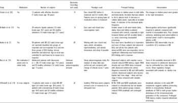

Car, carbamazepine; CNS, central nervous system; CT, computed tomography; Li, lithium; MZ, monozygotic; OCD, obsessive-compulsive disorder; REM, rapid eye movement.
Resting EEG
A large study by Clementz et al. that investigated resting EEG for 50 first-episode schizophrenia patients and 31 first-episode BD patients, together with their relatives, reported increased delta and theta and decreased alpha activity in both patient groups when compared with controls Reference Clementz, Sponheim, Iacono and Beiser(12). Although this study lacked specificity when it came to distinguishing between the two patient groups, the findings robustly differentiated individuals with and without psychiatric illnesses.
Patients with BD in a state of euthymia differ to manic or depressed patients in that their mood is stable, with no obvious highs or lows. However, differences in neural frequencies between euthymic bipolar patients and healthy volunteers persist Reference El-Badri, Ashton, Moore, Marsh and Ferrier(13), indicating that phenotypical traits such as resting EEG frequencies may be central to BD and not state dependent. In this study, greater power in all bands, i.e. beta, alpha, theta and delta, was found, especially in areas involved in visuospatial processing Reference El-Badri, Ashton, Moore, Marsh and Ferrier(13). There were no significant differences between medicated and unmedicated groups of BD subjects with euthymia, suggesting that the effects of medication did not impact on the EEG. In addition, results from neurocognitive tasks given to both patient and control groups yielded further cognitive deficits in the processing of visuospatial tasks in patients. The results suggest that despite patients being euthymic, there remain residual cognitive deficits in the visuospatial domain as revealed by EEG and neurocognitive data. Cognitive deficits in the euthymia state of BD have since been corroborated Reference Olley, Malhi and Bachelor(1).
The P300 ERP
When investigating specific waveforms that differ in BD, the P300 is one that has been widely studied (Reference Gerez and Tello14–Reference Muir, St Clair and Blackwood16). It has been associated with the orienting response and occurs as a result of the presentation of an infrequent or ‘target’ stimulus embedded among a series of frequently occurring background stimuli. It is often suggested that the P300 reflects a comparator process in the brain whereby an existing neuronal template is compared with incoming stimuli Reference Donchin(17). A large study of the P300 was conducted by Muir et al. that examined 96 patients with schizophrenia, 88 with BD, 46 with major depression and 213 healthy controls. The study employed an auditory oddball paradigm, which involved subjects silently counting the randomly presented, infrequently occurring, higher pitched tones among the more frequent lower pitched tones. It was found that a significantly increased latency for the P300 component existed in patients with schizophrenia and BD when compared with depressed patients and healthy controls. From this study, it was reported that the prolongation of the P300 latency could not be attributed to medication effects as it was also present in unmedicated patients. This increase in P300 latency in BD has been consistently reported in subsequent studies Reference Souza, Muir and Walker(15,Reference O’Donnell, Vohs, Hetrick, Carroll and Shekhar18). Moreover, reduced amplitude of the P300 component has also been reported Reference O’Donnell, Vohs, Hetrick, Carroll and Shekhar(18,Reference Hall, Rijsdijk and Kalidindi19).
It has been suggested that the amplitude of the P300 reflects resource allocation Reference Donchin(17,Reference Isreal, Wickens, Chesney and Donchin20) and that the latency of the P300 is an indicator of the speed of information processing or the time taken to categorise the infrequent stimulus Reference Kutas, McCarthy and Donchin(21). Both these characteristics of the ERP have been shown to be abnormal in BD. Such impairments go beyond the typical mood-related symptoms and exist as secondary deficits in information processing.
It is possible that the P300 changes seen in BD are related to structural brain changes. Few studies have demonstrated these functional-structural relationships in BD, yet it has been argued that temporal-parietal abnormalities affecting regions involved in language and audition are responsible for the reductions in amplitude Reference O’Donnell, Vohs, Hetrick, Carroll and Shekhar(18). There is some evidence that the reduced amplitude, which is also seen in schizophrenia, may be related to structural abnormalities in temporal areas Reference McCarley, Salisbury and Hirayasu(22). However, further imaging studies of BD and other mood disorders are required to explicate a structural cause for the changes seen in the P300.
Laterality
Another interesting finding from the Clementz et al.’s study of resting EEG was that those patients with BD had additional right hemisphere activity, suggesting non-dominant hemisphere involvement in regulation of elevated mood Reference Clementz, Sponheim, Iacono and Beiser(12). This is consistent with another study that revealed right hemisphere hyperfunctioning in mania as well as moderate hypofunctioning in depressed subjects and severe hypofunctioning in those with schizophrenia Reference David and Cutting(23). Koles et al., however, demonstrated left-sided hyperactivity in schizophrenia, right-sided hyperactivity in the depressed group and bilateral hyperactivity in those with BD Reference Koles, Lind and Flor-Henry(5); with the author arguing that right-sided hyperactivity in BD was a consequence of verbal cognitive activation. However, a study by Small et al. found that EEG abnormalities were less common in the right hemisphere than in the left Reference Small, Milstein, Malloy, Medlock and Klapper(24).
These results reveal the possibility of laterality in BD because of the persistent finding that the right side of the brain displays hyperactivity in patients with the disorder and may thus implicate this hemisphere in the regulation of mood. It has been suggested that the propensity for abnormally elevated mood during the manic phase of BD relates to increased noradrenergic arousal in the right hemisphere and hence the increased activity reported therein Reference Clementz, Sponheim, Iacono and Beiser(12). Another explanation that has been posited is that patients with BD have ‘sticky’ right hemisphere neural circuitry Reference Pettigrew and Miller(25). No matter what the root cause for right-sided hyperactivity in patients with BD, these findings provide supporting evidence for the hemispheric theory of duality that differentiates left (logical) vs. right (creative) hemispheres Reference Sperry(26). Moreover, an overactive non-dominant hemisphere may explain patient traits such as the intense artistic creativity, which has been demonstrated in some cases Reference Rothenberg(27). However, further studies are required to reinforce hemispheric dominance in BD.
Genetics
One of the earliest studies to investigate BD using EEG technology found a correlation between ‘abnormal’ EEG results and no familial history of the illness Reference Cook, Shukla and Hoff(28), a finding corroborated by a subsequent study Reference Small, Milstein, Malloy, Medlock and Klapper(24). Cook et al. defined an abnormal EEG as one involving generalised slowing, left temporal-parietal and occipital slowing, as well as presence of spike and slow waves Reference Cook, Shukla and Hoff(28). Further support for lack of genetic involvement in the aetiology of BD is demonstrated in another study Reference Clementz, Sponheim, Iacono and Beiser(12). EEG data obtained from patients and their first-degree relatives showed that patients with schizophrenia and BD had reduced alpha activity when compared with controls; a trait that was also found in the first-degree relatives of patients with schizophrenia but not in those of BD Reference Clementz, Sponheim, Iacono and Beiser(12). However, one study uncovered in the literature does suggest genetic causative factors for incidence of BD Reference Potash, Toolan and Steele(29). A large phenome database of BD uncovered strong familial traits such as history of psychiatric hospitalisation (odds ratio = 3.94), psychotic features (odds ratio = 2.01), co-morbid obsessive-compulsive disorder (odds ratio = 3.53) and absences from work because of mood disorder (odds ratio = 3.07) (p < 0.0001 for all).
The BD EEG literature would suggest that genetic factors do not appear to underpin the changes observed in electrical brain activity. This lack of association highlights the importance of environmental and stress-related factors in the development of BD. However, it should be noted that genetic heritability may still exist in non-familial cases of BD and that cases with a positive family history of BD may be illnesses otherwise acquired Reference Taylor and Abrams(30). In addition, a large study of phenotypic variables for BD does indicate familial traits of the disorder Reference Potash, Toolan and Steele(29). This particular study, arising from a significant cohort of 5721 subjects, suggests that genetics do play a role in the aetiology of BD. Therefore, the evidence outlined above, while principally one sided, suggests a variable aetiology of BD that could differ between patients, i.e. that some cases have a strong genetic component, but others are independent of genetic make-up as a causal factor. It is clear that further verification is required to confirm genetic involvement, and large samples of bipolar patients are needed to detect any environmental effects that may be the causal factors of the illness Reference McGuffin, Rijsdijk and Andrew(31).
Summary
The most common EEG findings in BD as uncovered in this review can be summarised as follows. Resting EEG data demonstrated increased theta and delta but decreased alpha band power in patients with the disorder Reference Clementz, Sponheim, Iacono and Beiser(12). Studies that investigated euthymic BD patients compared with healthy controls demonstrated differences in EEG results in areas associated with visuospatial processing Reference El-Badri, Ashton, Moore, Marsh and Ferrier(13), which the authors attribute to underlying disruptions in beta, alpha, theta and delta waves. In regard to ERPs, longer latency of the P300 has been demonstrated in several studies (Reference Souza, Muir and Walker15,Reference Muir, St Clair and Blackwood16,Reference O’Donnell, Vohs, Hetrick, Carroll and Shekhar18), and evidence of reduced amplitude has also been observed in patients with the illness Reference O’Donnell, Vohs, Hetrick, Carroll and Shekhar(18,Reference Hall, Rijsdijk and Kalidindi19). Lastly, hyperactivation of the right hemisphere has been reported by some studies in subjects with BD Reference Clementz, Sponheim, Iacono and Beiser(12,Reference David and Cutting23). The role of genetics in the aetiology of BD, however, still remains unclear (Reference Clementz, Sponheim, Iacono and Beiser12,Reference Small, Milstein, Malloy, Medlock and Klapper24,Reference Cook, Shukla and Hoff28,Reference Potash, Toolan and Steele29).
Methodological limitations in EEG studies of BD
The issue of how medication impacts on the EEG remains unresolved. Some studies have demonstrated effects on the EEG in response to neuroleptic therapy Reference Small, Milstein, Kellams, Miller, Boyko and Small(9,Reference Schulz, Mavrogiorgou, Schroter, Hegerl and Juckel32), with other studies reporting increased delta amplitudes as well as overall total power in patients taking lithium compared with carbamazepine Reference Small, Milstein and Malloy(6). A common dilemma in EEG research into BD is that patients with BD are dependent on medication as they need to remain stable, making it very difficult to recruit non-medicated or even de novo patients for EEG testing. Therefore, the possibility that lithium or other neuroleptic treatment may be responsible for the differing neural activity seen in patients cannot be excluded at this stage.
To be able to discriminate whether findings are a ‘trait’ (i.e. specifically BD) or ‘state’ feature of BD (i.e. mania or depression), a cross-section of many subjects in different phases of the illness need to be tested or longitudinal studies involving testing and retesting subjects over their own mood fluctuations would be required.
Another consideration that needs to be taken into account when examining EEG data are the number of electrodes used to record neural activity. A large study conducted by Muir et al. used only one recording electrode for simplicity Reference Muir, St Clair and Blackwood(16). The authors discussed that because of this methodological restriction, the two components of the P300 waveform, namely the P3a (which occurs frontally) and the P3b (which occurs centroparietally), could not be differentiated because of their regional specificity and that the longer latency (P3b) witnessed in psychiatric subjects may actually have been caused by the absence of the earlier (P3a) element. In comparison, evidence of a longer P300 latency in BD may be clearer in another study that employed a 32-channel EEG Reference O’Donnell, Vohs, Hetrick, Carroll and Shekhar(18). Furthermore, because of this more global measurement of neural activity, the authors were able to observe that the P300 component was largest at parietal sites and smallest at frontal lobe sites. They were therefore able to deduce that the temporal and parietal regions are likely to contribute to the generation of the two subtypes of the P300 ERP component.
Directions for future research
This review summarises studies that have investigated EEG activity in patients with BD, comparing them with healthy controls, unipolar depressed patients as well as with patients with schizophrenia. Although EEG studies of BD have demonstrated many confounds and features that need to be controlled for, they are still of significant importance and meaningful in developing a pattern of brain function in BD. Future studies need to consider the different phases of the illness (i.e. depression, mania and euthymia) separately as well as aim to control for medication and technical considerations, such as numbers of electrode channels. Finally, future studies need to have greater control of variables such as co-morbidity with other illnesses more stringently if the results are to be of clinical salience.
Acknowledgements
This research was supported by NHMRC Program Grant 510135.

