Introduction
Apoptosis is a physiological process occurring spontaneously in the majority of cell populations. One very important function of apoptotic processes during preimplantation development is elimination of the minority of cells with abnormal, detrimental or superfluous potential and the control of embryo cell numbers. Although the targeted cells appear to self-destruct from within, it is now increasingly recognized that the cascade of intra-cellular events that lead to cell elimination rarely occurs in a strictly endogenous context. It has been shown that different external impacts increase the incidence of apoptosis in preimplantation embryos (reviewed in Fabian et al., Reference Fabian, Koppel and Maddox-Hyttel2005).
Tumor necrosis factor α (TNFα) was originally identified as a protein that was cytotoxic for tumor cells; however, recent studies have demonstrated TNFα to be a pleiotropic cytokine that can exert a variety of effects including growth promotion, growth inhibition, inflammation, immunomodulation, cytotoxicity and apoptosis (reviewed in Argiles et al., Reference Argiles, Carbo and Lopez-Soriano1997).
Mammalian embryos are probably exposed to substantial amounts of TNFα before their implantation. In rodents, TNFα expression has been detected in oviductal and uterine tissues throughout the preimplantation period (Sanford et al., Reference Sanford, De and Wood1992; Hunt, Reference Hunt1993). In humans, the presence of TNFα has been demonstrated also in hydrosalpingeal fluid (Bedaiwy et al., Reference Bedaiwy, Falcone, Goldberg, Attaran, Sharma, Miller, Nelson and Agarwal2005).
Recent investigations have shown that the production of TNFα is upregulated in the reproductive tract of the diabetic pregnant rat and that this apoptotic inductor may be a contributing factor of early embryopathy (Pampfer et al., Reference Pampfer, Vanderheyden, McCracken, Vesela and De Hertogh1997).
The action of TNFα is mediated through its interaction with specific high-affinity cell surface receptors. The presence of TNFα receptors has been demonstrated in blastomeres (Whiteside et al., Reference Whiteside, Boucaut, Teh, Garcia-Aragon, Harvey and Herington2003), in trophectoderm cells (Ben-Yair et al., Reference Ben-Yair, Less, Lev, Ben-Yehoshua and Tartakovsky1997) and in embryonic stem cells (Wuu et al., Reference Wuu, Pampfer, Vanderheyden, Lee and De Hertogh1998). Mouse preimplantation embryos express only the p60 form of the TNFα receptor (Pampfer et al., Reference Pampfer, Wuu, Vanderheyden and De Hertogh1994). This expression is probably initiated as embryos proceed through the morula– blastocyst transition stage, as prior studies have shown a lack of TNF receptors at the cell surface of morulae (Lachapelle et al., Reference Lachapelle, Miron, Hemmings, Falcone, Granger, Bourque and Langlais1993). Furthermore, human and mouse embryos have been shown to release TNFα in vitro (Lachapelle et al., Reference Lachapelle, Miron, Hemmings, Falcone, Granger, Bourque and Langlais1993).
Actinomycin D is a potent inducer of apoptosis in a variety of cells in vitro and in vivo and it is used in cancer therapy. It binds to DNA and inhibits DNA replication, RNA synthesis and protein synthesis. It has been shown that high doses (>1 mg/ml) of actinomycin D block transcription of all RNA species, whereas low doses (<100 ng/ml) cause preferential inhibition of ribosomal RNA synthesis in somatic cells (Perry & Kelley, Reference Perry and Kelley1970; Rivera-Leon and Gerbi, Reference Rivera-Leon and Gerbi1997).
Experimental treatment of porcine embryos with actinomycin D at low doses evenly caused the selective inhibition of rRNA synthesis. Higher concentrations completely inhibited RNA synthesis in porcine embryos and bovine blastocysts (Tomanek et al., Reference Tomanek, Kopecny and Kanka1986; Pivko et al., Reference Pivko, Grafanau and Kubovicova2002). A typical nuclear morphology feature of blastomeres in bovine embryos treated with actinomycin D was chromatin marginalization and degeneration of cytoplasm, characteristic signs of apoptosis (Pivko et al., Reference Pivko, Grafanau and Kubovicova2002). Moreover, actinomycin D injected into the blastocoel of Xenopus laevis embryos extensively reduced the transcription of DNA and higher amounts of injected actinomycin D resulted in embryonic death (van der Velden et al., Reference Van Der Velden, Destree, Voorma and Thomas2000).
Although the ability of different apoptotic inductors to affect preimplantation embryo development has been proven previously, the detailed comparative study of their effects has not been performed so far. The aim of this research work was to determine the lowest concentrations and shortest treatment times of TNFα and actinomycin D capable of increasing cell death incidence in mouse blastocysts and to identify the specific biochemical and morphological features of induced apoptosis. Comparison of the evaluated parameters could bring information on the consequences of the presence of different environmental factors on blastomere survival and on the character of apoptotic processes in damaged blastomeres. These results could also broaden existing knowledge on the physiology of programmed cell death in preimplantation embryo.
Materials and Methods
Embryo recovery and culture
Female mice (ICR strain, Velaz, Prague, Czech Republic; 4 week old) underwent superovulation treatment with pregnant mare's serum gonadotropin (PMSG 5 UI i.p.; Folligon, Intervet International), followed 47 h later by administration of human chorionic gonadotropin (hCG 5 UI i.p.; Pregnyl). Females were mated with males of the same strain overnight. Mating was confirmed by identification of a vaginal plug. Females were killed by cervical dislocation. Embryos were recovered at the blastocyst stage (91–92 h after hCG treatment) by flushing the oviduct using a flushing–holding medium (FHM) (Lawits & Biggers, Reference Lawits and Biggers1993). The embryos were then pooled, washed five times in KSOM culture medium (Lawits & Biggers, Reference Lawits and Biggers1993) and prepared for subsequent treatment.
Induction of apoptosis
Isolated mouse blastocysts were randomly divided into experimental and control groups, washed three times and then transferred to 15, 20 or 25 µl drops of KSOM (1 embryo/1µl approximately) containing an apoptosis inductor or its solvent in equivalent amounts and cultured in a humidified atmosphere with 5% CO2 at 37 °C.
Two inductors of apoptosis were used: TNFα (Sigma–Aldrich) dissolved in aqua pro injectione and actinomycin D (Sigma–Aldrich) dissolved in 100% ethanol. For the assessment of dose-dependent effects of apoptosis inductors, blastocysts were cultured 24 h in KSOM containing following concentrations of TNFα: 5, 12.5, 25, 50, 100, 500, 750, 1000, 5000 and 10 000 ng/ml; and actinomycin D: 50 and 500 ng/ml. Selected doses of actinomycin D were already used in our previous studies (Baran et al., Reference Baran, Fabian, Rehak and Koppel2003; Fabian et al., Reference Fabian, Il'kova, Rehak, Czikkova, Baran and Koppel2004). For the assessment of time-dependent effects of apoptosis inductors, blastocysts were cultured 1 h, 6 h and 24 h in KSOM containing TNFα and actinomycin D at concentrations significantly inducing apoptosis but not affecting embryo growth (TNFα 100 ng/ml, actinomycin D 50 ng/ml). For assessment of the biochemical–morphological profile of induced apoptosis, additional 6 h, 24 h and 48 h cultures of blastocysts in KSOM with the addition of apoptosis inductors at effective concentrations were performed.
Embryo growth evaluation and cell death detection
After experimental culture, blastocysts were washed in KSOM and their nuclei were stained with Hoechst 33342 (20 µg/ml; Sigma–Aldrich; stains all cells, shows nuclear morphology) and propidium iodide (PI, 10 µg/ml; Sigma–Aldrich; stains dead cells only) for 5 min at 37 °C. Embryos were then washed in phosphate-buffered saline (PBS) with bovine serum albumin (BSA, Sigma–Aldrich), fixed in 4% paraformaldehyde (Merck) in PBS at room temperature for 1 h and optionally stored in 1% paraformaldehyde in PBS at 4 °C. Nuclei with degraded DNA were detected using a cell death-detection technique based on the TUNEL principle using fluorescein-conjugated dUTP (Gjorret et al., Reference Gjorret, Knijn, Dieleman, Avery, Larsson and Maddox-Hyttel2003; Fabian et al., Reference Fabian, Gjorret, Berthelot, Martinat-Botte and Maddox-Hyttel2005). Fixed blastocysts were washed in PBS (+BSA), permeabilized for 1 h in PBS with 0.5% Triton X-100 (Sigma–Aldrich) and again washed in PBS (+BSA). As positive controls, two to three additional untreated blastocysts were preincubated in DNase I (deoxyribonuclease I 100 U/ml; Invitrogen) for 30 min at 37 °C and then washed in PBS (+BSA). Positive controls and all experimental embryos were then incubated in 10 µl of terminal deoxynucleotidyl transferase and 90 µl of fluorescein-conjugated dUTP (TUNEL, In Situ Cell Death Detection Kit; Roche) for 60 min at 37 °C in the dark. Negative controls, represented by another two to three additional untreated blastocysts, were not incubated with the terminal transferase enzyme. After TUNEL reaction, all embryos were washed in PBS (+BSA), mounted on a slide, covered with a coverslip and observed under fluorescence microscope at ×400 magnification (BX50 Olympus).
The average number of nuclei per blastocyst was determined as the main indicator for embryo growth. According to their nuclear morphology (M), the presence of specific DNA fragmentation (T) and PI positivity/negativity (PI±), nuclei were classified as: normal (M–T–P–; oval, without morphological changes, without TUNEL labelling and able to exclude PI), apoptotic (M+T±P–; PI negative, with typical fragmented or condensed morphology, usually containing degraded DNA), secondary necrotic (M+T±P+; with typical apoptotic morphology, usually containing degraded DNA, reaching terminal stages of apoptotic process characterized by PI positivity) and necrotic (M–T±P+; PI positive, mostly without specific morphological changes and usually without TUNEL labelling). Occasionally observed mitotic configurations were also classified as normal nuclei (Fabian et al., Reference Fabian, Il'kova, Rehak, Czikkova, Baran and Koppel2004). The percentages of normal, apoptotic, secondary necrotic and necrotic blastomeres were calculated as the numbers of normal, apoptotic, secondary necrotic and necrotic nuclei relative to the total number of evaluated nuclei. The percentage of dead blastomeres represented the proportion of all apoptotic, secondary necrotic and necrotic blastomeres.
Biochemical–morphological profile of apoptosis
The co-incidence of morphological changes in nuclei, specific DNA degradation and the presence of caspase-3, the main enzyme of apoptotic cascade, was evaluated in embryos from additional cultures with effective concentrations of apoptotic inductors. After experimental culture, blastocysts were washed in PBS (+BSA) and fixed in 4% paraformaldehyde. Fixed embryos were washed in PBS (+BSA) and 0.3% Triton X-100, permeabilized in PBS (+BSA) with 1% Triton X-100 at room temperature for 2 h, rewashed several times in PBS (+BSA) and subjected to TUNEL-reaction with fluorescein-conjugated dUTP. TUNEL-reacted embryos were then subjected to immunohistochemical staining with a wash in PBS (+BSA), blocking in a buffer comprising PBS (+BSA) with 5% normal goat serum (Invitrogen) and 0.1% Triton X-100, followed by overnight incubation with a primary anti-active caspase-3 antibody (polyclonal rabbit cleaved caspase-3 (Asp175) antibody; Cell Signaling Technology). Specimens were then rewashed and incubated with a Texas Red-conjugated secondary antibody (goat anti-rabbit IgG; Jackson ImmunoResearch). Finally, specimens were washed in 0.3% Triton X-100 and PBS (+BSA), counterstained with Hoechst 33342 (20 µg/ml) and mounted under a coverslip. Immunohistochemical control staining was performed by either: (i) preabsorption of primary antibody with specific antigen (cleaved caspase-3 (Asp175) blocking peptide; cell signalling technology); (ii) substitution of primary antibody with non-immune rabbit IgG (rabbit immunoglobulin fraction – negative control; DakoCytomation); or (iii) omission of secondary antibody. Five untreated blastocysts were used for each control staining.
According to their nuclear morphology (M+, fragmentation or condensation), the presence or absence of specifically degraded DNA in nucleus or cytoplasm (T±, TUNEL labelling) and the presence or absence of active caspase-3 in cytoplasm (C±, immunohistochemical staining), apoptotic blastomeres were categorized into four groups: M+T–C–, M+T+C–, M+T+C+ and M+T–C+. The percentages of all types of apoptotic blastomeres were calculated as the numbers of these apoptotic blastomeres relative to the total number of evaluated blastomeres.
Statistical analysis
The results are expressed as mean values ±SD. The mean numbers of cells per blastocyst were subjected to analysis of variance (ANOVA) and differences between group means were determined by Dunnett's method. Statistical analysis was performed using Statistica (StatSoft). Standard chi-squared tests were used to detect differences in cell death incidence and five-way chi-squared tests were used to detect differences in the profiles of apoptotic cell death. Values p < 0.05 were considered as significant.
Results
Induced cell death in mouse blastocysts
Dose-dependent effect of apoptotic inductors
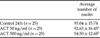
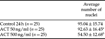
As shown in Fig. 1 and Table 1, the addition of actinomycin D at lower concentration (50 ng/ml) into culture media for 24 h significantly increased the percentage of dead blastomeres without affecting embryo growth when compared with control. The majority of dead blastomeres in such treated mouse blastocysts were apoptotic. Addition of a 10 times higher concentration of actinomycin D (500 ng/ml) caused extensive damage to blastocysts and arrested embryo development. Almost half of the blastomeres underwent apoptotic cell death and the majority of them reached the terminal stage of secondary necrosis. Moreover, the percentage of necrotic blastomeres was also increased.
Table 1 Dose-dependent effect of actinomycin D (ACT) on the development of mouse blastocysts cultured 24 h in vitro

a p > 0.05; bp < 0.001; n: the number of evaluated blastocysts.

Figure 1 Dose-dependent effect of actinomycin D (ACT) on the incidence of cell death in mouse blastocysts cultured 24 h in vitro. Graph shows the proportion of apoptotic, secondary necrotic and necrotic nuclei evaluated by fluorescence microscopy. M+: nuclei with typical fragmented or condensed morphology; M–: nuclei mostly without specific morphological changes; T±: TUNEL labelled/unlabelled nuclei; PI±: propidium iodide positive/negative nuclei; ***: p < 0.001.
As shown in Fig. 2 and Table 2, the addition of TNFα at concentrations 25 ng/ml and higher into culture media for 24 h significantly increased the percentage of dead blastomeres in mouse blastocysts when compared with control. The percentage of dead cells, predominantly of apoptotic origin, increased linearly in dose-dependent manner. However, even the highest used concentrations of TNFα (5000 and 10 000 ng/ml) did not cause as extensive damage to mouse blastocysts as actinomycin D at concentration 500 ng/ml. The negative effect of TNFα on embryo growth was also only modest. The significant decrease of average number of nuclei in mouse blastocyst was recorded only in TNFα at concentrations 750 ng/ml and higher.
Table 2 Dose-dependent effect of TNFα on the development of mouse blastocysts cultured 24 h in vitro

ap > 0.05; bp < 0.05; cp < 0.01; n: the number of evaluated blastocysts.

Figure 2 Dose-dependent effect of TNFα on the incidence of cell death in mouse blastocysts cultured 24 h in vitro. Graph shows the proportion of apoptotic, secondary necrotic and necrotic nuclei evaluated by fluorescence microscopy. M+: nuclei with typical fragmented or condensed morphology; M–: nuclei mostly without specific morphological changes; T±: TUNEL labelled/unlabelled nuclei; PI±: propidium iodide positive/negative nuclei; NS: p > 0.05; **: p < 0.01; ***: p < 0.001.

Figure 3 Time-dependent effect of actinomycin D (ACT) on the incidence of cell death in mouse blastocysts cultured in vitro for 1 h, 6 h and 24 h. Graph shows the proportion of apoptotic, secondary necrotic and necrotic nuclei evaluated by fluorescence microscopy. M+: nuclei with typical fragmented or condensed morphology; M–: nuclei mostly without specific morphological changes; T±: TUNEL labelled/unlabelled nuclei; PI±: propidium iodide positive/negative nuclei; NS: p > 0.05; ***: p < 0.001.

Figure 4 Time-dependent effect of and TNFα on the incidence of cell death in mouse blastocysts cultured in vitro for 1 h, 6 h and 24 h. Graph shows the proportion of apoptotic, secondary necrotic and necrotic nuclei evaluated by fluorescence microscopy. M+: nuclei with typical fragmented or condensed morphology; M–: nuclei mostly without specific morphological changes; T±: TUNEL labelled/unlabelled nuclei; PI±: propidium iodide positive/negative nuclei; NS: p > 0.05; ***: p < 0.001.
Time-dependent effect of apoptotic inductors
Addition of apoptotic inductors (actinomycin D 50 ng/ml or TNFα 100 ng/ml) into the culture media had no influence on cell death incidence in mouse blastocysts after 1 h culture when compared with controls (see Figs 3 and 4).
Actinomycin D treatment significantly increased the percentage of dead blastomeres already after 6 h culture (p < 0.001). Again, the highest increase was recorded in the proportion of apoptotic cells. TNFα treatment also caused slight increase in cell death, but its effect was not significantly different from controls after 6 h culture (p > 0.05).

Figure 5 The biochemical–morphological profile of apoptotic cells in mouse blastocysts treated with actinomycin D (ACT) at 500 ng/ml for 6 h. M+: blastomeres with typical fragmented or condensed morphology of nuclei; T±: blastomeres with TUNEL labelled/unlabelled nuclei; C±: blastomeres with/without the presence of active caspase-3 in cytoplasm. The average number of blastomeres is expressed as mean values ± SD; ***: p < 0.001.

Figure 6 The biochemical–morphological profile of apoptotic cells in mouse blastocysts treated with actinomycin D (ACT) at 500 ng/ml and TNFα at 100 ng/ml for 24 h. M+: blastomeres with typical fragmented or condensed morphology of nuclei; T±: blastomeres with TUNEL labelled/unlabelled nuclei; C±: blastomeres with/without the presence of active caspase-3 in cytoplasm. The average number of blastomeres is expressed as mean values ± SD; ***: p < 0.001.

Figure 7 The biochemical–morphological profile of apoptotic cells in mouse blastocysts treated with TNFα at 100 ng/ml and 5000 ng/ml for 48 h. M+: blastomeres with typical fragmented or condensed morphology of nuclei; T±: blastomeres with TUNEL labelled/unlabelled nuclei; C±: blastomeres with/without the presence of active caspase-3 in cytoplasm. The average number of blastomeres is expressed as mean values ± SD; ***: p < 0.001.
Results achieved after 24 h culture of mouse blastocysts in medium with the addition of apoptotic inductors were similar to those achieved in the previous experiment. Both actinomycin D at 50 ng/ml and TNFα at 100 ng/ml significantly increased cell death incidence (p < 0.001) without affecting embryo growth (p > 0.05) when compared with untreated controls (Table 3).
Table 3 Time-dependent effects of actinomycin D (ACT) and TNFα on the development of mouse blastocysts cultured in vitro for 1 h, 6 h and 24 h

ap > 0.05; bp < 0.05; n, the number of evaluated blastocysts.
Biochemical–morphological profile of induced apoptosis
As shown in Figs 5–7, the majority of apoptotic cells in control blastocysts cultured for 6 h, 24 h and 48 h showed both fragmented morphology of nuclei and specific DNA degradation (M+T+). A relatively high proportion of such cells (usually more than 1/3) also showed the presence of active caspase-3 in their cytoplasm (C+). The percentages of apoptotic cells showing only apoptotic morphology in nuclei (M+T–C–) and the percentages of cells showing apoptotic morphology in nuclei and the presence of active caspase-3 in cytoplasm but no TUNEL labelling (M+T–C+) were much lower.
The addition of high doses of actinomycin D (500 ng/ml) into culture media for 6 h and 24 h and of TNFα (5000 ng/ml) for 48 h generally increased the incidence of all types of apoptotic cells in blastocysts (actinomycin D 6 h p < 0.05, 24 h p < 0.001, TNFα 48 h p < 0.01). The correlative ratio of apoptotic cells was again characterized by the predominance of M+T+C– and M+T+C+ types, the rare occurrence of M+T–C– type and the relatively low proportion of M+T–C+ type. Profiles of such induced apoptosis were usually characterized by increased occurrence of active caspase-3 in the cytoplasm of apoptotic blastomeres.
The addition of TNFα at 100ng/ml into culture media for 24 h and 48 h did not affect the correlative ratio of apoptotic cell types (p < 0.05).
Discussion
Experimental culture of mouse blastocysts in medium with the addition of apoptotic inductors significantly increased the cell death incidence in embryos. The effects of apoptotic inductors were dose and time dependent.
The addition of inductors at lower concentrations (actinomycin D 50 ng/ml, TNFα 25 to 500 ng/ml) increased cell death incidence, usually without affecting embryo growth after 24 h culture. Higher concentrations of inductors (actinomycin D 500 ng/ml, TNFα 750 to 10 000 ng/ml) caused wider cell damage and also retarded or arrested blastocyst development after 24 h culture. As proved by evaluation of nuclear morphology, viability staining and TUNEL assay, the majority of dead cells in such treated blastocysts were apoptotic.
The direct effect of actinomycin D on mouse blastocysts has not been evaluated so far. Our previous studies showed that 15 h treatment of 4-cell mouse embryos with actinomycin D (same doses: 50 and 500 ng/ml) delayed their further development, extensively decreased the average number of their blastomeres, restrained blastocyst formation and significantly increased the percentage of cell death. Morphological and viability assessment proved the apoptotic origin of these dead cells (Baran et al., Reference Baran, Fabian, Rehak and Koppel2003; Fabian et al., Reference Fabian, Il'kova, Rehak, Czikkova, Baran and Koppel2004). Similar observations were presented by other authors and additionally confirmed the induction of apoptosis by TUNEL labelling (Ju et al., Reference Ju, Kwak, Lee, Kim, Kim, Kim, Choi, Jung, Lee, Do, Park and Choo2005). The assessment by electron microscopy of late bovine blastocysts exposed to a high dose of actinomycin D (1 µg/ml) also demonstrated the presence of typical signs of apoptosis in evaluated blastomeres (Pivko et al., Reference Pivko, Grafanau and Kubovicova2002). In contrast, it has been shown that 2-cell rabbit embryos exposed to actinomycin D (2 µg/ml) were prevented from dividing but did not degenerate (Bellier et al., Reference Bellier, Chastant, Adenot, Vincent, Renard and Bensaude1997).
It has also been shown previously that 24 h treatment of mouse and rat blastocysts with TNFα (50 ng/ml in mouse, 3000 U/ml in rat) delayed their morphological development and reduced the average number of cells (Pampfer et al., Reference Pampfer, Wuu, Vanderheyden and De Hertogh1994, Reference Byrne, Southgate, Brison and Leese1997; Wuu et al., Reference Wuu, Pampfer, Becquet, Vanderheyden, Lee and De Hertogh1999). Decrease in cell number was recorded predominantly in the inner cell mass (ICM). Lower doses of TNFα (0.5 and 5 ng/ml) decreased the average number of blastomeres only after longer exposure to the cytokine (48 h) (Pampfer et al., Reference Pampfer, Wuu, Vanderheyden and De Hertogh1994). In our experiments, the negative effect of similar doses of TNFα (5 to 100 ng/ml) on blastocyst growth was usually very low and not significantly different when compared with controls after 24 h culture. Significant decrease in the average number of cells was observed only after exposure of blastocysts to higher doses of TNFα (over 750 ng/ml) and after longer culture (48 h). Longer-lasting treatment of mouse zygotes (Byrne et al., Reference Byrne, Southgate, Brison and Leese2002) and mouse preimplantation embryos in cleavage stages of development (Kurzawa et al., Reference Kurzawa, Glabowski and Wenda-Rozewicka2001) with various concentrations of TNFα also reduced the average number of cells and blastocyst formation. In spite of minor differences between the outcomes of the presented studies, caused probably by the use of different experimental protocols and analysis systems, all data prove that the negative effect of TNFα on embryo development increases with concentration and time.
Previous studies also showed that exposure of mouse and rat blastocysts to TNFα resulted in significant increase in the frequency of TUNEL labelled nuclei and TUNEL labelled nuclei with typical apoptotic morphology (Pampfer et al., Reference Pampfer, Vanderheyden, McCracken, Vesela and De Hertogh1997; Wuu et al., Reference Wuu, Pampfer, Becquet, Vanderheyden, Lee and De Hertogh1999). In contrast to our findings, no difference was found in the incidence of nuclear fragmentation between control and treated blastocysts in these studies. However, inspection of the morphology of dying cells, performed in later studies, showed that 96 h incubation of early mouse embryos with TNFα at 50 ng/ml increased the frequency of both karyolysis (detected by TUNEL assay) and karyorhexis (fragmented nuclear morphology) (Byrne et al., Reference Byrne, Southgate, Brison and Leese2002). This contrast has been explained by possible longer activation of enzymes governing nuclear fragmentation compared with shortened activation of karyolyitic enzymes (Byrne et al., Reference Byrne, Southgate, Brison and Leese2002). Nowadays we may suggest that the incidence of induced nuclear fragmentation could also be influenced by other factors. Their identification is difficult, since the experimental protocols used in the presented studies differed in many various aspects, such as embryo species or strain, ovulation of mothers, culture conditions, the source of TNFα or cell death evaluation system.
Increased incidence of apoptosis following the exposure of bovine preimplantation embryos to TNFα has been documented too (Soto et al., Reference Soto, Natzke and Hansen2003).
In all our experiments, the negative effect of actinomycin D on blastomere survival and blastocyst growth was greater than the effect of TNFα. Furthermore, the addition of actinomycin D (50 ng/ml) into culture media increased cell death incidence even after 6 h culture. Exposure to a high concentration of actinomycin D (500 ng/ml) also increased the proportion of necrotic blastomeres in blastocysts. According to this, we may hypothesize that the cytotoxic mode of action of actinomycin D is probably much less specific than of TNFα.
The TNFα-induced cell death usually results from complex apoptotic cascade. TNFα binding with its specific receptor is followed by recruitment of adapter proteins such as TNFR1-associated death domain protein and Fas-associated death domain protein, with further recruitment of initiator procaspase-8 and procaspase-10. Depending on the cell type, mature caspase-8 either activates effector caspase-3 or a proapoptotic Bcl-2 family protein, Bid, which in turn activates the intrinsic death pathway. Activated Bid induces the release of cytochrome c from the mitochondria into the cytoplasm. In the cytoplasm, cytochrome c binds to apoptosis-protease activating factor-1 (Apaf-1), which in turn binds to procaspase-9, forming an apoptosome. Procaspase-9 is activated within the apoptosome and then activates caspases 3 and 7 (reviewed in Torchinsky et al., Reference Torchinsky, Markert and Toder2005).
The exact biochemical pathway triggered by actinomycin D has not been described so far. However, actinomycin D induced time- and dose-dependent apoptosis in various cell lines. Induced apoptosis was associated with cytochrome c release, the activation of caspase-3, inhibition of the expression of the anti-apoptotic proteins BCR-ABL and Bclxl, induction of the cleavage of the DNA repair protein – poly (ADP–ribose) polymerase (PARP), internucleosomal DNA fragmentation and the cleavage of cytoskeletal substrates gelsolin and alpha II spectrin (Yamazaki et al., Reference Yamazaki, Tsuruga, Zhou, Fujita, Shang, Dang, Kawasaki and Oka2000; Abdelhaleem, Reference Abdelhaleem2003; Williams et al., Reference Williams, Smith, Cianci, Morrow and Brown2003; Takeuchi et al., Reference Takeuchi, Hoshijima, Onuki, Nagasaka, Chowdhury, Kawase and Sakagami2005).
Previous experiments on various murine tumor cell lines showed that the cytotoxic effect of actinomycin D was usually stronger than the effect of TNFα (Lasek et al., Reference Lasek, Giermasz, Kuc, Wankowicz, Feleszko, Golab, Zagozdzon, Stoklosa and Jakobisiak1996). Moreover, actinomycin D was able to cause apoptosis in human pancreatic cancer cells, which were completely resistant to TNFα (Kleeff et al., Reference Kleeff, Kornmann, Sawhney and Korc2000).
The incidence of apoptosis caused by TNFα is evidently dependent on the concentration of its receptors in the population of target cells, whereas actinomycin D probably does not require such a specific mechanism for cell entrance. Furthermore, the relatively low inducing effect of TNFα, observed also in our experiments, could be influenced by the ability of treated cells to suppress the functional apoptotic pathway (Fabian et al., Reference Fabian, Rehak, Czikkova, Il'kova, Baran and Koppel2003). In contrast, the stronger damaging effect of actinomycin D could be caused by lower reversibility of the destructive process and also by the ability of actinomicin D to induce DNA damage directly, not only by the apoptotic pathway, as we hypothesized previously (Fabian et al., Reference Fabian, Rehak, Czikkova, Il'kova, Baran and Koppel2003). According to the presented data we may also suggest that the complex biochemical pathway triggered by TNFα requires a longer time to accomplish the stage with detectable signs of apoptosis than the damaging action of actinomycin D.
The results of our latest experiment show that the morphological and biochemical features of both spontaneous and induced apoptosis in untreated and treated mouse blastocysts are similar. The majority of evaluated apoptotic cells showed both fragmented nuclear morphology and the presence of specifically degraded DNA in the nucleoplasm, accompanied by simultaneous presence or absence of active caspase-3 in the cytoplasm. A minority of apoptotic cells showed only fragmented nuclear morphology or fragmented nuclear morphology with the presence of active caspase-3 in the cytoplasm. Other combinations of apoptotic signs were very rare and they were included into the already-determined classification groups according to the highest parallelism when they occasionally appeared in the evaluated material.
When compared with controls, blastocysts exposed to high concentrations of actinomycin D (500 ng/ml, 6 h and 24 h) and TNFα (5000 ng/ml, 48 h) showed increased incidence of all apoptotic cell types. These results prove that the biochemical pathways of apoptosis induced by both actinomycin D and TNFα include activation of caspase-3 and internucleosomal DNA fragmentation not only in somatic cells but also in preimplantation embryos.
In conclusion, we can say that the presence of different apoptotic inductors in the blastocyst environment influences the development and the survival of embryos and their cells in different ways. However, while specific outcomes of inductor action differ, some basic aspects of the process are similar. Apoptotic cascade triggered in embryonic cells by both actinomycin D and TNFα includes activation of caspase-3 in the cytoplasm, followed by activation of other enzymes responsible for internucleosomal DNA fragmentation in the nucleoplasm and typical apoptotic changes in nuclear morphology. The differences are that apoptotic features appear in a shorter time in actinomycin D treated blastomeres, whereas the apoptotic pathway triggered by TNFα is probably more specific and reversible. Moreover, we may hypothesize that the incidence of actinomycin D induced apoptosis depends mostly on its concentration in the cell environment, while the incidence of TNFα induced apoptosis is limited by the concentration of its receptors in the targeted population of cells. Increased incidence of induced necrosis and the ability to cause embryo arrest show that actinomycin D also affects other important cellular mechanisms besides apoptosis.
Our results characterizing the effects of TNFα and actinomycin D could be helpful for the selection of apoptotic inductors at appropriate doses and times in different models of apoptotic studies in preimplantation embryos. We have shown that the incidence of induced apoptosis in treated blastocysts could be regulated by different concentrations of apoptotic inductor and the typical features of apoptotic process could be detectable in relatively short times after induction.
Acknowledgements
We thank Dana Čigašová for her technical assistance and Andrew Billingham for the revision of English grammar. This work was supported by the Science and Technology Assistance Agency under contract No. APVT-51–006204.


