Introduction
The definition of substernal goitre must be anatomical. Goitre should be considered to be cervicothoracic (i.e. substernal) when its inferior part presents an expansion penetrating into the mediastinum, passing through the cervicothoracic isthmus and below the subclavian vessels.
A cervical goitre may be substernal when its inferior pole is not palpable in the maximal neck extension position (surgical position). However, this clinical impression may be insufficient, and the substernal position of the goitre should be confirmed by a radiological examination demonstrating the required anatomical features.
A retrospective study of 223 surgical patients was conducted in order to evaluate and compare the anatomical features of cervicothoracic goitres demonstrated by conventional chest radiography and cervicothoracic computed tomography (CT). An anatomical classification of substernal goitres is proposed. The surgical management of such goitres is also analysed.
Material and methods
This retrospective study was performed over a period of eight years from January 1997 to January 2005.
Population
Two hundred and twenty-three patients (49 men and 174 women (the latter being 78 per cent of cases); mean age 57 years, age range 29–85 years) underwent thyroid surgery for substernal goitre. All patients had benign, multinodular goitre. No patients with Grave's disease, cancer or thyroiditis were included. All patients underwent total thyroidectomy.
Method
The pre-operative assessment consisted of: hormonal assays (thyroid-stimulating hormone, tri-iodothyronine and thyroid hormone); thyroid ultrasound; chest radiograph in two planes; and cervicothoracic CT scan with intravenous contrast medium.
Extension of the goitre was evaluated on chest radiographs and on cervicothoracic CT scan. All radiological examinations were reviewed and compared by a senior ENT surgeon and a senior radiologist.
The classification of goitre extension into the mediastinum was performed in the longitudinal and vertical planes. The description of longitudinal extension considered both lobes of the thyroid gland and their position in relation to the large thoracic vessels. In addition, goitre extension was considered to be unilateral or bilateral, anterior or posterior, and sited between the trachea and the oesophagus or between the oesophagus and the vertebral plane. Extension in the vertical plane was considered below the subclavian vessels.
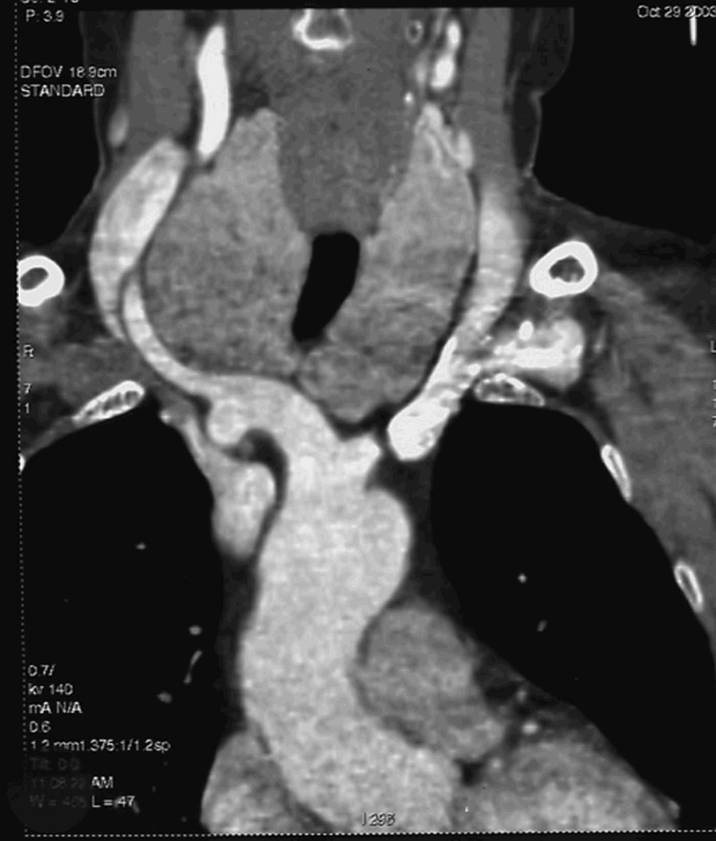
Goitres were classified as: type one (i.e. not reaching the top of the aortic arch); type two (reaching the aortic arch); type three (extending beyond the aortic arch without reaching the carina); and type four (reaching or extending beyond the carina) (see Figures 1–6).

Fig. 1 Computed tomography scan, frontal view, showing cervical goitre without extension into the mediastinum.

Fig. 2 Computed tomography scan, frontal view, showing cervical goitre with extension into the mediastinum.

Fig. 3 Computed tomography scan, axial view, showing right anterior and posterior extensions into the mediastinum.

Fig. 4 Computed tomography scan, axial view, showing crossed extension between the trachea and the oesophagus.

Fig. 5 Computed tomography scan, axial view, showing goitre extension beyond the top of the aortic arch.

Fig. 6 Computed tomography scan, axial view, showing goitre extension reaching the carina.
Surgical management was planned according to: the size of the cervical incision; the need for sternotomy; the need to section the infrahyoid muscles; identification of the parathyroid glands; and identification of the recurrent laryngeal nerve.
Post-operative assessment comprised assessment of serum calcium concentration and laryngeal nerve function.
Results
The ultrasound examination demonstrated all the nodules in the thyroid gland, but did not adequately assess mediastinal extension of the goitre.
The radiological examinations demonstrated the following. Widening of the superior mediastinum was seen on the chest radiograph in 96 per cent of cases. In 4 per cent of cases, the anomalies seen on the chest radiograph were minor, and the chest radiograph was even considered to be normal in four cases (1.79 per cent of cases); however, the CT scans of these cases showed a diving cervical goitre with large, posterior, mediastinal expansion, and this was confirmed during thyroid surgery. Tracheal deviation was found on the chest radiograph in 21 per cent of cases, but the CT scan revealed that the tracheal deviation was associated with compression in 53 per cent of cases. Tracheal compression was visualised on CT scan in 48 per cent of all cases.
Radiological examination of longitudinal mediastinal expansion of the goitres revealed the following. Left-sided anterior (i.e. prevascular) expansion was seen in 76 per cent of cases and right-sided anterior expansion in 58 per cent of cases. Left-sided posterior (i.e. retrovascular) expansion was seen in 24 per cent of cases and right-sided posterior expansion in 16 per cent of cases. Multiple or complex expansions were: bilateral and anterior in 37 per cent of cases; bilateral and posterior in 5.5 per cent of cases; anterior and posterior on the left side in 24 per cent of cases; anterior and posterior on the right side in 10.3 per cent of cases; and crossed in 13 per cent of cases.
The vertical goitre expansion was type one in 59 per cent of cases, type two in 30 per cent, type three in 10 per cent and type four in 1 per cent.
Chest radiography showed a cervicothoracic goitre in 96 per cent of cases, but without any precision concerning the extension of the goitre and the tracheal compression.
Computed tomography findings correlated with surgical findings in 100 per cent of cases.
The surgical procedure was cervical in 98.65 per cent of cases.
Only three sternotomies were performed, in cases of huge, type four goitres, and all before 1999. In two of these cases, the sternotomy was decided on before surgery. In one case, the decision was taken during surgery, because of the occurrence of a serious arterial haemorrhage at the inferior pole of the thyroid gland, probably corresponding to a thyroid ima artery.
The average length of the skin incision was 8.4 cm; it was less than 10 cm in all cases, ranging from 6 to 10 cm. The skin incision was enlarged laterally during surgery in 23 per cent of cases in order to release a very large goitre. Horizontal section of the infrahyoid muscles was performed in 32 per cent of cases.
The post-operative complications were as follows. Definitive inferior laryngeal nerve palsy occurred in five cases (2.24 per cent of all cases) with type three and four goitres. Definitive hypoparathyroidism occurred in 5.3 per cent of cases (up to three years' follow up). Post-operative cervical infection occurred in three cases (1.35 per cent of all cases) with type three goitres. Post-operative bleeding occurred in five cases (2.24 per cent of cases).
Discussion
Frequency
The frequency of substernal goitre reported in the medical literature varies considerably, ranging from 3.5 to 15 per cent of all surgical goitres. Table IReference Maruotti, Zannini, Viani, Voci and Pezzuoli1–Reference Flati, De Giacomo, Porowska, Flati, Gaj and Talarico7 shows some of the reported frequencies. However, the frequency is difficult to determine because of the variable definition of substernal goitre.
Table I Substernal goitre reported in the literature

Definition
There is no clear and precise definition of substernal goitre. SiragusaReference Siragusa, Gelarda, Geraci, Albanese and Di Pace4 emphasised the importance of a definition especially considering the different medisatinal extensions in the longitudinal and vertical planes. Netterville et al. Reference Netterville, Coleman, Smit, Smith, Day and Burkey3 considered a goitre to be substernal when the majority of the goitre was situated in the mediastinum; however, this is not a precise definition. Flati et al. Reference Flati, De Giacomo, Porowska, Flati, Gaj and Talarico7 and Hili et al. Reference Hili, Mayer, Carini, Cantelli and Modigliani2 considered that mediastinal retrosternal extension should represent at least 50 per cent of any goitre labelled as substernal.
We contend that these definitions are not sufficiently precise, and that the definition of substernal goitre should be based on anatomical findings.
A goitre should be considered to be substernal when it passes through the cervicothoracic isthmus below the subclavian vessels. In 1985, Borrely et al. Reference Borrely, Grosdidier and Hubert8 proposed a clinical and surgical classification according to longitudinal and vertical planes, combining double vertical and sagittal directions of mediastinal goitre extension, as follows: right, left, anterior, posterior, and multiple or crossed extensions in the mediastinum.
We have adapted this classification into four types of substernal goitre, on the basis of CT findings.
Chest radiograph vs cervicothoracic computed tomography scan
The anatomical features of substernal goitre can be confirmed by CT scan, which more accurately visualises goitre development than chest radiography. Moreover, in 4 per cent of our cases, the chest radiograph was normal but the CT scan showed a cervicothoracic goitre.
Axial CT slices must be strictly parallel to the cervicothoracic isthmus in order to avoid including the lungs and the lower extremity of the neck above the manubrium on the same slice. Reconstructed sagittal sections may also help to demonstrate that the goitre reaches but does not extend beyond the cervicothoracic isthmus.
A pre-operative CT scan can also visualise the anatomical relations between the goitre and each mediastinal component, the level of extension, and the presence or absence of tracheal or oesophageal compression. Computed tomography is always much more reliable than chest radiography, particularly in the case of isolated posterior extension or anteroposterior tracheal compression, which may not be seen on radiography. Chest radiographs may only indicate the presence of a large substernal goitre when widening of the superior mediastinum is observed at the top of the radiograph. Chest radiography is also quite imprecise concerning tracheal compression.
Computed tomography may also reveal retropharyngeal goitre extensions, which can modify the resonating cavities, leading to dysphonia.
The calcifications visible on CT scanning of the thyroid gland are of limited interest. These were found in 66 per cent of benign goitre cases in our series and therefore cannot be considered a sign of malignancy.
The contribution of CT scanning is not limited to academic classification of substernal goitre. It can also predict possible surgical difficulties and define the surgical strategy, according to the mediastinal position of the goitre.
In the case of posterior, large or low mediastinal extension, the superior thyroid pedicle must be ligated first and the recurrent laryngeal nerve must be identified in the superior part of the thyroid as it enters the larynx (this was performed in 41 per cent of our cases). This is because the inferior laryngeal nerve can be ‘laterally superficialised’ during surgery, especially on the right side, and can be damaged when performing blunt dissection of the lateral lobe or ‘digitoclasy’.
Type one substernal goitre can be treated surgically in the same way as noncervicothoracic goitre. Sternotomy may be necessary for goitres located very low in the mediastinum (i.e. type four). In our series, 11 per cent of goitres descended below the aortic arch, but planned sternotomy was performed in only two cases.
Substernal goitre: literature review
In our study, goitres were most frequently prevascular with unilateral or bilateral extension. Multiple extensions were frequently observed, particularly bilateral anterior and posterior extension. Fifty-nine per cent of goitres had a limited extension in the superior mediastinum, 30 per cent reached the aortic arch and 11 per cent extended beyond the aortic arch.
BlondeauReference Blondeau9 found that thoracic extension could be easily evaluated by chest radiography, ultrasonography or scintigraphy in 86 per cent of a series of 585 substernal goitres. Computed tomography was helpful in 14 per cent of this series (comprising 26 crossed extensions, 26 thoracic goitres and five malignant tumours). However, this series, published in 1994, is relatively old.
In Vadasz and Kotsis'Reference Vadasz and Kotsis10 series of 175 substernal goitres, 79 per cent extended below the aortic arch and 21 per cent were intrathoracic. Hedayati and McHenryReference Hedayati and McHenry11 reported prevascular extension in 94 per cent of cases and retrovascular extension in 6 per cent of cases in a series of 116 substernal goitres. In a series of 237 substernal goitres reported by Torre,Reference Torre, Borgonovo, Amato, Arezzo, Ansaldo, De Negri, Ughe and Mattioli12 59.5 per cent were anterior goitres, 11.5 per cent were posterior goitres and 29 per cent were complex goitres, according to the classification of Borrely et al. Reference Borrely, Grosdidier and Hubert8
Maruotti et al. Reference Maruotti, Zannini, Viani, Voci and Pezzuoli1 analysed the chest radiographs for 151 substernal goitre cases. They found mediastinal widening with tracheal deviation in 84 per cent of cases, extension to the right in 55 per cent, extension to the left in 37 per cent and bilateral extension in 6 per cent.
Hili et al. Reference Hili, Mayer, Carini, Cantelli and Modigliani2 subdivided substernal goitres into three categories: largely intrathoracic (i.e. more than 80 per cent of the goitre volume); partially intrathoracic (i.e. between 50 and 80 per cent); and other (i.e. less than 50 per cent of the volume). In this series, 97 goitres were entirely intrathoracic and 68 reached the aorta arch.
Al Suliman et al.,Reference Al-Suliman, Graversen and Blichert-Toft13 in their series of 195 goitres, compared the sensitivities of various types of investigation. They found sensitivies of 52 per cent for physical examination, 33 per cent for scintigraphy and 5 per cent for chest radiography.
• This study attempted to analyse and compare chest radiographs and cervicothoracic computed tomography (CT) scans performed before thyroid surgery in order to locate and predict thoracic extension of substernal goitres
• Anterior or prevascular extension of the goitre were the most frequent modes of extension, being found in 76 per cent of cases, especially on the left side
• Thorough examination of CT scans is essential to identify and classify substernal goitres and to decide the best surgical procedure
• A cervical surgical procedure is almost always sufficient and safe for the surgical management of substernal goitres
BelardinelliReference Belardinelli, Gualdi, Ceroni, Guadalaxara, Polettini and Pappalardo14 compared CT and magnetic resonance imaging (MRI) and concluded that MRI more accurately visualised mediastinal goitre extension. We performed MRI in only three of our patients, but the anatomical findings were reliable. According to Rodriguez et al.,Reference Rodriguez, Hernandez, Pinero, Ortiz, Soria and Ramirez15 the best imaging modalities in term of sensitivity are CT scan (100 per cent), chest radiography (75 per cent), scintigraphy (19 per cent) and ultrasonography (15 per cent).
Conclusion
The definition of substernal goitre should be based on anatomical findings. A goitre must be considered to be substernal when the inferior pole passes through the cervicothoracic isthmus below the subclavian vessels.
Pre-operative cervicothoracic CT scanning can be used to assess and classify substernal goitres. The anatomical findings demonstrated by CT can also help to guide the surgical strategy and to predict possible surgical difficulties.
We propose an anatomical classification of four types of substernal goitre. The surgical management of small, benign, multinodular, substernal goitre (type one) is very similar to that of benign, cervical goitres. The surgical management of larger substernal goitre (i.e. types three and four) requires craniocaudal thyroidectomy after first identifying the recurrent laryngeal nerve as it enters the larynx. Sternotomy is now only necessary in exceptional cases.


