


Introduction
Haemangiomas are benign tumours originating in the vascular tissue of the skin, mucosa, muscles, glands and bone.Reference Takeda, Takenaka and Hashimoto1 Lobular capillary haemangioma (also known as pyogenic granuloma) is a rapidly growing, benign, fibrovascular lesion with extensive endothelial proliferation, of unknown aetiology.Reference Jones, Nguyen and Tabaee2–Reference Choudhary, MacKinnon, Morrissey and Tan4 The occurrence of such lesions during pregnancy has resulted in the popular term ‘pregnancy tumour’.Reference Jones, Nguyen and Tabaee2 These lesions grow rapidly. They have a proclivity for the face, fingers and toes, with almost 60 per cent occurring in the head and neck region.Reference Lim, Singh, Prasad, Chan, Lam and Ratnam5, Reference Simic, Vlahovic and Subarevic6 The oral gingiva is the commonest site, although the lesion is also likely to occur on the lips, tongue, buccal mucosa and palate.Reference Choudhary, MacKinnon, Morrissey and Tan4, Reference Lance, Schatz, Nach and Thomas7 The lesion rarely occurs subcutaneously, intravenously or in the gastrointestinal tract.Reference Zarrinneshan, Zapanta and Wall8 Nasal haemangiomas are rare, arising mostly from the soft tissues of the nasal cavity, and occurring more frequently on the septum (65 per cent), lateral wall (18 per cent) and vestibule (16 per cent).Reference Takeda, Takenaka and Hashimoto1
Pyogenic granuloma gravidarum occurs as an oral or nasal lesion. Its incidence during pregnancy ranges from less than 2 per cent to approximately 5 per cent.Reference Jones, Nguyen and Tabaee2, Reference Choudhary, MacKinnon, Morrissey and Tan4 Most of these lesions are small, and they normally involute spontaneously after childbirth.Reference Choudhary, MacKinnon, Morrissey and Tan4
The management of a pregnant woman with such a lesion may be complex, and depends on the severity of symptoms and the status of the pregnancy.Reference Jones, Nguyen and Tabaee2 If massive haemorrhage is encountered during the third trimester, consideration should be given to the gestational age of the fetus and the possible consequences on the outcome of the pregnancy. Uncontrolled haemorrhage may warrant medication to accelerate fetal lung maturation, induction of labour or caesarean section.Reference Jones, Nguyen and Tabaee2
We present a case of a pregnant woman with a rapidly growing mass within the nasal cavity, which was imaged by computed tomography (CT) and magnetic resonance imaging (MRI) at two weeks post partum, and which required pre-operative, super-selective embolisation and subsequent removal under general anaesthesia.
Case report
A 29-year-old, pregnant woman was referred by her gynaecologist to our department at 38 weeks' gestation with a two-month history of left-sided nasal obstruction. She had a history of recurrent epistaxis with nasal obstruction. There was no other history of trauma or nasal irritation. Her past medical history was otherwise unremarkable, and her pregnancy had been uneventful.
Anterior rhinoscopic examination revealed a large, grey to pink, polypoidal mass completely blocking the left anterior nasal cavity (Figure 1). The posterior extension of the mass could not be established by endoscopy.
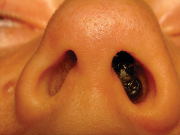
Fig. 1 Clinical photograph showing the lobular capillary haemangioma completely obstructing the left anterior nasal cavity.
An initial biopsy, performed at another hospital, was suggestive of lobular capillary haemangioma.
As there was a concern that epistaxis may occur during delivery, we discussed the patient's case with her obstetrician, together with the radiologist and the hospital anaesthesia service. It was decided to follow the patient until delivery.
Two days before delivery, treatment with methylprednisolone (1 mg per kg) was commenced.
The delivery went smoothly, without epistaxis.
Two weeks after delivery, CT and MRI of the nose and sinuses were performed. The CT revealed a sessile mass near the head of the inferior turbinate, filling the anterior part of the left nasal cavity and extending close to the alar cartilage (Figure 2). No bony erosion was detected, suggesting a benign nature. The MRI revealed a well vascularised, 2.2 × 1.2 cm mass obstructing the left nasal cavity (Figure 3).

Fig. 2 Axial computed tomography scan demonstrated a soft tissue opacity extending from the left nasal vestibule to the head of the inferior turbinate, without bony erosion or invasion of the paranasal sinuses. R = right; P = posterior

Fig. 3 Axial magnetic resonance imaging scan showing a well circumscribed mass obstructing the left nasal cavity.
During the first month post partum, the lesion reduced slightly in size. However, a few weeks later, following cessation of corticosteroid treatment, it began to grow again. Due to the lesion's continued post-partum growth, it was decided that the patient required surgical intervention.
Pre-operative, super-selective embolisation was performed one day before surgery. An angiogram confirmed the hypervascularity of the lesion, which was supplied by a very large transverse facial artery and by branches of the sphenopalatine artery (Figure 4).

Fig. 4 Super-selective angiograms showing the haemangioma supplied by a large transverse facial artery and by branches of the sphenopalatine artery.
At operation, a mass of bleeding tissue was endoscopically excised from its mucosal attachment points, using bipolar cautery. The mass was attached to the lateral wall of the nasal cavity, adjacent to the head of the inferior turbinate. The mass was soft, mobile and nonpulsatile, and measured 2.5 × 1.5 × 1 cm. Histopathological examination (Figure 5) confirmed the provisional diagnosis from the initial biopsy.

Fig. 5 Photomicrograph of the lesion (surgical specimen), showing stromal sclerosis and thin-walled ectatic blood vessels in a lobular arrangement. (H&E; original magnification ×4)
The patient reported immediate relief of her symptoms. She had an uneventful post-operative course, with no recurrence after six months of follow up.
Discussion
The term pyogenic granuloma was first used in 1904 to describe a single mass of granulomatous tissue. The first case of pyogenic granuloma of the nose was described in 1940.Reference Frank and Blahd9 In 1980, Stacey Mills suggested the term lobular capillary haemangioma, arguing that pyogenic granuloma was a misnomer as the tumour was neither pyogenic nor granulomatous and had well described histological characteristics.Reference Kapella, Panosetti, Rombaux, Delos and Weynand3
Pyogenic granuloma, otherwise known as granuloma gravidarum, granuloma pyogenicum, lobular capillary haemangioma, pregnancy tumour, pregnancy granuloma and telangiectatic polyp, is a common lesion of the skin and mucous membranes.Reference Lance, Schatz, Nach and Thomas7, Reference Zarrinneshan, Zapanta and Wall8 In order of decreasing frequency, most authors list the gingiva, lips, tongue, buccal mucosa and palate as the most commonly involved locations.Reference Jones, Nguyen and Tabaee2 The tumour is more commonly found as a skin lesion in males and as a mucosal lesion in females.Reference Zarrinneshan, Zapanta and Wall8 It is characterised by granulation tissue which is similar to that found in any healing wound, but which is more exuberant and confined to a localised area. Extreme endothelial proliferation and the formation of numerous vascular spaces are the most constant histological features.Reference Lance, Schatz, Nach and Thomas7 The lesion has a characteristic lobular arrangement of capillaries within a network of fibrovascular tissue at its base, and this has led to growing use of the term lobular capillary haemangioma.Reference Jones, Nguyen and Tabaee2, Reference Nichols, Gaffey, Mills and Weiss10
The lesion may develop at any age, but the incidence is greatest in the third decade, with a slight female predominance.Reference Kapella, Panosetti, Rombaux, Delos and Weynand3, Reference Nichols, Gaffey, Mills and Weiss10, Reference Puxeddu, Berlucchi, Ledda, Parodo, Farina and Nicolai11 After the age of 40 years, the sex ratio appears to be equal.Reference Kapella, Panosetti, Rombaux, Delos and Weynand3
Lobular capillary haemangiomas show an unusual predilection for women in their reproductive years.Reference Nichols, Gaffey, Mills and Weiss10 The lesion may develop at any point during pregnancy, but mainly occurs during the last two trimesters.Reference Jones, Nguyen and Tabaee2, Reference Zarrinneshan, Zapanta and Wall8 Lobular capillary haemangioma has been associated with oral contraceptive use. These clinical features suggest that this tumour may be hormone-sensitive.Reference Nichols, Gaffey, Mills and Weiss10
The pathogenesis of lobular capillary haemangioma is poorly understood. No specific aetiological agent has been identified, and the lesion is considered to be an inflammatory overgrowth rather than a true neoplasm. However, as it can grow rapidly and bleed, it may be mistaken for a malignant tumour.Reference Lance, Schatz, Nach and Thomas7, Reference Scott and van Hasselt12 The tumour's pathogenesis has been postulated to involve hormonal influences, trauma, viral oncogenes, underlying microscopic arteriovenous malformations and angiogenic growth factor production.Reference Zarrinneshan, Zapanta and Wall8, Reference Puxeddu, Berlucchi, Ledda, Parodo, Farina and Nicolai11
Lesions which develop in pregnancy, or with oral contraceptive or retinoid therapy, show regression after parturition or withdrawal of the causative drug, respectively.Reference Zarrinneshan, Zapanta and Wall8 During pregnancy, levels of oestrogen and progesterone are markedly elevated, and this could stimulate endothelial proliferation within mucosal surfaces in the oral and nasal cavities.Reference Choudhary, MacKinnon, Morrissey and Tan4 However, studies assessing the number of oestrogen and progesterone receptors within lobular capillary haemangioma tissue samples have found similar numbers of receptors in samples from men, pregnant women and nonpregnant women.Reference Jones, Nguyen and Tabaee2, Reference Choudhary, MacKinnon, Morrissey and Tan4, Reference Nichols, Gaffey, Mills and Weiss10 Nevertheless, we cannot exclude the possibility that oestrogen and progesterone receptors are expressed transiently at an early stage of lesion development. It seems that the higher levels of circulating hormones associated with pregnancy are more important to lobular capillary haemangioma pathogenesis than the number of hormone receptors.Reference Jones, Nguyen and Tabaee2
Of the reported traumatic factors, habitual nose-picking is considered to be the most significant, followed by nasal packing.Reference Puxeddu, Berlucchi, Ledda, Parodo, Farina and Nicolai11 A traumatic aetiology during pregnancy is often a result of local irritation, within the general proinflammatory hormonal environment of pregnancy.Reference Jones, Nguyen and Tabaee2
Lobular capillary haemangioma usually develops at the site of a pre-existing injury, and evolves rapidly over a period of some weeks to a maximum size of 2 cm. It typically regresses, over several months, to become a fibroma.Reference Zarrinneshan, Zapanta and Wall8 Involution coincides with increased apoptosis of endothelial and stromal cells.Reference Simic, Vlahovic and Subarevic6
• Lobular capillary haemangioma (pyogenic granuloma) is a rapidly growing, benign, fibrovascular lesion with extensive endothelial proliferation, of unknown aetiology
• It is commonest in women of reproductive age; its incidence in pregnant women ranges from <2 per cent to approximately 5 per cent
• Nasal haemangiomas are rare
• Management of nasal haemangioma in pregnancy may be complex, and depends on symptom severity and pregnancy status
• In pregnant women, lobular capillary haemangioma usually regresses in one to two months post partum; surgical excision may be required if the lesion persists
Epistaxis is the major symptom reported, followed by nasal obstruction and protrusion of the lesion from the nose.Reference Jones, Nguyen and Tabaee2, Reference Kapella, Panosetti, Rombaux, Delos and Weynand3, Reference Zarrinneshan, Zapanta and Wall8
On CT, lobular capillary haemangioma appears as a unilateral mass of soft tissue density which usually fills the nasal cavity, while MRI shows T2 hyperintensity and spontaneous T1 hypointensity.Reference Kapella, Panosetti, Rombaux, Delos and Weynand3, Reference Puxeddu, Berlucchi, Ledda, Parodo, Farina and Nicolai11 Some rare cases of bony erosion due to compression have been described.Reference Kapella, Panosetti, Rombaux, Delos and Weynand3 Angiography typically shows increased vascularity in the area of the tumour, with feeder vessels but no large draining veins.Reference Takeda, Takenaka and Hashimoto1
Corticosteroids are the first-line treatment for nasal haemangioma; they give excellent results in 30 per cent of cases, and retard growth in a further 40 per cent.Reference Simic, Vlahovic and Subarevic6 Recently, propranolol has proven effective in inducing regression of growing haemangiomas by causing vasoconstriction. Several reports have confirmed prompt haemangioma response to propranolol, with no major side effects.Reference Simic, Vlahovic and Subarevic6 Our patient was treated with corticosteroids but not propranolol, to avoid side effects for her baby.
Although lobular capillary haemangiomas occurring in pregnant women usually regress in one to two months post partum, surgical excision may be required if the lesion fails to resolve.Reference Zarrinneshan, Zapanta and Wall8 The mainstay of treatment is complete surgical excision of the entire lesion with complete desiccation of the base, with or without pre-operative embolisation.Reference Takeda, Takenaka and Hashimoto1, Reference Kapella, Panosetti, Rombaux, Delos and Weynand3, Reference Zarrinneshan, Zapanta and Wall8 In our patient's case, the initial biopsy (performed in another hospital) was haemorrhagic. Therefore, to facilitate removal and to reduce the risk of intra-operative bleeding, we decided to embolise the lesion before surgery.
Recurrence of lobular capillary haemangioma is usually due to incomplete excision.Reference Zarrinneshan, Zapanta and Wall8 Following surgical excision, complete diathermy desiccation of the lesion bed decreases the risk of recurrence.Reference Kapella, Panosetti, Rombaux, Delos and Weynand3
In pregnant women, surgical treatment is usually deferred until after childbirth, and is reserved for lesions that fail to regress completely.Reference Choudhary, MacKinnon, Morrissey and Tan4 However, lesions which bleed excessively during pregnancy should also be excised.Reference Zarrinneshan, Zapanta and Wall8
Promising results have been reported for various new treatments, e.g. laser therapy, alitretinoin gel and sclerosing agents.Reference Zarrinneshan, Zapanta and Wall8
Conclusion
Haemangiomas are commonly occurring, benign, vascular proliferations of unknown pathogenesis. Microtrauma and pregnancy are the most commonly proposed aetiological factors. These lesions most commonly affect pregnant women, although in such cases the tumour is clinically and histologically indistinguishable from lobular capillary haemangioma occurring in men and nonpregnant women. Because these lesions can grow rapidly and bleed, they can be mistaken for malignant tumours. A diagnosis of lobular capillary haemangioma should be considered in any pregnant woman with a mass within the mouth or nasal cavities.
Although these lesions generally regress spontaneously after pregnancy, anxiety about malignancy and distress due to nasal obstruction and epistaxis often mean that excision is the optimal course of management. If the lesion has not regressed spontaneously after delivery, complete surgical excision, with or without pre-operative embolisation, is the treatment of choice.
Acknowledgments
We thank S Norrenberg for supplying the photomicrograph, and M Lubicz for supplying the angiogram.


