


Mastitis is the most economically important disease in dairy herds worldwide, and it is caused by more than 100 bacterial species, even if several risk factors, including host susceptibility are relevant in the epidemiology of the disease.
Non-specific (innate) immune response plays a major role in defending the udder from bacterial invasion (Zecconi & Smith, Reference Zecconi and Smith2003; Rainard & Riollet, Reference Rainard and Riollet2006). Udder immune response has similar characteristics to the lung immune response. Indeed, innate cellular [polymorphonuclear neutrophils (PMN) and macrophages] and soluble immune defences [cytokines, lysozyme, lactoferrin, defensins and N-acetyl-β-d-glucosaminidase (NAGase)] are all involved in preventing bacterial adhesion, multiplication and in bacteria killing (Zecconi & Smith, Reference Zecconi and Smith2003; Burton & Erskine, Reference Burton and Erskine2003). Moreover, recent investigations suggest that mammary gland epithelial cells (MGEC) could have a large and important role as a source of soluble components of immune defences (Piccinini et al. Reference Piccinini, Binda, Belotti, Daprà and Zecconi2007; Rainard & Riollet, Reference Rainard and Riollet2006).
Despite many attempts to find other ways to control/prevent mastitis (i.e. vaccine) antimicrobial therapy is still the most used and effective means of curing clinical and subclinical mastitis. However, drug concentrations are largely lower and treatments have a shorter duration in food-producing animals in comparison with the ones applied in human medicine. Therefore, efficacy of therapy is dependent not only on the antimicrobial activity but also on the positive interactions with host innate immune response. Surprisingly, information on these interactions is rather scarce (Nickerson et al. Reference Nickerson, Paape and Dulin1985; Zecconi et al. Reference Zecconi, Piccinini, Bronzo and Casula1996; Hoeben et al. Reference Hoeben, Burvenich and Heynemann1997; Zecconi & Piccinini, Reference Zecconi and Piccinini2001).
The availability of stabilized cell lines such as BME-UV developed by Zavizion et al. (Reference Zavizion, van Duffelen, Schaeffer and Politis1996) allows the exploring of the interactions between bacteria and MGEC, including the innate immune response and opens the way to assess also the role of antimicrobials on both bacteria and MGEC (Didier & Kessel, Reference Didier and Kessel2004; Fitzgerald et al. Reference Fitzgerald, Meade, McEvoy, Lillis, Murphy, Machugh and Baird2007). Thus, a simple in-vitro model applying BME-UV cells as a substrate, Staphylococcus aureus isolates as challenge and tylosin as treatment was developed. Staph. aureus is the major contagious pathogen worldwide (Zecconi & Piccinini, Reference Zecconi, Piccinini, Kaske, Scholz and Holtershinken2002; Zecconi, Reference Zecconi2007) and it can adhere and invade MGEC (Lammers et al. Reference Lammers, Nuijten, Kruijt, Stockhofe-Zurwieden, Vecht, Smith and van Zijderveld1999; Dego et al. Reference Dego, van Dijk and Nederbragt2002). Macrolides are antimicrobials characterized by having a 12- to 16-member lactone ring and they are classified according to the number of atoms of the ring. Tylosin is a natural macrolide of the 16-ring class, isolated from Streptomyces fradiae, and it is a weak base and highly soluble in lipids. Macrolides such as tylosin inhibit protein synthesis by binding to 50S ribosomal subunits and are bacteriostatic (Giguere, Reference Giguere, Giguere, Prescott, Baggot, Walker and Dowling2007). Macrolides penetrate the cellular membrane and accumulate within the cells, as shown also for tylosin (Scorneaux & Shryock, Reference Scorneaux and Shryock1999). There is increasing evidence of their anti-inflammatory and immunomodulating properties, at least for the molecules used in human medicine (Zalewska-Kaszubska & Gorska, Reference Zalewska-Kaszubska and Gorska2001; Labro, Reference Labro2004a,Reference Labrob; López-Boado & Rubin, Reference López-Boado and Rubin2008). Among macrolides, tylosin is one of the most used molecules for mastitis therapy both in lactation and in the drying-off period (Ziv, Reference Ziv1980b; Bolourchi et al. Reference Bolourchi, Hovareshti and Tabatabayi1995; Zecconi et al. Reference Zecconi, Piccinini and Guarini1999; Bonnier et al. Reference Bonnier, Dore, Amedeo and Guerin-Faublee2006). Some information on the interactions between tylosin and leucocytes is available, but not for other macrolides used in mastitis therapy (Zecconi et al. Reference Zecconi, Piccinini, Bronzo and Casula1996; Chin et al. Reference Chin, Lee, Murrin, Morck, Merrill, Dick and Buret2000; Zecconi & Piccinini, Reference Zecconi and Piccinini2001; Cao et al. Reference Cao, Dong, Shen, Wu, Wu, Du, Wang, Qi and Li2006). Based on this information we selected tylosin to assess its potential activities on Staph. aureus-infected MGEC.
The aim of this paper was to assess the potential effects of antimicrobials on the immune system when invading bacteria are present, by applying a simple experimental model based on the BME-UV cell line, Staph. aureus as a challenge and a macrolide as antimicrobial. The BME-UV cell line was selected because it expresses immunological and inflammatory molecules; Staph. aureus because it is an intra- and extra-cellular mastitis pathogen and tylosin because it has an intra- and extra-cellular activity against Gram-positive mastitis pathogens.
Materials and Methods
Bacteria characteristics
Ten Staph. aureus isolates from subclinical mastitis cases in dairy cows of different herds were considered. Isolates were selected from our strain collection based on their genetic characteristics and antimicrobial susceptibility. Indeed, they have different virulence gene patterns (efb, spa, cna) and leukocidin genes (pvl, hla, lukDE, lukM) as described elsewhere (Zecconi et al. Reference Zecconi, Binda, Borromeo and Piccinini2005; Zecconi et al. Reference Zecconi, Cesaris, Liandris, Daprà and Piccinini2006). The minimum inhibitory concentration (MIC) for tylosin was assessed for each isolate by means of the agar dilution method following the guidelines of the Clinical and Laboratory Standards Institute (NCCLS, 2000; NCCLS, 2002). Tylosin for these assays and for the experiment was provided by Elanco, Eli Lilly (Italy).
Epithelial cells
A clonal cell line (BME-UV) established from udder primary epithelial cell synthesizing several milk components (Zavizion et al. Reference Zavizion, van Duffelen, Schaeffer and Politis1996) was used as an in-vitro model. Previous reports show that this cell line can express several cytokines when stimulated by bacteria or toxins (Didier & Kessel, Reference Didier and Kessel2004; Fitzgerald et al. Reference Fitzgerald, Meade, McEvoy, Lillis, Murphy, Machugh and Baird2007).
Experimental design
BME-UV cells were exposed to different Staph. aureus isolates in the logarithmic growth phase at a final concentration of 105 CFU, assessed spectrophotometrically, and incubated in a CO2 incubator at 37°C for 2 h to allow cell invasion by bacteria. Each isolate was assessed separately. Then, tylosin at a concentration of 1/3 of the MIC (1/3×), the MIC (1×) and 3-times the MIC (3×) of the respective isolate, was added to the infected cell line and incubated at 37°C for 14 h in a CO2 incubator. The antimicrobial was used at different concentrations to assess the presence of a dose-dependent effect and the influence of viable bacteria count. At the end of the incubation time, supernatant and cells were collected to assess bacteria count and for biochemical and molecular assays.
Sample preparation
At the end of the incubation period cell monolayer supernatants were pipetted into sterile plastic tubes and 1000 μl used for bacteria counts, while 1000 μl was stored at −20°C for biochemical assays. To lyse extracellular bacteria, 40 μl of lysostaphin (Sigma-Aldrich, Italy) in 500 μl of HBSS (Sigma-Aldrich, Milan, Italy) was added to each sample and incubated at 37°C for 30 min to destroy Staph. aureus adhering to cells. Then, the cell monolayer was trypsinized and the cell suspension was centrifuged at 700 g for 10 min. Pellet samples used for cytokine assays were stored at −80°C, while pellet samples used for enzyme assays and bacteria counts were suspended in 500 μl of saline solution and stored at −20°C.
Bacteria count
Extracellular and intracellular Staph. aureus counts were performed by the standard dilution method. One-hundred μl of sample dilutions (up to 10−3) were plated on blood agar plates and incubated at 37°C for 18 h.
Cytokine assays
Total RNA was extracted from cells with RNAqueos kit (Ambion Inc., Austin TX, USA), while reverse transcription was performed with Quanti Tect kit (Qiagen, D). Real-time PCR systems for bovine GAPDH and the cytokines were run in triplicate with probes described in Table 1 and commercially available master mix for Sybr green (Power Sybr Green Master Mix, Applied Biosystems, Foster City CA, USA). Real-time Q-RT-PCRs were performed by using the Opticon 2 detection system (BioRad, Milan, Italy) and expression rate was calculated with Rest 2005 software (Pfaffl et al. Reference Pfaffl, Horgan and Dempfle2002). Cytokine expression was assessed as expression rate compared with a reference represented by BME-UV cells not exposed to Staph. aureus and not treated with tylosin. A preliminary trial showed that without bacterial challenge and in the presence of tylosin, changes in cytokine expression rates were not detectable. Therefore, a unchallenged tylosin-treated sample was not included in the experiment.
Table 1. Description of primer used in amplification of bovine GAPDH gene and cytokine genes

† Designed from NM_174088 Bos taurus sequence
Biochemical assays
Lysozyme (LYZ) was assessed in duplicate by a fluorescence-based procedure (EnzChek Lysozyme Kit, Invitrogen, Carlsbad CA, USA). The method is based on lysis of Micrococcus lysodeycticus labelled with fluorescine to such a degree that fluorescence is quenched. LYZ activity is measured by changes of fluorescence on a microplate fluorimeter at 355 nm exc and 460 nm em (Ascent, Thermo Labsystem, FL) against a standard curve obtained for each test with a range of 8–500 units. One unit of LYZ is defined as the quantity of enzyme that produces a decrease in turbidity of 0·0001 OD units per min at 450 nm measured at pH 7·0 (25·8°C) using 0·3 mg/ml. NAGase (NAG) was assessed in duplicate by the procedure described by Kitchen et al. (Reference Kitchen, Middleton and Salmon1978) and expressed as units (pmol of 4-methylubelliferon released per min at 25·8°C catalysed by 1 ml of milk) on a microplate fluorimeter at 355 nm exc and 460 nm em (Ascent, Thermo Labsystem, FL).
Statistical analysis
Staph. aureus virulence patterns were clustered by the UPGAMA method and clusters were defined by the presence of a relatedness <80% between isolates as described by Piccinini et al. (Reference Piccinini, Cesaris, Daprà, Borromeo, Picozzi, Secchi and Zecconi2008).
Statistical analysis on response variables was performed by GLM procedure of SAS software (SAS rel 9.2, Cary NC, USA) with bacteria counts, cytokine expression rates and enzyme concentrations as response variables and treatment, bacteria and virulence factors clusters as independent variables.
Results
MIC values of Staph. aureus isolates were in the range 0·625–5·0 mg/l. All the isolates were positive for hla gene, while for the other genes different patterns were observed. However, statistical analysis did not show any significant influence of isolates on the response variables. Therefore, they were not considered further.
Antibacterial activity
All Staph. aureus isolates were found intracellularly, independently of the exposure to antimicrobial treatment. Analysis of antimicrobial activity measured in the supernatant of the cell cultures (Table 2) showed a numerical linear decrease of bacteria concentration as tylosin concentration increased, with significant differences among 1/3× and the other two treatment levels (1× and 3×).
Table 2. Extracellular and intracellular Staphylococcus aureus mean counts (±sd) of BME-UV cells exposed to different levels of tylosin (see text for details of concentrations) for 14 h
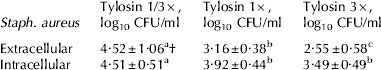
† Mean values in a row with different letters are statistically different (P<0·05)
The same pattern was observed for intracellular killing of Staph. aureus (Table 2) with an increasing killing rate as tylosin concentration increased. Differences among treatment groups were always significant (P<0·05).
Cytokine expression
Analysis of IL-1 expression rate (Table 3) showed a significant reduction of IL-1 in tylosin-treated samples when compared with the untreated sample, without any significant difference among treatment groups.
Table 3. Relative mean expression rate (±sd) of cytokines in BME-UV cells exposed to Staphylococcus aureus and to different levels of tylosin (see text for details) compared with untreated and unexposed controls

† Mean values in a row with different letters are statistically different (P<0·05)
Analogously, the expression rates observed for IL-6 (Table 3) were significantly (P<0·05) reduced in tylosin-treated cells when compared with matching untreated controls, with significant differences among treatment groups.
When TNF expression rate was considered (Table 3) the pattern observed was reversed in comparison with IL-1 and IL-6 with rather small increases of expression rates in treated samples, in comparison with untreated controls. However, none of the differences observed was statistically significant. The same pattern was observed for IL-8 (Table 3) but with a large increase of expression rate in treated samples, without significant differences.
Lysosomal enzymes
When lysosomal enzymes were considered, a significant increase (P<0·05) in intracellular NAG (Table 4) was observed in tylosin-treated cells, when compared with untreated control. However, no differences were observed among treatment groups. Extracellular NAG values were much lower than intracellular ones and without significant differences among groups.
Table 4. Mean intracellular and extracellular concentration (±sd) of NAGase and lysozyme in BME-UV cells exposed to Staphylococcus aureus and to different levels of tylosin (see text for details)

† Mean values in a row with different letters are statistically different (P<0·05)
Lysozyme concentration was significantly lower both intracellularly and extracellularly, when compared with untreated controls (Table 4). In both cases the differences between control and respective treatment groups were statistically significant, while significant differences among treatment groups were not observed.
Discussion
In the past, leucocytes were considered the main source of molecules involved in inflammatory and immunological responses. It has been shown that other cells could be an important source of inflammatory and immunological mediators. Indeed, lung epithelial cells could modulate the inflammatory response in the airways and modulate cell recruitment through producing chemokines, cytokines, receptors and adhesion molecules (López-Boado & Rubin, Reference López-Boado and Rubin2008). Similarly, udder epithelial cells could produce both cytokines and immunomodulating molecules (Didier & Kessel, Reference Didier and Kessel2004; Rainard & Riollet, Reference Rainard and Riollet2006; Fitzgerald et al. Reference Fitzgerald, Meade, McEvoy, Lillis, Murphy, Machugh and Baird2007; Piccinini et al. Reference Piccinini, Binda, Belotti, Daprà and Zecconi2007). Thus, the role of MGEC cannot be ignored in performing mastitis pathogenesis and therapy studies, and particularly when the interactions between bacteria and host are of interest.
Antimicrobials are still the most used tool to control and cure clinical and subclinical mastitis worldwide, and their efficacy is usually associated with their direct effect on bacteria. However, the efficacy of antimicrobial therapy for mastitis should not only be related to direct antimicrobial activities but also to its interactions with the host immune system. This is particularly true when bovine mastitis treatment is considered, because the quantity and the length of the treatments are respectively lower and shorter than optimal levels, to limit the cost of the treatment and milk withdrawal time (Ziv, Reference Ziv1980a; Wagner & Erskine, Reference Wagner, Erskine, Giguere, Prescott and Desmond Baggot2006).
Although there are plenty of papers on antimicrobial activity in mastitis treatment very few addressed the interactions with the immune system (Nickerson et al. Reference Nickerson, Paape and Dulin1985; Zecconi et al. Reference Zecconi, Piccinini, Bronzo and Casula1996; Hoeben et al. Reference Hoeben, Burvenich and Heynemann1997; Zecconi & Piccinini, Reference Zecconi and Piccinini2001). These kinds of studies are indeed difficult and expensive; thus, the availability of in-vitro models to assess both the interactions between bacteria and epithelial cells and the immune/inflammatory response could be helpful to assess in a holistic way antimicrobial activity. We applied an in-vitro model, designed to mimic as closely as possible what happens in a mammary gland when a new infection occurs and milk leucocytes are few and not activated. Therefore, MGEC were used as a substrate, different Staph. aureus isolates able to invade cells represented the challenge and the incubation time of 14 h resembles the interval between milkings, when both bacteria grow and antimicrobial works. In this model a macrolide, tylosin, was used as a prototype to assess interactions of antimicrobials with MGEC because macrolides are known to be active both intra- and extra-cellularly and to have immune/anti-inflammatory effects in man.
All the Staph. aureus isolates were shown to be able to invade epithelial cells in a very short time and to multiply, even when a sub-optimal dose of antimicrobial was applied. An influence of isolate characteristics on cellular response to the invasion and on antimicrobial treatment was expected, but the statistical analysis did not show any significant results. These unexpected results could be explained by the relatively high dose of inoculum used (105 CFU) or by a weak relationship between virulence pattern considered to classify isolates and their competence to invade cells.
The present results confirmed that tylosin had no effects on cell viability and functions. Moreover, a statistically significant antimicrobial activity against both intracellular and extracellular Staph. aureus in BME-UV cells was observed, confirming previous data on milk leucocytes (Zecconi et al. Reference Zecconi, Piccinini, Bronzo and Casula1996). Therefore, our data suggest that when intracellular bacteria are involved, antimicrobials with intracellular activity could be helpful in improving bacteria clearance.
Previous studies show that macrolides, including tylosin can have an anti-inflammatory/immunomodulating effect by inhibiting the prostanoid pathway and by down-regulating pro-inflammatory cytokines production, such as TNF, IL-1, IL-6, 6-keto-PGF1a, and NO (Ianaro et al. Reference Ianaro, Ialenti, Maffia, Sautebin, Rombola, Carnuccio, Iuvone, D'Acquisto and Di Rosa2000; Cao et al. Reference Cao, Dong, Shen, Wu, Wu, Du, Wang, Qi and Li2006). In our study, we observed a significant down-regulation of IL-1 and IL-6, while TNF and Il-8 expression rates numerically increased, but differences were not significant. Previous studies were performed using mainly leucocytes, and observing these effects also in epithelial cells supports the presence of the anti-inflammatory/immunomodulating activity of tylosin. It is worth noticing that the absence of a down-regulation of chemotactic chemokine IL-8, which would affect the cellular response, can be considered a positive outcome.
To our knowledge, this is the first paper assessing the concentration of two lysosomal enzymes, lysozyme and NAGase, in Staph. aureus-stimulated MGEC. Even though both enzymes have a similar substrate as a specific target for their activity, the results obtained suggest different secretion mechanisms. This was not completely unexpected because differences were also observed in NAG and LYZ concentrations in bovine milk of healthy animals (Piccinini et al. Reference Piccinini, Binda, Belotti, Daprà and Zecconi2007). In the present study, extracellular NAG levels were not affected by treatment, but a significant increase was observed intracellularly. Both intracellular and extracellular LYZ levels were significantly decreased in tylosin-treated cells, without differences among treatment group. Both enzymes have acetyl-glucosamine as a target. This molecule is a component of bacterial cell walls, but it is also heavily involved in several biochemical pathways in Golgi apparatus and lysosomes (Cooper & Hausman, Reference Cooper and Hausman2007). It is well known that LYZ activity plays a role in the host immune defences by killing ingested bacteria in the phagolysosomes and by the control of colonization through exocytosis (Zecconi & Smith, Reference Zecconi and Smith2003). In this latter case, the killing activity is related to the damage of bacteria cell walls rich in acetyl-glucosamine, and to other enzymic means not yet completely investigated (Ganz, Reference Ganz2004). NAGase is a glycosidase known to be produced in tubular epithelial cells. In this latter case, tubular dysfunction led to an increased release of this enzyme, and therefore, it is considered as a marker of inflammation in kidney diseases (Bazzi et al. Reference Bazzi, Petrini, Rizza, Arrigo, Napodano, Paparella and D'Amico2002). It is also detectable in milk and its levels are correlated with stage of lactation in healthy cows (Piccinini et al. Reference Piccinini, Binda, Belotti, Daprà and Zecconi2007) or inflammation (Kitchen et al. Reference Kitchen, Seng Kwee, Middleton and Andrews1984). The patterns observed in the present study support the suggestion that this enzyme is much more involved in intracellular biochemical pathways (Cooper & Hausman, Reference Cooper and Hausman2007) than in a direct antibacterial activity, even if its release could be enhanced by cellular dysfunctional status (Bazzi et al. Reference Bazzi, Petrini, Rizza, Arrigo, Napodano, Paparella and D'Amico2002) or bacterial stimuli (Kelly & Carchman, Reference Kelly and Carchman1987).
The different concentration pattern of the two enzymes confirms that tylosin treatment reduces the inflammatory response, as suggested by the significant decrease of LYZ, without affecting cell functionality and integrity, as suggested by the absence of extracellular NAG and by the increase of intracellular NAG, which was probably induced by bacterial stimuli (Kelly & Carchman, Reference Kelly and Carchman1987).
The results of this study show that low levels of tylosin could have a significant influence on immune and inflammatory molecules expression and concentrations as suggested by the presence of these activities at sub-optimal therapeutical levels and by the absence of any linear relationship among these parameters, tylosin concentrations and bacteria counts.
Conclusions
Successful mastitis cure is the result of achieving the optimal efficiency of both innate immune defences and therapeutic activity, by means of killing bacteria without eliciting an excessive inflammatory response. Therefore, antimicrobials for mastitis therapy should be selected not only on the basis of bacterial sensitivity, but also for their positive interaction with the innate immune response of the mammary gland. This study showed that an in-vitro model based on Staph. aureus challenge on MGEC could be helpful in assessing the interactions between invading bacteria and cells, intracellular/extracellular activity of antimicrobials and the influence of these latter on epithelial cell immune and inflammatory response.


