Introduction
Fibroepithelial polyps of the adult pharynx are rare.Reference Mangar, Jiang and Lloyd1 To our knowledge, this case represents the first English language report of a fibroepithelial polyp arising from the tonsil.
Case report
A 33-year-old, Asian, male taxi driver presented to our ENT clinic with a three month history of difficult breathing, snoring and disturbed sleep. He also complained of feeling something in his mouth, especially after a bout of coughing, when he would have the sensation of something resting on his tongue. This would affect his swallowing and cause him some discomfort. He had no other medical problems, was not taking any regular medications, and suffered no allergies. He smoked 20 cigarettes a day, but did not consume alcohol.
On examination, the patient was mildly stertorous but comfortable at rest. Oropharyngeal examination revealed a finger-like, polypoidal mass arising from the left tonsil (Figure 1).

Fig. 1 Fibroepithelial polyp of left tonsil.
On flexible nasendoscopy, the mass was seen to hang posteriorly near the vallecula. Coughing dislodged it forwards onto the patient's tongue, impairing speech and swallowing.
The rest of the ENT examination was normal.
A tonsillectomy was performed using ‘cold steel’ dissection, and the specimen was sent for histological analysis (Figure 2).

Fig. 2 Operative specimen of left tonsil with attached fibroepithelial polyp.
Macroscopic evaluation of the specimen revealed a single piece of tonsillar tissue measuring 3 × 2 × 1 cm, bearing an encapsulated, exophytic, firm, grey-white nodule measuring 3 × 1.5 cm. Microscopic evaluation showed reactive tonsillar tissue with an overlying, epithelial-covered, fibrofatty lesion, representing a fibroepithelial polyp (Figure 3). There was no evidence of dysplasia or malignancy.

Fig. 3 Photomicrograph showing reactive tonsillar tissue with an overlying, epithelially covered, fibrofatty lesion representing a fibroepithelial polyp ( H&E; ×100).
The patient was discharged and made a full recovery. He suffered no further complications or disturbed sleep.
Discussion
Fibroepithelial polyps are also known as acrochordons or, if large in size, soft fibromas or pedunculated lipofibromas. Their aetiology remains largely unknown.Reference Eads, Chuang, Fabré, Farmer and Hood2 They are benign lesions with an extremely low incidence of malignancy.Reference Eads, Chuang, Fabré, Farmer and Hood2 Fibroepithelial polyps are thought to have a prevalence of approximately 12 per 1000 population, with a male predisposition.Reference Bouquot and Gunlach3
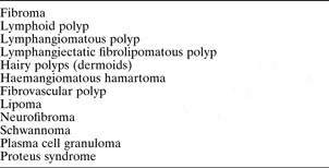
A wide variety of benign tonsillar lesions is described in the literature, all rare (Table I). Most are thought to represent a type of tonsillar hamartoma.
Table I Benign tonsillar lesions reportedReference Nayak, Murthy, Gopalakrishna and Padhee4–Reference Harada, Kashiwagi, Morimatsu, Kameyama and Takahashi15

• Fibroepithelial polyps and other benign tonsillar lesions are rare; when they do arise, management is usually straightforward
• Although malignancy is rare, excision biopsy is always advisable
• Fibroepithelial polyps can cause upper airway obstruction and should be managed as a medical emergency, with securing the airway of paramount importance
Although usually not life-threatening, fibroepithelial polyps of the pharynx may present as an acute medical emergency, causing upper airway obstruction. Management involves first securing the airway.Reference Mangar, Jiang and Lloyd1
Conclusion
Fibroepithelial polyps and other benign tonsillar lesions are rare. When they do arise, management is usually straightforward. Although malignancy is rare, excision biopsy is always advisable. Fibroepithelial polyps can occasionally cause upper airway obstruction and should be managed as a medical emergency, with securing the airway of paramount importance. Pathologically, tonsillar fibroepithelial polyps may represent a type of hamartoma.