


INTRODUCTION
In order to study the biodiversity of free-living marine nematodes in the China Sea, sediment samples were collected in many sites from the intertidal to the sublittoral region of the Yellow Sea, the East China Sea and the South China Sea from January 2003 to October 2007. More than 240 species have been identified from these habitats (Huang et al., Reference Huang, Zhang and Liu2006; Huang & Wu, Reference Huang and Wu2010; Haung & Zhang, Reference Huang and Zhang2010a, Reference Huang and Zhangb). The present paper describes three new species from this region which belong to the genus Micoletzkyia Ditlevsen, 1926.
MATERIALS AND METHODS
Undisturbed meiofauna samples were obtained using a syringe (2.6 cm diameter), and they were fixed with 5% formalin in filtered seawater. In the laboratory, samples were stained with 0.1% rose Bengal for 24 hours (Higgins & Thiel, Reference Higgins and Thiel1988). All the samples were washed to remove the formalin and sieved over two mesh sizes (500 µm and 42 µm) in order to separate the macrofauna (500 µm) from the meiofauna (42 µm). Heavier sediment particles were removed using centrifugation in Ludox-TM with a specific gravity adjusted to 1.15 (Jonge & Bouwman, Reference Jonge and Bouwman1977). Each sample was washed into a lined Petri dish and the meiofauna was sorted under a stereoscopic microscope up to higher taxonomic levels. Nematodes were transferred into a 9:1 (V:V) solution of 50% alcohol–glycerol in block cavity to slowly evaporate alcohol and then mounted in glycerol on permanent slides (McIntyre & Warwick, Reference McIntyre, Warwick, Holme and McIntyre1984). The descriptions were made from glycerin mounts using interference contrast microscopy. Drawings were made with a camera lucida. Type specimens have been deposited in the type collections of the museum of Qingdao Institute of Oceanology, Chinese Academy of Sciences.
Abbreviations are as follows: L, total body length; max. b.d., maximum body diameter (M); a, body length/maximum body diameter; a.b.d., anal body diameter; b, body length/pharynx length; c, body length/tail length; c′, tail length/a.b.d.; c.b.d., corresponding body diameter; Spic, spicule length along arc. The a, b, c ratios are from de Man (1888).
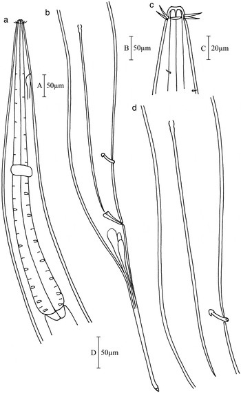
Fig. 1. Micoletzkyia longispicula sp. nov. (a) Lateral view of male pharynx region; (b) lateral view of male tail end, showing spicules, gubernaculum, precloacal supplement, caudal glands; (c) lateral view of male head end, showing cephalic setae, buccal cavity and amphideal fovea; (d) lateral view of male posterior part, showing spicule and precloacal supplement.

Fig. 2. Micoletzkyia longispicula sp. nov. (a) Lateral view of male head end, showing cephalic capsule and excretory pore; (b) lateral view of juvenile head end, showing cephalic setae; (c) lateral view of male cloacal region, showing gubernaculum and precloacal supplement; (d) lateral view of male posterior part, showing spicule anterior portion.
TYPE MATERIAL
Only one male was discovered and studied. Holotype: ♂1 on the slide 36005 (2–5) 091.
TYPE LOCALITY AND HABITAT
Subtidal muddy sediment in the Yellow Sea. Station 36005, located at 36°N 122°30′E, water depth 40 m, water temperature at the sediment–water interface 9.9°C, salinity 33.6.
ETYMOLOGY
This species is named after the long spicules.
MEASUREMENTS

DESCRIPTION
Body long spindle-shaped, gradually tapering towards both extremities, more pronounced in anterior region. Cuticle smooth. Head set off, globular, with weak cephalic capsule.
Six lips, each with a tiny inner labial papilla. Outer labial and cephalic sensilla arranged in one circle of ten setae, about 15 µm long. Buccal cavity small and simple, unarmed. Pharynx cylindrical, with wide basal part. Amphideal fovea pocket-shaped with elliptical opening, 10 µm wide (0.7 c.b.d.) in males. Secretory–excretory pore situated 130 µm from anterior end, or 19% of pharynx length from head end. Nerve ring about mid-way pharynx. Tail conico-cylindrical with relatively long distal filiform part, 428 µm or 6.5 a.b.d. long. No terminal setae. Three caudal glands confined to tail region.
Spicules elongated and straight, 461 µm or 7 a.b.d. long, pear-shaped proximally and pointed distally. Gubernaculum conical, 51 µm long, without apophysis. Single tubular supplement 37 µm long, headlike proximally, 153 µm (2.5 a.b.d.) in front of cloacal opening, and about one-third of spicule from cloacal opening. Reproductive system diorchic with two outstretched testes.
Differential diagnosis
Micoletzkyia longispicula sp. nov. is characterized by amphideal fovea pocket-shaped with elliptical opening; spicules elongated and straight, 7 a.b.d. long, pear-shaped proximally and pointed distally; gubernaculum conical, without apophysis, a single tubular precloacal supplement, cephalated proximally.
The new species is close to M. magna Vitiello, Reference Vitiello1970. Micoletzkyia magna Vitiello, however, is much longer in body length (8073 µm versus 6075 µm); the spicules are longer too (545 µm versus 461 µm) and not pear-shaped proximally; and supplement is not headlike proximally. Micoletzkyia longispicula sp. nov. is similar to M. elegans Ditlevsen, 1926, but the spicules of M. elegans are relatively shorter (318 µm or 4.42 a.b.d.); and the position of supplement is equal level with the proximal end of retracted spicules. This new species differs from the other two new species by relatively longer spicules with pear-shaped proximal end, gubernaculum without apophysis, and precloacal supplement cephalated proximally.

Fig. 3. Micoletzkyia filicaudata sp. nov. (a) Lateral view of male pharynx region, showing head, nerve ring, excretory pore; (b) oblique view of male cloacal region, showing spicules, gubernaculum and precloacal supplement; (c) lateral view of male head end, showing cephalic seta, buccal cavity and amphideal fovea; (d) lateral view of male tail end, showing anus and tail filiform part.

Fig. 4. Micoletzkyia filicaudata sp. nov. (a) Lateral view of male head end, showing cephalic capsule; (b) lateral view of male head end, showing cephalic setae; (c) ventral sublateral view of male posterior part, showing spicule and precloacal supplement; (d) ventral sublateral view of male cloaca region, showing spicules and gubernaculum.
TYPE MATERIAL
Only one male was discovered and studied. Holotype: ♂1 on the slide NH130-138(2-5)1.
TYPE LOCALITY AND HABITAT
Subtidal muddy sediment in the South China Sea. Station D13-4, located at 20°59′N 110°46′E, water depth 45 m, water temperature at the sediment–water interface 27.5°C, salinity 33.7.
ETYMOLOGY
This species is named after the long filiform tail.
MEASUREMENTS

DESCRIPTION
Body is finely spindle-shaped, gradually tapering towards both extremities. Cuticle is smooth. Head is globular, constricted a short distance behind cephalic setae, with weak cephalic capsule.
Six lips, each with a tiny inner labial papilla. Outer labial and cephalic sensilla arranged in one circle of ten setae, 16 µm long. Buccal cavity is small and simple. Pharynx cylindrical, basal part not broadened. Amphideal fovea pocket-shaped with elliptical opening, 6 µm wide (0.5 c.b.d.), 8 µm from anterior border of fovea to anterior end of body. Secretory–excretory pore situated 120 µm from head end, or midway between anterior end of body and nerve ring. Nerve ring about half-way down pharyngeal length, or 51% of pharynx length from anterior end.
Tail is conico-cylindrical with five sixths of distal filiform. 486 µm or 8.1 a.b.d. long. No terminal seta. Three caudal glands confined to tail region.
Spicules elongated and straight, 262 µm or 4.4 a.b.d. long, cephalate proximally and pointed distally. Gubernaculum is tubular, with dorso-caudal apophysis. Single tubular precloacal supplement 24 µm long, 104 µm (1.7 a.b.d.) in front of cloacal opening. Reproductive system is diorchic with two outstretched testes.
Differential diagnosis
Micoletzkyia filicaudata sp. nov. is characterized by slender body with relatively long filiform tail, spicules elongated and straight (4.4 a.b.d.), cephalate proximally and pointed distally. Gubernaculum is tubular, with dorsal apophysis.
The new species is close to M. magna Vitiello, Reference Vitiello1970. Micoletzkyia magna Vitiello, however, is much bigger in body size (L: 8073 µm, a: 71.4 versus 4430 µm, 54.7); the spicules are longer too (545 µm versus 262 µm) and gubernaculum without dorsal apophysis. Micoletzkyia filicaudata sp. nov. is similar to Micoletzkyia longispicula sp. nov. in some respects. But in the latter, spicules are relatively longer and tail is shorter, spicules with pear-shaped proximal end, gubernaculum without apophysis, and precloacal supplement headlike proximally. The new species differs from Micoletzkyia nanhaiensis sp. nov. by longer tail (8.1 a.b.d. versus 4.7 a.b.d.), longer spicules (4.4 a.b.d. versus 2.5 a.b.d.) with pointed distal end, and precloacal supplement distant from cloacal opening (1.7 a.b.d. versus 0.4 a.b.d.).

Fig. 5. Micoletzkyia nanhaiensis sp. nov. (a) Lateral view of male pharynx region, showing head, nerve ring, excretory pore and pharynx; (b) lateral view of male head end, showing cephalic seta and buccal cavity; (c) lateral view of male posterior end, showing spicules, gubernaculum, precloacal supplement and tail.

Fig. 6. Micoletzkyia nanhaiensis sp. nov. (a) Lateral view of male head end, showing cephalic capsule and cephalic setae; (b) lateral view of male tail end, showing tail and filiform part; (c) lateral view of male cloaca region, showing spicules, gubernaculums and precloacal supplement; (d) lateral view of male cloacal region, showing gubernaculums.
TYPE MATERIAL
Only one male was discovered and studied. Holotype: ♂1 on the slide NH28-36(5-8)1.
TYPE LOCALITY AND HABITAT
Subtidal muddy sediment in the South China Sea. Station D20-1, located at 18°35′N 110°17′E, water depth 30 m, water temperature at the sediment–water interface 26.7°C, salinity 33.5.
ETYMOLOGY
This species is named after the sea area where the specimens were collected, South China Sea.
MEASUREMENTS

DESCRIPTION
Body is finely spindle-shaped, gradually tapering towards both ends. Cuticle is smooth. Head set off, globular, with weak cephalic capsule.
Four cephalic setae and 6 outer labial setae arranged in one circle, about 10 µm long. Buccal cavity is small and simple. Pharynx is cylindrical, broadened gradually towards the base, no terminal bulb. Amphideal fovea not observed. Secretory–excretory pore situated 150 µm from anterior end of body, or 22% pharynx length from head end. Nerve ring situated 268 µm from anterior end, or 39% of pharyngeal length.
Tail is conico-cylindrical with relatively long distal filiform. 290 µm or 4.7 a.b.d. long. No terminal seta.
Spicules are slender and curved, 152 µm or 2.5 a.b.d. long, cephalate proximally and chapiter-shaped distally. Gubernaculum with dorsal apophysis, 22 µm long. Single tubular precloacal supplement 22 µm long, 26 µm in front of cloacal opening. Reproductive system is diorchic with two outstretched testes.
Differential diagnosis
Micoletzkyia nanhaiensis sp. nov. is characterized by curved spicules, cephaloid proximally and chapiter-shaped distally; gubernaculum with relatively large dorsal apophysis; precloacal supplement near to cloacal opening. The new species is close to M. mucronata Vitiello, Reference Vitiello1970. Micoletzkyia mucronata Vitiello, however, with short tail (2.3 a.b.d. versus 4.7 a.b.d), and very short posterior cylindrical portion; spicules are longer than 400 µm and pointed distally, not chapiter-shaped. Micoletzkyia nanhaiensis sp. nov. differs from the other two new species by short spicules with chapiter-shaped distal end, gubernaculum with large dorsal apophysis and precloacal supplement very near to cloacal opening (not enough 0.5 a.b.d.).
DISCUSSION
Micoletzkyia was established in the family Phanodermatidae by Ditlevsen in 1926 based on the following features: body length 3–9 mm; head attenuated and set-off; cephalic capsule weak; tubular supplement present. Up to now, nine species of Micoletzkyia have been recorded: M. anomala Wieser, 1953 (2 female type specimens, 1 female in Mawson), M. austrogeorgiae Allgen, 1954 (1 female types, 1 female in Mawson), M. elegans Ditlevsen, 1926 (1 male), M. falklandiae Allgen, 1954 (1 juvenile female), M. magna Vitiello, Reference Vitiello1970 (1 male), M. mucronata Vitiello, Reference Vitiello1970 (1 male, 2 juveniles), M. nudicapitata Allgen, 1959 (1 male), M. parelegans Allgen, 1954 (1 male, 1 juvenile), M. sedata Gagarin, 2010 (1 male, 9 females). Of them, M. anomala Wieser, 1953 and M. austrogeorgiae Allgen were known only from the female. Micoletzkyia falklandiae was known from a single juvenile only by Allgen in 1954. So, we consider them as invalid species. A key to all valid species of Micoletzkyia is given.
KEY FOR NINE VALID SPECIES OF MICOLETZKYIA DITLEVSEN, 1926
1. Body length shorter than 4 mm, precloacal supplement absenceM. nudicapitata Allgen
— Body length close to or longer than 4.5 mm, precloacal supplement presence… … … … … … … … … … … …2
2. Gubernaculum with dorsal apophysis… … … … … … 3
— Gubernaculum without dorsal apophysis… … … … …5
3. Tail longer than 8 a.b.d., with long posterior filiform; spicule longer than 260 µmM. filicaudata sp. nov
— Tail shorter than 5 a.b.d. long… … … … … … … …4
4. Tail 2.3 a.b.d. long, with short posterior cylindrical portion; spicule longer than 400 µmM. mucronata Vitiello
— Tail 4.7 a.b.d. long, with long posterior filiform; spicule shorter than 160 µmM. nanhaiensis sp. nov.
5. Tail with short posterior thinned portion, no filiform … … … … … … … … … … … … … M. parelegans Allgen
— Tail with long posterior filiform … … … … … … …6
6. Cephalic setae shorter than 1 h.d. … … … … … … … 7
— Cephalic setae longer than 1 h.d. … … … … … … …8
7. Cephalic setae 0.4 h.d., spicule 7.8 a.b.d… … … … … … … … … … … … … … … … … …M. magna Vitiello
— Cephalic setae 0.8 h.d., spicule 4.4 a.b.d… … … … … … … … … … … … … … … … … M. elegans Ditlevsen
8. Spicule 6.8 a.b.d., gubernaculum 51 µm… … … … … … … … … … … … … … … … M. longispicula sp. nov.
— Spicule 5 a.b.d., gubernaculum 23 µm … … … … … … … … … … … … … … … … … … M. sedata Gagarin
ACKNOWLEDGEMENTS
This work was supported by the National Natural Science Foundation of China (No. 30770250), the Knowledge Innovation Program of the Chinese Academy of Sciences (No. KZCX2-YW-417) to K. Xu, and the Open Research Cruise offshore China by RV ‘KE XUE SAN HAO’, Institute of Oceanology, Chinese Academy of Sciences (IOCAS) in July 2008. The authors are very grateful to Dr Kuidong Xu for supplying specimens and Ms Zhongying Shan for her slide making. Two anonymous referees are thanked for their valuable comments and revision of the paper.


