


Introduction
Chondrodermatitis nodularis chronica helicis was first described by Winkler in 19153 and later by Forester in 1918.Reference Forester4 It is a benign painful condition affecting prominent parts of the pinna. Most often these lesions develop on the most protuberant part of the ear, i.e. the helix and antihelix.Reference De Ru, Lohuis, Saleh and Vuyk5 They commonly present as a painful erythematous nodule often covered with a scale or crust with an underlying central depression.Reference Wade6 The nodules attain a maximum size in a few months and if left untreated remain unchanged indefinitely.Reference Kennedy, Burns, Breathnach, Cox and Griffiths7 In our centre we treated all the patients with chondrodermatitis nodularis chronica helicis using a specially designed pillow.
Materials and methods
The cohort of patients was collected from those who attended our ENT out-patient department from September 2003 to September 2004. These patients were referred either by a general practitioner or dermatologist. They were all seen by an ENT consultant with a special interest in facial skin lesions. They were clinically diagnosed with chondrodermatitis nodularis chronica helicis based on the history of a painful, tender lesion on the pinna, which was benign in appearance. All the patients diagnosed with chondrodermatitis nodularis chronica helicis were treated conservatively using a specially designed doughnut pillow (Figure 1).
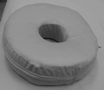
Fig. 1 Doughnut pillow.
The ENT and orthodontic departments at Wrexham Maelor Hospital designed the pillow jointly. It is made up of a circular foam cushion with a hole cut in the centre. A washable pillow cover with zip fastener was also provided. The patients were advised to customise their pillow by adjusting its thickness to suit their individual needs. They were advised to use the pillow to sleep, making sure that the affected ear fitted into the hole in the middle of the pillow (Figure 2).

Fig. 2 Patients are advised to make sure that the affected ear is fitted into the hole in the middle of the pillow.
The patients were followed up a year after dispensing the pillow. At follow-up visits they were asked about the usage of the pillow and the time taken for symptomatic relief if any. They were encouraged to discuss any problems encountered during the use of the pillow.
Results and analysis
A total of 23 patients (14 female and nine male) were clinically diagnosed with chondrodermatitis nodularis chronica helicis between September 2003 and September 2004. Nineteen had lesions on the helix and four on the antihelix. Thirteen were affected on the left side and 10 on the right. The median follow up period was 12 months (range 11–14 months). At the end of one year, among the 23 treated with a doughnut pillow, 13 (56 per cent) were pain free. The median time required to become pain free was 21 days (range 2–90 days).
Among the 13 who were pain free, 12 had no problems with the pillow and hence used it regularly. One patient said the pillow hurt his neck, but he still carried on using it.
Among the 10 who were not pain free at the end of one year, three had no problems with the pillow and hence used it regularly. The remaining seven did not use the pillow regularly, the reasons given being neck pain, stiff neck and the fact that the pillow kept dislodging.
Discussion
Chondrodermatitis nodularis chronica helicis is usually unilateral but bilateral incidence of 3–7 per cent has been reported.Reference Schuman and Helwig8, Reference Eberius9 Most patients are Caucasian men over 50 years of age.Reference Schuman and Helwig8, Reference Eberius9 Although the diagnosis can be made fairly easily in most cases it is worth considering early squamous cell carcinoma and basal cell carcinoma in the differential diagnosis. The aetiology and pathogenesis of chondrodermatitis nodularis chronica helicis remains unknown. Several factors have been implicated including trauma,Reference Yaffee10 degeneration,Reference Newcomer, Steffen, Sternberg and Lichtenstein11 actinicReference Goette12 and cold injury.Reference Wilkinson, Rook, Wilkinson and Ebling13 Many authors have suggested that pressure is a significant factorReference De Ru, Lohuis, Saleh and Vuyk5, Reference Wade6, Reference Schuman and Helwig8, Reference Yaffee10 in the aetiopathogenesis.
Over the years different surgical and non-surgical modes of treatment have been described for chondrodermatitis nodularis chronica helicis. They all have different success rates varying from 27 per cent to 90 per cent. Lawrence reported a 27 per cent success from combined topical and intralesional corticosteroid.Reference Lawrence14 Cox achieved a success rate of 33 per cent with a single intralesional injection of triamcinolone.Reference Cox and Denham15 A success rate of 87 per cent has also been reported following conservative management using a pressure-relieving prosthesis.Reference Moncrieff and Sassoon16 The success rate following surgical management varies from 66 per cent to 90 per cent depending on the technique used and the site of the lesion.Reference De Ru, Lohuis, Saleh and Vuyk5, Reference Lawrence14, Reference Moncrieff and Sassoon16, Reference Hudson-Peacock, Cox and Lawrence17
Studies have shown that 77–99 per cent of the patients with these lesions sleep on the same side as their lesion.Reference Schuman and Helwig8, Reference Lawrence14 It has also been shown that patients may develop lesions on the contralateral side after altering their sleeping posture, as the first ear is too painful to sleep on.Reference Hudson-Peacock, Cox and Lawrence17 The treatment of chondrodermatitis nodularis chronica helicis using a doughnut-shaped pillow was based on the principal of relieving pressure on the affected ear. As seen in our results, all the patients who were pain free at the end of one year used the pillow regularly. Among those who were not pain free at the end of one year, 70 per cent did not use the pillow on a regular basis. This further supports the possibility of pressure being a major contributing factor in the progression of the disease.
The use of the pillow on a regular basis is very important for the treatment to be successful. The common reasons given for not using the pillow were neck pain and neck stiffness. We suggest that identifying patients with existing neck problems before dispensing the pillow could prove valuable. If they are found to have neck problems, then a different mode of treatment could be tried. If the patients develop neck pain after using the pillow, the pillow itself can be readjusted to suit the patient or a different treatment altogether can be planned. Follow up is advisable after about a month of dispensing the pillow to assess its success and address any problems.
In our study we achieved an overall success rate of only 56 per cent by using the doughnut-shaped pillow. However, 70 per cent of the patients who were not pain free did not use the pillow regularly. We believe with regular use of the pillow and regular follow up to minimise non-compliance with the treatment, a higher success rate can be achieved. The treatment of chondrodermatitis nodularis chronica helicis using a doughnut-shaped pillow is very cost effective. It only costs about £18 to dispense the pillow. If used regularly by the patient, it can be a very cost effective way of treating chondrodermatitis nodularis chronica helicis.
Conclusion
Chondrodermatitis nodularis chronica helicis can be treated using a doughnut-shaped pillow if the pillow is used regularly. Early follow up is essential to identify the development of non-compliance with the treatment. A careful screening to identify existing neck problems could further reduce non-compliance with the treatment.


