Published online by Cambridge University Press: 01 March 2004
The factors responsible for the reactivation of a Neospora caninum latent infection are unknown, but it is postulated that the maternal immune response could be altered during pregnancy. The immune response was investigated in N. caninum chronically infected mice during successive pregnancies as well as in non-pregnant infected mice and mice infected when pregnant. Vertical transmission was demonstrated in chronically infected mice after the first pregnancy but the rate of fœtal infection fell after further pregnancies. Non-pregnant chronically infected mice showed a marked specific proliferative response and an IgG2a isotype preferential secretion. During the course of the first pregnancy, no significant modification of the immune response was recorded. After 2 successive pregnancies, the specific cellular response showed a significant fall whereas Th2 cytokine mRNA expression was noted. At the same time, IgG1 secretion increased to reach the IgG2a level. At the third delivery, a partial restoration of the proliferative response was observed. The reactivation of N. caninum chronic infection during pregnancy does not seem to be consecutive to an immunodepression. Nevertheless, pregnancy could favour parasite multiplication in utero after an occasional spontaneous release of bradyzoites.
Neospora caninum is an apicomplexan parasite. Fœtal infection in cattle can lead to abortion, stillbirth or the birth of either clinically affected or unaffected calves. However, most of the latter are chronically infected. Indeed, the vertical transmission of the parasite is very efficient (80–90%) and can occur in chronically infected cows through successive pregnancies or from congenitally infected animals for several generations (Paré, Thurmond & Hietala, 1997; Thurmond & Hietala, 1997; Schares et al. 1998). This feature highlights the likely involvement of the foeto–maternal relationship and the maternal immune response in the control of neonatal neosporosis. N. caninum, in chronically infected cows, can revert to rapid tachyzoite multiplication and dissemination which is responsible for a very efficient vertical transmission. The factors responsible for this reactivation are unknown. The immune response, which limits the parasite dissemination, could be altered during pregnancy. As the immune control of N. caninum infection requires a Th1-type cellular response (Khan et al. 1997; Tanaka et al. 2000), a Th2 cytokine bias as observed in pregnant mice (Athanassakis & Iconomidou, 1996) and women (Raghupathy et al. 2000; Saito, 2000) could be responsible for the reactivation of N. caninum in pregnant cattle. Studies in Leishmania major-infected mice demonstrated that parasite load and lesion severity increased in pregnant animals which produced less IFN-gamma but higher levels of IL-4, IL-5 and IL-10 (Krishnan et al. 1996a). In order to develop therapeutic or prophylactic strategies to control neosporosis it is necessary to understand the immunological mechanisms, which are responsible for the reactivation of chronic infection. Cows inoculated 6–9 weeks before insemination produced healthy uninfected calves (Williams et al. 2000; Innes et al. 2001); these results suggest that adult cows experimentally infected with tachyzoites fail to establish a chronic infection which is amenable to reactivation unlike what is observed in naturally infected animals (Guy et al. 2001). The availability of immunological tools and inbred strains of mice led to the extensive use of different murine models to study the immune response to N. caninum (Khan et al. 1997; Tanaka et al. 2000; Ritter et al. 2002). In N. caninum infected mice, the effect of pregnancy has been studied once. Long & Baszler (2000) demonstrated that IL-4 neutralization before pregnancy concomitant with the inoculation of an avirulent strain of N. caninum decreased congenital transmission after a challenge during pregnancy. This fœtal protection was associated with significantly lower in vitro secretion of IL-4, lower IL-4 mRNA levels and higher levels of IFN-gamma. The purpose of the present study was to investigate immune modulation accompanying reactivation of N. caninum chronic infections in pregnant mice.
The NC-1 isolate of N. caninum (Dubey et al. 1988) was used for the inoculation of mice and for the preparation of antigenic extract. The tachyzoites were passaged once in mice and the parasites used for inoculation were passaged in vitro less than 10 times in order to maintain virulence as described by Long & Baszler (2000). The tachyzoites were maintained in Vero cells grown in RPMI-Stabilix medium supplemented with 100 UI/ml penicillin, 100 μg/ml streptomycin, non-essential amino acids, sodium pyruvate (Biomedia, Boussens, France) and 2% horse serum. In order to separate the parasites from the cell debris, the suspension was extruded through a 27-G needle and centrifuged at 200 g for 10 min to separate cell clusters. The supernatant fraction containing tachyzoites was centrifuged at 2000 g for 20 min, the pellet was resuspended in phosphate-buffered saline (PBS) and after a centrifugation at 2000 g for 20 min, the parasites were counted in a Neubauer chamber. For the preparation of antigen extract, tachyzoites were treated by 3 freeze-thawing cycles followed by sonication (Innes et al. 1995). The protein concentration was measured by the bicinchoninic acid (BCA) method (Sigma, St Louis, USA); then the solution was aliquoted and stored at −20 °C until used. Toxoplasma gondii tachyzoites (RH strain) were propagated on Vero cells as described for N. caninum tachyzoites and were processed in the same way to produce antigen.
Six to eight-week-old female CBA/Ca mice were purchased from B and K (Hull, UK) and fed a rodent mix and water ad libitum. To investigate the immune factors accompanying a possible reactivation of chronic infections during pregnancy and to compare with acute infection in naive pregnant and non-pregnant mice, 3 groups of females were used: a group of non-pregnant acutely infected mice (group A), a group of mice infected when pregnant (group B) and a third of pregnant chronically infected mice (group C). In order to obtain chronically infected mice (i.e. harbouring cysts), the animals received 2 mg of methylprednisolone acetate (Moderin, Pharmacia-Upjohn, Puurs, Belgium) 7 days before and on the day of a subcutaneous inoculation with 5×10⁶N. caninum tachyzoites. The mice were left for 4–6 months infection and then mated. The day on which a vaginal plug was observed was considered as day 0 of pregnancy. Four mice were sacrificed on days 0, 10, 18 and after delivery of their first pregnancy. Groups of chronically infected mice were mated on the day of their first delivery and 4 mice were sacrificed at the delivery of their second pregnancy. The other mice were mated again on the day of the second delivery and 4 mice were sacrificed at the delivery of the third pregnancy. The mice which were not successfully mated on the day of delivery, were discarded from immunological studies but their litter were analysed to settle the vertical transmission rate. The non-pregnant age-matched mice were inoculated subcutaneously with 5×10⁶ tachyzoites and 4 mice were sacrificed on days 0, 3, 8, 11, 14 and 18 post-infection whereas pregnant mice were given the same inoculum on day 8 to 10 after a successful mating. Four of these mice were sacrificed on days 0, 8 and 11 post-infection.
The spleen of each animal was aseptically collected and disrupted through a wire mesh. The cells were washed twice in DMEM culture medium and diluted in RPMI culture medium containing 50 μM 2-mercaptoethanol. Cell concentration was adjusted at 1×10⁶ cells/ml and 2·5×105 cells in 250 μl were plated in quadruplicates into 96-well microplates. The wells contained either 20 μl of NC (N. caninum), TG (T. gondii) or Vero antigens (100 μg/ml), PBS or Concanavalin A (ConA, 50 μg/ml). The plates were incubated for 3 days; then the cells were pulsed for 24 h with 0·5 μCi/well of [methyl-3H] thymidine, harvested onto nitrocellulose filters and the incorporated radioactivity was measured with a β-counter (Beckman Instruments, Fullerton, USA) for 3 min. The results were expressed as mean count per min (CPM) of stimulated wells minus mean CPM of control wells (ΔCPM).
Maxisorp (Nunc, Rocksilde, Denmark) microplate-wells were coated with 100 μl of NC and Vero cell antigen solutions containing either 15 μg or 10 μg/ml for IgM and IgG measurements respectively. Plates were incubated for 1 or 7 h at 37 °C or at room temperature (RT) respectively. To block non-specific antibody binding, the plates were coated with casein hydrolysate solution overnight at RT. Sera were diluted 1[ratio ]100 and incubated at 37 °C for 1 h. After washing, 100 μl of biotinylated rat antibody (0·5 μg/ml) to mouse IgM, IgG1 and IgG2a (Serotec, Oxford, United-Kingdom) were added for 60 min at 37 °C. After washing, streptavidin-bound peroxidase (Amersham, Piscataway, USA) was added at a dilution of 1[ratio ]1000 and left to incubate for 1 h. The reaction was then visualized using TMB (3,3′, 5,5′-tetramethyl-benzidine) as substrate. The reaction was allowed to develop for 10 min (IgG1 and IgG2a) or 15 min (IgM). The 450 nm OD of each well was determined using an electronic plate reader. For each assay, a positive control standard was run. The OD values obtained with control wells (Vero cell antigens) were subtracted from the values recorded for NC coated wells. Each OD value was converted to a titre. A titre was considered as positive when above the mean of negative control ±3 standard deviations. Results from each plate were related to the positive standard.
The brain, liver, heart and lung were pooled from each foetus and pup except in the case of 8 to10-day-old foetuses, which were processed whole. Whenever feasible, placentas were collected. The tissues were snap-frozen in liquid nitrogen and conserved at −80 °C until analysis. The samples were homogenized in lysis buffer containing 50 mM Tris–HCl, pH 8, 100 mM NaCl, 50 mM ethylenediaminetetraacetic (EDTA), 0·8% (w/v) sodium dodecyl sulfate (SDS) and 0·2 mg/ml proteinase K. The samples were incubated for 16–20 h at 55 °C with occasional vigorous vortexing. After a phenol[ratio ]chloroform[ratio ]isoamyl alcohol (25[ratio ]24[ratio ]1) and chloroform[ratio ]isoamyl alcohol (24[ratio ]1) extraction, DNA was precipitated in 100% ethanol, washed once in 70% ethanol and the pellet was resuspended in 200 μl of a 10 mM Tris–HCl, 1 mM EDTA buffer (TE). DNA concentration was adjusted to 50–100 μg/ml and aliquots of 5 μl were used in each PCR. PCR were performed on 50 μl of the final mixture containing 5 μl of reaction buffer, 3 μl of MgCl2, 1 μl dNTPs, 0·25 μl Taq (PCR Core System 1, Promega, Madison, USA), 1 μl of extraction solution, 25 pmol of each primer and water. The first amplification was performed using Np7 (5′ GGGTGAACCGAGGGAGTTG 3′) and Np4 (5′ CCTCCCAATGCGAACGAAA 3′) primers in a Peltier-Thermal-Cycler (MJ Research, Biozym, Landgraaf, Netherlands). Thirty-five cycles (denaturation at 95 °C, annealing at 57 °C and extension at 72 °C for 1 min during each cycle) were carried out. The second amplification was carried out using Np7-Np6 (5′ CAGTCAACCTACGTCTTCT 3′) primers using 2 μl of the solution obtained after the first step. PCR mixture and amplification cycles were identical except annealing which was performed at 56 °C for 1 min. PCR products were analysed by electrophoresis on 2% agarose gels. Positive control (purified from DNA NC-1 tachyzoites) and negative controls (DNA from control mice and absence of DNA) were included in each PCR run. In order to check DNA quality, negative samples were subjected to a PCR using β-actin primers.
For each animal, total RNA from splenocytes was extracted using TRIzol reagent (Gibco-BRL, Life Technologies, Grand Island, USA) as described in the TRIzol protocol. The cells were lysed by multiple pipetting. Following precipitation with isopropanol and subsequent ethanol washes, the RNA pellets were resuspended in diethyl pyrocarbonate-treated water. Rnase-free Dnase (Promega, Madison, USA) was used to remove genomic DNA. Total RNA was quantified using spectrophotometry at 260 nm. One μg of total RNA was reverse transcribed in a total volume of 20 μl containing 0·5 μg of oligo(dT)15 as the primer, 5 μl of reaction buffer, 1 μl dNTP mix, 20 U RNasin®, 4·8 μl of MgCl2 and 1 μl of reverse transcriptase (Improm-II™ Reverse Transcription System, Promega, Madison, USA). The RT procedure consists of an initial step of incubation of the RNA with oligo(dT) at 70 °C for 5 min. After mixing the different reagents, the samples were incubated at 42 °C for 1 h and then at 70 °C for 15 min. The cDNA was stored at −20 °C until PCR amplification. The final PCR mixture contained 2 μl cDNA, 4 μl PCR buffer, 3 mM MgCl2, 10 mM dNTP, 0·25 μl Taq DNA polymerase (5 U/μl) (PCR Core Systems, Promega, Madison, USA), 50 pM concentration of (each) sense and antisense primers and sterile water to 50 μl. Cytokine-specific primers were synthesized by Gibco-BRL, Life-Technologies according to sequence design previously described (Ulett, Ketheesan & Hirst, 2000). The amplification was performed in a thermocycler as follows: after 2 min at 95 °C, 33 cycles each of denaturation at 94 °C for 1 min, annealing at 60 °C for 1 min and extension at 72 °C for 1 min were performed. The PCR using IL-4 primers required 35 cycles of denaturation, annealing at 50 °C and extension, the other parameters being unchanged. A final extension at 72 °C for 4 min was included. The number of cycles was chosen to lie in the linear portion of the curve for each set of primers. The PCR products were visualized by UV illumination after electrophoresis through a 2% agarose gel containing ethidium bromide. A 1 kb ladder (1 Kb Plus DNA ladder®, Gibco-BRL, Life-Technologies, Grand Island, USA) was used as a DNA size marker. A PCR using β-actin primers was performed on each individual sample as an internal positive-control standard and to allow cytokine product ratio comparison. RNA from mitogen-stimulated mouse splenocytes was used as positive control for β-actin and cytokine PCR. Negative control consisting in water sample was tested with each set of primers as well as individual RNA pre-RT for β-actin primers. To quantify cDNA bands, the ethidium bromide stained agarose gels were photographed using a DC265 Zoom digital camera (Kodak, Rochester, New York), scanned and analysed using Quantity One software program (Bio-Rad Laboratories, Richmond, USA). The results were expressed as a ratio between the band signal for a given cytokine and the signal for actin, and then ratios were calculated relative to actin positive control.
Spleen cells (5×10⁶) were resuspended in PBS-FCS (5%) and a total of 1×10⁶ cells was incubated for 30 min at 4 °C with appropriate dilutions of rat anti-mouse monoclonal antibodies to CD4+ (YTS191.1), CD8+ (YTS169.4) or B cells (IBL-2) marquers (Serotec, Oxford, UK). After washing, the cells were incubated for 30 min at 4 °C with F(ab′) IgG goat anti-rat R-Phycoerythrin (RPE) conjugated. The cells were washed, suspended in PBS and kept at 4 °C in the dark until flow cytometry (FACStar PLUS™, Becton Dickinson, San Jose, USA). For each analysis, 10000 events were recorded. The percentage of stained cells was determined for each monoclonal antibody and a sample stained with RPE conjugate only was used as control.
For each mouse, the brain, heart and liver were removed, fixed in 10% neutral buffered formalin and 4 μm sections were stained with haematoxylin-eosin. Two distant sagital sections of each organ were examined. Lesions were graded using the following system: (1) no lesions; (2) slight (some small inflammatory foci, perivascular cuffing, no necrosis); (3) mild (numerous larger inflammatory foci, no necrosis); (4) moderate (inflammatory foci with necrosis concerning up to 5% of the parenchyma); (5) severe (over 5% of the parenchyma).
The cell proliferation, IgM, IgG1 and IgG2a responses were analysed using one-way analysis of variance (ANOVA). The Kruskal–Wallis test was used wherever the distributional assumptions required for an ANOVA were felt to be unrealistic. The Kruskal–Wallis and Mann–Whitney U-tests were used to compare mean lesion scores and cell subpopulation data. All statistical analyses were considered significant at the P<0·05 level.
Following inoculation all acutely infected mice remained clinically normal. A few mice from the chronically infected group showed hair coat ruffling, weight loss and/or neurological signs (head tilting and hind limb paresis) after inoculation under the immunosuppressive regimen. Two mice out of 42 mice died in the group. At mating, all animals but 1 had recovered. Some mice, which were found to be barren when sacrificed 8–10 days after mating, were discarded.
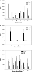
The non-pregnant acutely infected mice (group A) showed a marked decrease of the mitogenic response to ConA from 3 days post-inoculation (p.i.) (P<0·05) onwards; the lowest proliferation was observed on day 8 p.i. but the response had returned to pre-infection values on day 18 p.i. (Fig. 1A). On day 11 p.i. a lymphoproliferative response to both NC-antigen and TG-antigen was recorded. The proliferative response to NC-antigen increased to reach high values on days 14 and 18 whereas the response to TG-antigen remained somewhat lower. The pregnant acutely infected mice (group B) were unable to mount a specific proliferative response to both NC and TG antigens (Fig. 1B). The mitogenic response to ConA was dramatically reduced on day 8 p.i. (P<0·05) but was restored on day 11 p.i.

Fig. 1. Spleen cell proliferative response from (A) group A (non-pregnant acutely infected), (B) group B (pregnant acutely infected) and (C) group C (pregnant chronically infected) mice to concanavalin A (Con A), Neospora caninum (NC) and Toxoplasma gondii (TG) antigens; ΔCPM refers to the differences between stimulated and unstimulated cells and is expressed as a means for each group of mice.
The chronically infected mice (group C) showed an elevated response to NC-antigen several months after a primary infection (Fig. 1C); this response remained elevated throughout the first pregnancy (P>0·05) and until delivery, at which time a slight but insignificant decrease was observed. Mice, which were allowed to carry a second pregnancy, exhibited a marked decrease of the specific response to NC-antigen (P<0·05). After the third pregnancy, the specific cellular response increased although remaining lower than levels measured at day 10 and 18 of the first pregnancy (P<0·05). The response to Con A was variable but no significant differences were recorded (P>0·05). A proliferative response to TG-antigen was observed before mating and remained quite stable (P>0·05) although quite low until the end of the experiment.
The possible impairment of IFN-gamma expression in pregnant chronically infected mice was examined. Furthermore its possible relationship with IL-10 and IL-4 expressions was also studied. The results are shown in Table 1. In group A mice, the amount of IFN-gamma mRNA was already elevated on 3 days p.i., reached its peak on day 11 p.i. and returned to pre-infection level at the end of the experiment. Interferon-gamma was expressed in all mice tested but individual variations were quite high especially on day 8 p.i. The expression of the Th2 cytokines IL-10 and IL-4 was first detected on day 3 p.i. The relative amount of IL-10 was elevated on day 8 p.i. but IL-10 mRNA was undetectable thereafter. Large amounts of IL-4 mRNA were recorded in the majority of mice on day 11 p.i. In group B mice, the amount of IFN-gamma mRNA before infection was much lower than those in group A mice at the same time, although mRNA was detected in all group B mice. Thereafter its pattern was very similar to that observed in acutely infected non-pregnant mice. Before pregnancy, higher levels of IFN-gamma mRNA were expressed in chronically infected mice than in naive mice. The level fell until day 18 of pregnancy although individual variations were marked. At delivery high levels were recorded in all animals. Mice from group C, which were allowed to carry a second or a third pregnancy, had intermediate IFN-gamma levels. IL-10 and IL-4 mRNA were not detected during the first pregnancy except on day 0 in 1 animal out of 4. Both reappeared at the first delivery and equal levels were then expressed whereas IL-4 levels were higher than those of IL-10 at the second delivery.
Table 1. Relative amount of IFN-gamma, IL-10 and IL-4 mRNA as detected by RT-PCR on spleen cells from individual animals (Results are expressed as the ratio between a given cytokine and β-actin. Group A: acutely infected non-pregnant mice; group B: acutely infected pregnant mice; group C: chronically infected pregnant mice. Day 0: day of infection (groups A and B); day after successful mating (group C). Values between parentheses indicate the proportion of mice expressing the specific mRNA. Results are expressed as means±S.D. N.D., Not detected.)

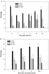
Non-pregnant acutely infected mice showed a marked decrease of the CD4+ and CD8+ subpopulations from day 3 to day 14 p.i. (P<0·05) (Fig. 2A). The level of CD4+ cells returned to pre-infection values on day 18 p.i. whereas CD8+ levels remained lower until the end of the experiment, although there was a tendency to return to the pre-infection level. In contrast, the percentages of B cells were increased on days 3 and 11 p.i. (P<0·05) but had returned to pre-infection value by day 18 p.i. Chronically infected mice showed no significant modulation of cell population during the first pregnancy despite a rise of CD4+ and B cells from day 0 to day 18 of pregnancy (Fig. 2B).

Fig. 2. CD4+, CD8+ and B cell subpopulations in spleen cells in (A) group A (non-pregnant acutely infected) and (B) group C (chronically infected pregnant) mice respectively. Day 0: day of infection (group A) or day of successful mating (group C). Results are expressed as the means of all mice in each group. N.D., No data.
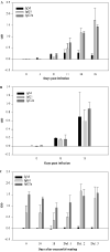
In acutely infected non-pregnant mice, parasite specific-IgM were detected 3 days after inoculation in non-pregnant mice whereas IgG were present on day 8 p.i. (Fig. 3A). IgM levels rose until day 11 p.i. (P<0·05) and then remained fairly stable until the end of the experiment whereas the levels of specific IgG1 and IgG2a were still on the rise. No preferential secretion of IgG1 and IgG2a antibodies was demonstrated. In acutely infected pregnant mice (group B), specific-antibody production was demonstrated 8 days p.i. IgM secretion rose until day 11 p.i. to reach a level comparable to those of IgG1 and IgG2a isotype (Fig. 3B). In mice chronically infected for several months, there was a predominant secretion of the IgG2a isotype (Fig. 3C). A non-significant decrease of the antibody levels was observed through the first pregnancy. At the first delivery levels had returned to pre-mating values (P<0·05). After the second delivery, IgG1 secretion increased to reach IgG2a level and the 2 isotypes remained equally high at the third delivery.

Fig. 3. Neospora caninum-specific serum levels of IgM, IgG1 and IgG2a response in (A) acutely infected non-pregnant mice (group A), (B) acutely infected pregnant mice (group B) and (C) chronically infected pregnant mice (group C). OD values refer to the difference between NC and Vero cell antigen-coated wells and are expressed within each group and for each day as a mean for all animals.
Vertical transmission efficiency from naive mice inoculated at 8–10 days of pregnancy reached 90%, 18 pups out of 20 being positive when tested by a semi-nested PCR. Placental tissues collected on day 8 p.i. were also positive. The mice chronically infected for 4–6 months before their first mating were able to transmit tachyzoites to their progeny although the rate of vertical transmission was very low within a litter (Table 2). Furthermore, not all mice transferred the infection. In 10-day-old foetuses (first pregnancy) 6 out of 8 were found positive, although only 1 out of 8 of the 18-day-old foetuses were positive. Six of the 11 mice from which the progeny had been examined after the first delivery had infected at least 1 of their pups. The rate of congenital infection after 2 or 3 pregnancies was very low. Indeed, from 12 (second delivery) and 5 females (third delivery) 2 mice in each case were shown to transmit the infection to 1 of their pups (2/54 and 2/45).

In the non-pregnant acutely infected group, mice exhibited perivascular cuffing and some scattered foci of encephalitis from 8 days p.i. onwards. The lesions became larger throughout the study with some necrosis. The mean lesion score increased until day 11 p.i. (P<0·05) and then remained stable. Heart lesions appeared more severe with several foci of necrosis and mineralization from 3 days p.i. onwards. The liver of these mice exhibited large foci of mild inflammation but no necrosis. In group B mice, lesions were less pronounced, as no inflammation was observed in brain sections and inflammatory foci in the liver were very sparse. However, sections collected on day 8 p.i. revealed some necrotic foci with mineralization. Some mice from group C had large mineralized necrotic foci devoid of inflammatory cells whereas others had no lesions. Such necrotic lesions were observed throughout the successive pregnancies but neither a significant increase in lesion mean score nor a macrophage or lymphoid cell infiltration were demonstrated. In liver, the lesions were very slight with scattered inflammatory cell foci. However, after the third delivery 1 animal showed extensive necrosis in this organ which was accompanied by a very low level of inflammatory cell infiltration (Fig. 4). In heart sections, despite the presence of mineralized necrotic foci in mice examined before and during the first pregnancy, animals sacrificed after a second or a third pregnancy showed a higher number of necrotic foci, accompanied by scarce inflammatory cells and sometimes parasite-like structures (Fig. 5). However, these differences were not significant (P>0·05).

Fig. 4. Large area of necrosis in the liver of a chronically infected mouse after its third pregnancy. Inflammatory cells are very rare. Haematoxylin-eosin staining.

Fig. 5. Tissue section from the heart of a chronically infected mouse examined after 3 successive pregnancies. Lesion of necrosis with mineralization and parasite-like structures. Haematoxylin-eosin staining.
The results of the present study indicate that in CBA/Ca mice inoculated 4–6 months before pregnancy a chronic N. caninum infection can be reactivated. Indeed the presence of N. caninum DNA in some foetuses and pups during the first pregnancy was demonstrated. This is most probably due to the reactivation of the parasite and the reemergence of tachyzoites from tissue cysts. However, during a successful pregnancy in chronically infected mice a significant alteration of the maternal immune response was not observed. During a second and a third pregnancy in the same type of mice there was an elevated specific antibody response, a decreased specific lymphoproliferative response and a low IL-10 and IL-4 mRNA expression. However, these difference were not accompanied by an increased rate of vertical transmission. Pregnancy is associated with the development of a Th2 cytokine bias at the foeto–maternal interface. This bias protects the fœtus against any potentially deleterious Th1-type cellular response (Krishnan et al. 1996b; Jenkins et al. 2000). In this context, maternal and fœtal tissues act jointly by secreting numerous immunomodulatory molecules such as steroid hormones (Choi et al. 2000; Ito et al. 2001; Miyaura & Iwata, 2002), cytokines, prostaglandins and many others (Hunt & Johnson, 1999; Shurin et al. 1999). This Th2 cytokine local pattern would have a systemic effect (Wegmann et al. 1993; Krishnan et al. 1996a) and thus could weaken resistance to intracellular pathogens which are controlled by Th1-type immune responses. It was demonstrated that IL-12, IFN-gamma and CD4+ cells are critical factors in the control of N. caninum infections (Khan et al. 1997; Tanaka et al. 2000; Ritter et al. 2002). However, a clear Th1/Th2 dichotomy was not the rule as variable levels of Th2 cytokines were produced also (Long, Baszler & Mathison, 1998; Eperon et al. 1999). In the present study, naive acutely infected non-pregnant and pregnant mice developed a specific humoral response from day 8 p.i. onwards. Additionally, in non-pregnant animals a specific lymphoproliferative response was recorded on day 11 p.i.; it was preceded by an increase of the IFN-gamma mRNA level on day 3 p.i. In the same group (group A) CD4+, CD8+ and B cells exhibited marked variations with a fall in the T cell subpopulations from day 3 p.i. onwards and a relative simultaneous rise of B cells. Similar results were reported by Khan et al. (1997) who monitored the cellular response and the cytokine production during the acute phase of a N. caninum infection in A/J mice. In the present study, acutely infected pregnant mice mounted a negligible specific lymphoproliferative response on day 11 p.i. In both groups of acutely infected mice a transient but significant fall in the proliferative response to Con A, a non-specific mitogen, was recorded. In parallel IL-10 and IL-4 mRNA expression was demonstrated. These cytokines have an immunomodulatory effect and promote the development of a Th2-type immune response (O'Garra, 1998; Joss et al. 2000). However, other factors such as nitric oxide (NO) might be responsible for an immunodepression during N. caninum infection (Khan et al. 1997). In mice chronically infected for 4–6 months, only a few foci of necrosis were still present in the brain of some mice. These chronically infected animals had a marked immune response characterized by a preferential production of IgG2a antibodies, a strong lymphoproliferative response to NC and TG antigens and higher expression of IFN-gamma mRNA when compared to naive mice. In mice chronically infected for 6 weeks, Long et al. (1998) demonstrated that the resistance to infection varied according to the murine strain used and was dependent on the Th1[ratio ]Th2 cytokine ratio. The present study failed to reveal any significant modulation of the specific maternal immune response during the first pregnancy of chronically infected mice (Figs 1C and 2B and Table 1). In these animals the specific lymphoproliferative response, the CD4+ CD8+ cell subpopulations and the IFN-gamma mRNA expression were unchanged. In mice chronically infected with T. gondii a simultaneous depletion of CD4+ and CD8+ T lymphocytes is required to reactivate the infection (Gazzinelli et al. 1992; Yap, Pesin & Sher, 2000). Nevertheless, N. caninum DNA was detected in 10- and 18-day-old fœtuses as well as in 3-day-old pups. The absence of immunodulation together with a high rate of vertical transmission was described in chronically infected cows (Guy et al. 2001). However, in this study peripheral blood lymphocytes were used and this might not reflect the modifications around the cysts at tissue level. Furthermore, in chronically infected mice the administration of 17 β oestradiol, progesterone and/or corticosterone was unable to reactivate the infection (Kobayashi et al. 2002). All the results suggest that neither the pregnancy associated hormones nor the modulation of the immune response during pregnancy are responsible for the reactivation of N. caninum chronic infections. Under these conditions the vertical transfer from mother to offspring in chronically infected animals could be the result of an occasional rupture of cysts which could occur naturally during the course of a chronic infection. This would allow a few tachyzoites to reach the uterus via the vascular system and to take advantage of a local Th2 bias to multiply. The absence of increase in the circulating antibodies, in contrast to what is observed in cattle (Stenlund et al. 1999; Guy et al. 2001), would suggest that in such cases the amount of antigenic material was too low to activate a specific immune response. Alternatively, tissue cysts could be localized at the genital tract level and thus take a direct advantage of a local immune bias. In this respect, Peters et al. (2001) observed tissue cysts in the muscles of cattle and dogs. The percentage of positive foetuses born to chronically infected mice remained low in comparison with the rate of vertical transmission in pregnant acutely infected mice. This is in agreement with the results of Cole et al. (1995) who observed, in mice inoculated during their first pregnancy, a rate of 25% after a second gestation and an absence of transfer after a third or a fourth gestation. These authors suggested a depletion of the maternal parasite reservoir. Indeed, several research teams (Liddell et al. 1999; Quinn et al. 2002) pointed out that the age of the pups and consequently the length of time left for parasite multiplication had a marked effect on the sensitivity of the techniques used for the detection of vertical transmission. In the present study the use of 3-day-old pups revealed a low rate of vertical transmission during a second or a third pregnancy. Although the specific proliferation was not monitored during the second and third pregnancy in chronically infected mice, the delivery 2 was characterized by a marked reduction of ΔCPM to NC-antigen (P<0·05). An increased secretion of specific IgG1 antibodies, which suggests a bias towards a Th2-like response, accompanied this. However, after the third delivery there was a partial restoration of the specific lymphocyte response. Numerous necrotic foci and parasite-like structures surrounded by very rare inflammatory cells were observed in heart sections. In parallel, IL-10 and IL-4 mRNA were expressed. The secretion of IL-10 and steroid hormones could explain the absence of inflammation around the lesions through their inhibitory effects on cell proliferation and activity (Joss et al. 2000; Roberts, Walker & Alexander, 2001). The results observed after successive pregnancies would suggest that immunomodulation could be dependent to the duration of the exposure to the pregnancy climate. In this respect, the long period of pregnancy in bovine could explain the very high rate of vertical transmission. In conclusion, in N. caninum chronically infected mice the reactivation of the infection was not consecutive to an immunosuppression or a marked Th1-Th2 bias. These mice were unable to fully protect their offspring from a vertical transfer for at least 3 pregnancies. Other components of the immune response such as specific antibodies might play a major protective role as demonstrated in B cell-deficient mice infected with T. gondii (Kang, Remington & Suzuki, 2000). Finally the intervention of local events taking place around tissue cysts i.e. at the brain level should be investigated.
We thank Dr Cassart from the Department of Morphology and Pathology for her help and her expertise in the interpretation of histopathological sections, and Bruno Detry from the Department of Infectious and Parasitic Diseases for their contribution to the cell population analysis (FACS) and Dr O'Brien (Dublin, Ireland) for useful comments on the manuscript. Chantal Rettigner is a recipient of a studentship from F.R.I.A. (Fonds pour la Formation à la Recherche dans l'Industrie et dans l'Agriculture, Belgium). This work was supported by grant S-6036 from the Ministry of Small enterprises, Traders and Agriculture, Belgium and by a grant from the Fonds pour la Recherche Scientifique Médicale (FRSM 3.4533).

Fig. 1. Spleen cell proliferative response from (A) group A (non-pregnant acutely infected), (B) group B (pregnant acutely infected) and (C) group C (pregnant chronically infected) mice to concanavalin A (Con A), Neospora caninum (NC) and Toxoplasma gondii (TG) antigens; ΔCPM refers to the differences between stimulated and unstimulated cells and is expressed as a means for each group of mice.

Table 1. Relative amount of IFN-gamma, IL-10 and IL-4 mRNA as detected by RT-PCR on spleen cells from individual animals

Fig. 2. CD4+, CD8+ and B cell subpopulations in spleen cells in (A) group A (non-pregnant acutely infected) and (B) group C (chronically infected pregnant) mice respectively. Day 0: day of infection (group A) or day of successful mating (group C). Results are expressed as the means of all mice in each group. N.D., No data.

Fig. 3. Neospora caninum-specific serum levels of IgM, IgG1 and IgG2a response in (A) acutely infected non-pregnant mice (group A), (B) acutely infected pregnant mice (group B) and (C) chronically infected pregnant mice (group C). OD values refer to the difference between NC and Vero cell antigen-coated wells and are expressed within each group and for each day as a mean for all animals.

Table 2. Neospora caninum vertical transmission in acutely infected (group B) and chronically infected (group C) mice

Fig. 4. Large area of necrosis in the liver of a chronically infected mouse after its third pregnancy. Inflammatory cells are very rare. Haematoxylin-eosin staining.

Fig. 5. Tissue section from the heart of a chronically infected mouse examined after 3 successive pregnancies. Lesion of necrosis with mineralization and parasite-like structures. Haematoxylin-eosin staining.