Introduction
Following resection of neoplasms of the oral cavity, a variety of reconstructive options are available which permit good functional and cosmetic results. Microvascular free flaps, such as the radial forearm and anterolateral thigh flaps, have replaced bulky, impliable flaps such as the pectoralis major and latissimus dorsi flaps. However, microvascular flaps prolong operating time, are relatively contraindicated in smokers, hypertensives and vasculopaths, and require costly haemodynamic monitoring to optimise results. The radial free forearm flap also has significant donor site morbidity. The expertise and equipment required to undertake free flap surgery with acceptable success rates are generally unavailable in the developing world.
The buccinator musculomucosal flap is an axial-pattern flap which can be based either on the buccal or facial arteries (Figure 1).Reference Bozola, Gasques, Carriquiry and Cordosa de Oliveira1, Reference Carstens, Stofman, Hurwitz, Futrell, Patterson and Sotereanos2 It is flexible and versatile and, unlike most free flaps, provides mucosal, as opposed to skin, cover. The donor site can usually be closed primarily without causing deformity or scarring.

Fig. 1 Blood supply of the buccinator muscle.
At Groote Schuur Hospital, there is an excellent microvascular reconstructive service which undertakes approximately 30 free flap reconstructions per year, following resection of cancers of the head and neck. However, we also use the buccinator flap because of its convenience, reduced operating time, and favourable functional and cosmetic results, and when microvascular surgery support is not available.
The aims of this study were to review the Groote Schuur Hospital experience with the buccinator myomucosal flap, and to present its anatomy, surgical technique and clinical applications.
Materials and methods
A retrospective review was performed of all patients who had had buccinator musculomucosal flaps created at Groote Schuur Hospital between 1999 and 2004. Patients who could be contacted were recalled to assess flap sensation (two-point discrimination compared with the opposite side of the mouth, and light touch), and trismus as a consequence of donor site scarring.
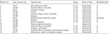
Fourteen patients (nine men and five women) between the ages of 40 and 68 years (mean age 57 years) had had a buccinator musculomucosal flap created during the study period. The indications for surgery are summarised in Table I. Twelve flaps were utilised to close defects following excision of oral cavity squamous cell carcinomas. All flaps were created at the time of resection, and all patients received post-operative radiotherapy. There were six cancers of the tongue (four T2 and two T3 lesions (tumour–node–metastasis (TNM) classification)), three of the floor of the mouth (all T1), and three of the tonsil or palate (all T2).
Table I Patient summary

No = number; F = female; M = male; T = tumour; N = node; Y = yes
Patient number seven had had an adenoid cystic carcinoma of the hard palate, which had been treated with neutron therapy. She developed a local recurrence 10 years later, for which a wide local excision was undertaken. A buccinator musculomucosal flap was utilised to repair the palatal defect. Patient 10 had a nasal septal perforation. He had had a cleft palate repair in infancy and subsequently sustained severe facial fractures in a motor vehicle accident. Following nasal reconstruction with rib cartilage, he had a large, symptomatic nasal septal perforation. As there was no suitable local tissue with which to repair the defect, a buccinator musculomucosal flap, based on the distal end of the facial artery, was utilised to repair the septal perforation.
Results
The outcome of the buccinator myomucosal flaps was assessed retrospectively by chart review and by recall of those patients who could be contacted (Table II).
Table II Outcome of buccinator myomucosal flaps

No = number; NT = not tested
Complete flap failure occurred only in patient seven (7 per cent), who had had palatal repair following neutron therapy. Scarring and vascular injury to the recipient site from the neutron therapy was a likely contributing factor to failure. One patient had suffered very minimal distal flap necrosis for which no additional surgery was needed, as the wound had healed by secondary intention with a good functional outcome. There was one case of partial flap dehiscence, which had healed with no further intervention. Donor sites were closed primarily with absorbable sutures. There was one case of dehiscence of the donor site closure, which had healed by secondary intention. No other flap complications were noted.
Only seven patients could be recalled to assess long-term results regarding flap sensation and trismus (Table II). Both light touch sensation and two-point discrimination were present, although reduced, in 71 per cent (five of seven) of the flaps. The average two-point discrimination of the flap was 21.6 mm, compared with 12.8 mm for normal buccal mucosa. No patient had trismus as a result of scarring at the donor site.
Discussion
The buccinator muscle is a thin, quadrilateral cheek muscle (Figure 1). It originates from the outer surfaces of the alveolar processes of the maxilla and mandible, overlying the three molar teeth. Posteriorly, it arises from the pterygomandibular raphe and inserts anteriorly into the orbicularis oris muscle. Laterally, it is related to the ramus of the mandible, the masseter and medial pterygoid muscles, the buccal fat pad and the buccopharyngeal fascia. Medially, it is covered by the submucosa and mucosa of the cheek. It is part of the pharyngeal-buccal-orbicularis sphincter system and functions to facilitate whistling, sucking, propelling food during mastication and voiding the buccal cavity.
The buccal, facial and posterosuperior alveolar arteries make up the main blood supply to the muscle (Figures 1, 2 and 3). The buccal artery is a branch of the internal maxillary artery and supplies the posterior half of the muscle. It courses anteroinferiorly under the lateral pterygoid muscle to reach the posterior half of the muscle, where it anastomoses with the posterior buccal branch of the facial artery.Reference Zhao, Li, Yan, Li, Yang and Mu3 The facial artery hooks around the lower border of the mandible at the anterior edge of the masseter muscle, and supplies numerous branches to the buccinator muscle (Figure 1), the largest of which is the posterior buccal, which supplies the posterior half of the muscle. The facial artery gives off one to three inferior buccal branches to supply the inferior half of the muscle, and then continues in an anterosuperior direction to give off three to five small, anterior buccal branches to the anterior half of the muscle. The posterosuperior alveolar artery, a branch of the internal maxillary artery, provides two small branches to the muscle, which enter the muscle posterosuperiorly, and the infraorbital artery gives off a few small branches which enter it anterosuperiorly. All these arteries form an extensive vascular anastomosis on the lateral surface of the muscle and within its fibres.

Fig. 2 Buccinator myomucosal flaps. (1) Posteriorly based flap; (2) anteriorly based flap; (3) superiorly based flap.

Fig. 3 Left buccinator flap planning. Thick arrow = buccinator musculomucosal flap; curved arrow = position of buccal artery; curved stippled arrow = position of facial artery
Venous drainage occurs via the pterygoid plexus (and internal maxillary vein). This lies posterior, superior and superficial to the buccinator and drains into the buccal vein via the deep facial vein. Anteriorly, the deep facial vein drains into the facial vein proper.
Sensory innervation is via the long buccal nerve, a branch of the maxillary nerve, which courses with the buccal branch of the internal maxillary artery. The motor supply to the buccinator muscle is the temporal and cervical divisions of the facial nerve, which form a plexus near the buccal fat pad.
The parotid duct has an important anatomical relationship to the muscle and pierces the buccinator opposite the second upper molar, slightly above the centre of the muscle.
Buccal mucosal flaps have been utilised to repair a variety of defects of the nasal septum, palate, midface, orbit and conjunctiva.Reference Tezel4–Reference Rayner8 Modifications to improve the vascularity of the mucosal flaps were reported by Maeda et al., who also described buccal ‘musculomucosal’ flaps for repair of cleft palate.Reference Maeda, Ojimi, Utsugi and Ando9 Various techniques have been described for the creation of the buccinator musculomucosal flap, mainly based on arterial supply. The buccinator musculomucosal flap can be based posteriorly, anteriorly or superiorly.
The limits of the flap are the parotid duct superiorly, the oral commissure anteriorly and the pterygomandibular raphe posteriorly. Inferiorly, the limit is dependent on the size of tissue required, but a flap as big as 7 × 5 cm can be raised.
Posteriorly based flaps
Figure 2 shows the posteriorly based buccinator musculomucosal flap. Bozola et al. first described an axial musculomucosal flap based posteriorly on the buccal artery.Reference Bozola, Gasques, Carriquiry and Cordosa de Oliveira1 Once the buccal artery is identified by Doppler ultrasound, the buccal mucosa and the buccinator muscle are incised to the level of the buccopharyngeal fascia, and the flap elevated in an anterior to posterior direction in the loose areolar plane between the buccinator muscle and the buccopharyngeal fascia. The buccopharyngeal fascia is preserved because it prevents herniation of the buccal fat pad and avoids injury to branches of the facial nerve. Small branches of the facial artery are ligated, as are anterior venous tributaries from the pterygoid plexus. The dissection proceeds posteriorly until just anterior to the pterygomandibular raphe, where the main neurovascular bundle enters the flap. The donor site is closed primarily. Care is taken that the pedicle does not interpose between the molar teeth, as this may interfere with mastication. Relevant molars might need to be extracted or an island flap created; alternatively, the vascular pedicle may be divided after a delay of a few weeks.
Modifications of this procedure include isolation of the pedicle to create an island flap, in order to facilitate rotation,Reference Licamelli and Dolan10 and creation of a ‘buccinator myomucosal neurovascular island pedicle flap’ based on the buccal artery, the buccal venous plexus and nerves innervating the muscle. The mucosa at the posterior end of the flap is divided from the underlying muscle and freed of its insertion from the pterygomandibular raphe.Reference Zhao, Li, Yan, Li, Yang and Mu3 Then the flap is passed through a short tunnel under the pterygomandibular ligament.
Anteriorly based flap
The anteriorly based flap (Figure 2) is based anteroinferiorly on the inferior buccal branches of the facial artery,Reference Carstens, Stofman, Hurwitz, Futrell, Patterson and Sotereanos2, Reference Zhao, Zhang, Li, Li, Xiao and Fan11, Reference Stofman, Carstens, Berman, Arena and Sotereanos12 the main trunk of which may be identified with a Doppler probe to establish its position. The mucosa and the buccinator muscle are incised superiorly and the facial artery and vein ligated. The dissection continues in a plane lateral to the vessels, as the flap is raised from front to back while branches of the facial artery are ligated (Figures 4 and 5).

Fig. 4 Left anteriorly based buccinator flap elevated and ready for insertion in anterior floor of mouth defect (arrow).

Fig. 5 Left anteriorly based buccinator flap inserted in floor of mouth defect (arrow).
Superiorly based flap
Zhao et al. described the superiorly based ‘buccinator myomucosal reversed-flow arterial island flap’ (Figure 2).Reference Zhao, Li, Yan, Li, Yang and Mu3 It is based on the distal end of the facial artery and its anterior buccal branches. The course of the artery is outlined by Doppler ultrasound, and dissection starts at the inferior margin of the flap with incision of the mucosa and buccinator muscle. The facial artery is ligated inferiorly and the flap is elevated in a superior direction. The arc of rotation is centred between the oral commissure and medial canthus.
The buccinator flap has proved to be very reliable. Our only clinically significant failure was in a patient who had previously received neutron therapy; this failure was probably related to a compromised microvascular blood supply, either to the flap or the recipient site.
Light touch sensation and two-point discrimination were intact in 71 per cent of patients, although sensation was reduced. However, even reduced sensation is advantageous in terms of oral function, when compared with insensate flaps. Recovery of sensation within the early post-operative period has been reported (following creation of anteriorly and posteriorly based buccinator flaps for tongue reconstruction). When a flap is posteriorly based, such sensory recovery can be attributed to the fact that the buccal nerve is included in the flap.Reference Licamelli and Dolan10, Reference Zhao, Zhang, Li, Li, Xiao and Fan11 We have also noted very early post-operative recovery of sensation.
All donor sites could be closed primarily (Figure 6). Even when harvesting a large flap, primary closure could usually be achieved due to the mobility and elasticity of the remaining buccal mucosa and muscle. Should the wound dehisce, it heals with minimal scarring, as was shown by the absence of trismus in our patients.

Fig. 6 Donor site defect closed primarily following raising of anteriorly based buccinator flap (arrows).
• The buccinator myomucosal flap is an axial-pattern flap based on the buccal or facial artery, and is used in the reconstruction of small and moderately sized defects of the oral cavity and oropharynx
• The authors report use of this flap, presenting the flap anatomy, the surgical technique utilised in elevatation, and the complications and applications of its use in reconstruction
• The study included 14 patients, of whom seven were reviewed to assess flap sensation and overall morbidity
• Seventy-one per cent of patients had sensation in the flap, albeit reduced compared with the opposite side
• Primary closure of the donor site was achieved in all cases, and the complication rate in raising the flap was low, with only one flap failure
• The buccinator myomucosal flap was found to be a reliable flap with good functional outcome and low morbidity
The buccinator musculomucosal flap is a versatile flap that can be used to reconstruct a variety of defects. It is remarkably elastic and malleable, and can be stretched to conform to complexly shaped defects. The procedures we performed are summarised in Table I. The arc of rotation of posteriorly based flaps will allow it to reach velar, palatine and lateral pharyngeal sites. We have used it to reconstruct defects of the tonsil bed, soft and hard palate, tonsillolingual sulcus, and retromolar trigone. It has also been used in primary cleft palate repair.Reference Bozola, Gasques, Carriquiry and Cordosa de Oliveira1 We used the anteriorly based buccinator musculomucosal flap to reconstruct the lip, and for repair of floor of mouth defects and reconstruction of the anterior and lateral tongue. It is particularly useful for anterior floor of mouth and tongue defects as it provides a normal mucosal covering and preserves mobility of the anterior tongue. It has also been described for closure of oro-antral and palatal fistulae, and for alveolar reconstruction.Reference Carstens, Stofman, Hurwitz, Futrell, Patterson and Sotereanos2 The superiorly based buccinator musculomucosal flap has been described as a means of closing defects in the anterior hard palate, alveolus, maxillary antrum, nasal cavity, upper lip and lower lip, as well as the orbit.Reference Zhao, Li, Yan, Li, Yang and Mu3, Reference Zhao, Zhang, Li, Li, Xiao and Fan11, Reference Dupoirieux, Plane and Penneau13 We employed this technique to close a large nasal septal perforation.
The buccinator musculomucosal flap has distinct advantages over free flap reconstruction. It is ideally suited to surgical units that lack expertise in microvascular free tissue transfer. Even in units that have this option, the considerable advantages of this form of reconstruction make it a good choice for many oral cavity and oropharyngeal defects. The operating time needed to raise and inset the buccinator musculomucosal flap is 45 minutes to one hour, which is considerably less than that required for both free flaps and distant axial flaps. Buccinator musculomucosal flaps also require a shorter hospital stay and are associated with low donor site morbidity. The buccinator musculomucosal flap is technically simple and does not require microvascular expertise. The flap has great versatility, as is evident from the variety of defects reconstructed. It is also very useful in patients with vascular disease, in whom there may be an increased risk of free flap failure. It is ideally suited for reconstruction following resection of T1, T2 and smaller T3 oral cavity and oropharyngeal lesions, at the time of excision of the primary tumour. However, it is not suited for larger defects for which more bulk is required; in these cases, distant axial or free flaps would be better options.
Conclusions
The buccinator myomucosal flap is a versatile flap useful for reconstruction of a variety of small and moderately sized defects of the oral cavity and oropharynx. It has a well described and constant blood supply, which makes it a dependable flap with little risk of flap failure. It has significant advantages over many other forms of oral reconstruction, including: technical simplicity; short operating time; lack of external scars; minimal donor site morbidity; donor and recipient sites in the same operative field; replacement of mucosa with mucosa; sensory innervation, which aids oral rehabilitation, mastication and speech; and flap reliability. Limitations include the amount of tissue available for reconstruction, and the fact that the anteriorly based flap cannot be used if the facial artery has been taken at neck dissection.