Tetralogy of Fallot is the most common cyanotic congenital heart lesion, comprising up to 7% of all congenital heart defects.Reference Perry, Neill and Ferencz 1 Surgical repair was first performed in 1954, and patients have excellent long-term survival following intracardiac repair.Reference Nollert, Fischlein and Bouterwek 2 However, these patients have numerous long-term complications including severe pulmonary regurgitation, right ventricular dysfunction, arrhythmias, and increasingly recognised, aortic root dilation.
Progressive aortic root dilation has been observed in a subset of adult patients with tetralogy of Fallot.Reference Niwa, Siu and Webb 3 Several factors that are associated with progressive aortic root dilation in patients with tetralogy of Fallot include male gender, pulmonary valve anatomy – atresia or absence – and right aortic arch.Reference Niwa, Siu and Webb 3 Interestingly, the last two features are commonly seen in patients with 22q11.2 deletion syndrome.Reference Goldmuntz 4 – Reference Marino, Digilio and Grazioli 6
Approximately 8–23% of patients with tetralogy of Fallot have 22q11.2 deletion syndrome.Reference Momma, Kondo and Ando 5 , Reference Amati, Mari and Digilio 7 – Reference Takahashi, Kido and Hoshino 9 Aortic root dilation is seen in patients in the context of several genetic syndromes and has been described with 22q11.2 deletion syndrome in the absence of conotruncal defects.Reference John, McDonald-McGinn and Zakai 10 However, to our knowledge the association of aortic root dilation in patients with tetralogy of Fallot with 22q11.2 deletion syndrome has never been tested. Adult patients with tetralogy of Fallot have been observed to have complications related to progressive aortic root dilation including aortic valve regurgitation requiring aortic valve replacement, aortic aneurysm formation, and aortic dissection.Reference Dodds, Warnes and Danielson 11 – Reference Kim, Seo and Kim 14 Identifying additional risk factors for aortic root dilation in tetralogy of Fallot, including the presence of a genetic syndrome, would allow for better risk stratification of patients and improve upon clinical care. We hypothesised that 22q11.2 deletion syndrome was associated with aortic root dilation in tetralogy of Fallot.
Methods
Patient selection
A total of 171 patients with tetralogy of Fallot, in the age group of 6–18 years, with echocardiograms performed from 2003 to 2009 were identified from an existing research database. Deletion status was determined for the entire cohort by either fluorescence in situ hybridisation or PCR-based multiplex ligation-dependent probe amplification.Reference Goldmuntz, Clark and Mitchell 8 , Reference Miller, Adam and Aradhya 15 Echocardiograms were performed on 142 patients as part of a cross-sectional study on genotype and clinical outcome, and recent standard-of-care echocardiograms were identified for an additional 29 cases. The charts of all patients were reviewed for demographics, height, weight, pulmonary valve morphology, aortic arch anatomy, and surgical information. Recorded surgical parameters consisted of previous palliative surgery, age at palliation and complete tetralogy of Fallot repair (ventricular septal defect closure), and length of time since complete repair. No case had undergone earlier aortic root or aortic valve repair or replacement. This study was approved by the Institutional Review Board at Children's Hospital of Philadelphia.
Echocardiographic assessment
Transthoracic echocardiograms were performed with a Sonos 5500 or an IE 33 ultrasound machine (Phillips Medical Systems, Andover, Massachusetts, United States of America) at a single centre using standard views. Two-dimensional aortic root measurements were recorded by two observers (A.S.J. and J.R.) for the 142 echocardiograms performed as part of the cross-sectional studies and one observer (A.S.J.) for the 29 standard-of-care echocardiograms. Measurements were taken in the parasternal long-axis view at three sites: the aortic valve annulus, the level of the sinuses, and the sinotubular junction. All measurements were taken in systole from the outer edge to the outer edge. Z-scores ([measured dimension-mean/standard deviation]) were derived from normal data developed by Dr Steven Colan at Children's Hospital of Boston on healthy children. A Z-score of 2 corresponds to two standard deviations above the mean.
Aortic insufficiency was graded as trivial, mild, moderate, or severe as assessed by colour flow Doppler by the same observer. Longitudinal assessments were made by retrospective echocardiogram review in 29 patients for whom pre-operative echocardiograms were available.
Statistical analysis
Continuous variables were expressed as mean and standard deviation or median and interquartile range when appropriate. Frequencies with proportions were determined for categorical variable, which were furthered examined through the χ 2-test. Fisher exact test was used when appropriate to account for small sample size distribution. The associations between deletion status, aortic insufficiency, and aortic root measurements were examined using Student's t-test. Variables that were statistically significant in the univariate analysis or reported to be significant in prior studies were then examined in a multivariate regression analysis. All statistical analyses were conducted using SAS, version 9.2 (SAS Institute, Cary, North Carolina, United States of America).
Results
Patient characteristics
Patient characteristics, anatomic details, and surgical approach are summarised in Table 1. A total of 27 cases (16%) had a 22q11.2 deletion syndrome. The median age of the total cohort was 12.2 years, with no significant difference between the population with the deletion and that without the deletion.
Table 1 Subject demographics.

Significant results are given in italic letters
*Ventricular septal defect closure
**Includes Blalock–Taussig shunt, Central aortopulmonary shunt
***Unifocalisation and/or fenestrated ventricular septal defect patch
****Includes the time between the initial shunt and staged repair to complete repair
Aortic arch anatomy was available in 166 patients: 69 (42%) patients had abnormal arch anatomy as defined by right aortic arch with mirror image branching (n = 61), right aortic arch with aberrant left subclavian artery (n = 4), or left aortic arch with aberrant right subclavian artery (n = 4).Reference Edwards 16 , Reference Allen, Driscoll and Shaddy 17 Aortic arch anomalies occurred in 67% of patients with 22q11.2 deletion as compared with 35% of patients with no deletion (p = 0.002). Tetralogy of Fallot subtypes with valvar pulmonary atresia and absent pulmonary valve were more prevalent in the population with 22q11.2 deletion; however, this was not statistically significant (p = 0.06).
All patients had complete surgical repair including ventricular septal defect closure and relief of right ventricular outflow tract obstruction. There was no significant difference in the mean age of complete repair between the patients with 22q11.2 deletion (median 0.4 years [0–3.0 years]) and the patients without the deletion (median 0.3 years [0–4.9 years]), including the subset with pulmonary atresia. Staged repairs included those with unifocalisation of major aortopulmonary collaterals, right ventricular-to-pulmonary artery conduit, and/or fenestrated ventricular septal defect patch placement before ventricular septal defect closure. There was a higher percentage of patients with initial aortopulmonary shunts (33% versus 12%, p = 0.004) in the subset with 22q11.2 deletion. In addition, there were more patients with staged repairs (11% versus 5%, p = 0.15), although this was not statistically significant.
Aortic root dilation and aortic insufficiency in the total cohort
Aortic root dimensions expressed as Z-scores are displayed in Table 2. A higher percentage of patients with 22q11.2 deletion had a Z-score >3 at the level of the sinus diameter as compared with the subset without the deletion (45% versus 24%, p = 0.02). Owing to the fact that so few patients were observed to have dilation at the sinotubular junction, the remainder of the study focused on dilation at the level of the aortic annulus and the level of the sinuses. In all, 82% of the cohort had no significant aortic insufficiency – trivial or less. A total of 28 patients (16%) had mild aortic insufficiency, whereas one patient had moderate aortic insufficiency. There was no statistically significant difference in aortic dimensions between those patients with none/trivial aortic insufficiency and those with mild/moderate aortic insufficiency.
Table 2 Extent of aortic dilation within the total cohort.

Significant results are given in italic letters
*Sinotubular junction measurements available in 142 patients
Univariate analysis: risk factors for aortic root dilation in tetralogy of Fallot
Patients with a 22q11.2 deletion had larger aortic root dimensions at both the aortic annulus and the sinus diameter, although these changes did not meet statistical significance. Patients with both 22q11.2 deletion and concomitant arch anomaly demonstrated a greater increase in aortic root dimensions, although again these changes did not meet statistical significance (Table 3).
Table 3 Univariate analysis examining 22q11.2 deletion and aortic arch anatomy and aortic root size.

AAA = aortic arch anomaly
Results presented as mean ± standard error. Significant results are given in italic letters
*Includes patients without the deletion with normal arch anatomy and patients with isolated 22q11.2 deletion OR aortic arch anomaly
Further subset analysis was performed on each of the tetralogy of Fallot subtypes. No significant difference in aortic root dimension was seen in the subsets with pulmonary stenosis or absent pulmonary valve. Analysis of the subset with tetralogy of Fallot and pulmonary atresia showed that patients with a 22q11.2 deletion and concomitant aortic arch anomaly demonstrated a statistically significant dilation at the level of the aortic annulus (Z-score 4.6 versus 2.3, p = 0.05). There was also increased dilation at the level of the sinuses, although this was not statistically significant (Z-score 4.4 versus 2.7, p = 0.06).
Multivariate regression analysis
Previously reported risk factors of male gender, pulmonary atresia, and aortic arch anomalies in addition to 22q11.2 deletion and age were included in the analysis (Table 4). Results are expressed as a net change in Z-score at the aortic annulus and sinus diameter.
Table 4 Multivariate analysis examining risk factors for aortic Z-scoresFootnote *.

AAA = aortic arch anomaly
* Sinotubular junction values were not significant
** Z-score values expressed as change in Z-score ± standard error
*** Controlled for age, pulmonary valve anatomy, and male gender
**** Change in Z-score observed with a 1 year increase in age
Controlled for pulmonary valve anatomy, male gender, 22q11.2 deletion + AAA
The presence of both aortic arch anomalies and 22q11.2 deletion was associated with an increase of 1.33 in the Z-score for the aortic annulus (p = 0.006) and an increase of 0.82 in the Z-score at the level of the sinus (p = 0.05). Neither aortic arch anomaly nor 22q11.2 deletion in isolation yielded a statistically significant result at either the aortic annulus or sinus diameter level. As previously reported, male gender and pulmonary atresia were also independent risk factors for dilation at both the annulus and the level of the sinuses (Table 4).
Age and aortic root dilation
The multivariate analysis of the full cohort revealed that older children had slightly smaller aortic dimensions. An increase in age by 1 year between two patients correlated with a decrease in both aortic annular and sinus diameter Z-scores by 0.13 and 0.1, respectively (see Table 4).
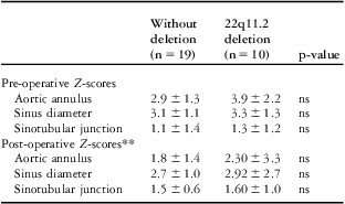
Pre-operative newborn (<4 months of age) echocardiograms were available in 29 cases – 19 without the deletion and 10 with the deletion – (see Table 5). In this limited subset with pre-operative measurements, the relative aortic dimensions are higher in the pre-operative setting, and decrease following repair. However, the subset with the deletion appears to have larger aortic dimensions both before and following surgical repair.
Table 5 Pre-operative and post-operative measurements of aortic root dimensionsFootnote *.

* Pre-operative echocardiograms available in 29 cases
** Most recent aortic dimensions
Discussion
Owing to the fact that over 85% of patients with repaired tetralogy of Fallot survive to adulthood, long-term complications including aortic root dilation have been increasingly observed in the adult population with tetralogy of Fallot. We have previously shown that isolated aortic root dilation is seen with 22q11.2 deletion syndrome in the absence of conotruncal defects; however, there are no studies examining whether 22q11.2 deletion is associated with aortic root dilation in patients with tetralogy of Fallot.Reference John, McDonald-McGinn and Zakai 10
This study is the first to show that a higher percentage of patients with 22q11.2 deletion syndrome had aortic root dilation at the level of the aortic sinuses as compared with patients without the deletion, a statistically significant difference. When examining the absolute Z-scores, the presence of 22q11.2 deletion syndrome and a concomitant aortic arch anomaly is an independent risk factor for aortic annular and aortic sinus dilation in children with tetralogy of Fallot, especially in the subset of patients with pulmonary atresia. Neither aortic arch anomaly nor 22q11.2 deletion syndrome was independently associated with a significant increase in aortic root dilation Z-scores in this paediatric age group. However, patients with 22q11.2 deletion with normal aortic arch anatomy had mild dilation at the level of the aortic annulus and the sinuses.
Similar to earlier studies, pulmonary atresia and male gender were also independent risk factors for aortic annular and aortic sinus dilation. The presence of these features might explain why a greater percentage of patients had Z-scores between 2 and 3 at the level of the sinuses in the subset without the deletion. To further ensure that the aortic root dilation was not due to the higher percentage of patients with pulmonary atresia within the 22q11.2 deletion subset, we performed a subset analysis on those cases with pulmonary atresia (Table 2). This analysis yielded a statistically significant increase in Z-scores at the level of the aortic annulus (p = 0.05) and near statistical significance at the sinuses (p = 0.06) in the subset of patients with pulmonary atresia and concomitant 22q11.2 deletion syndrome and aortic arch anomalies. Although the aortic annular Z-score increase of 1.33 seen in patients with both aortic arch anomaly and a 22q11.2 deletion may not be a substantial increase, the presence of multiple risk factors results in an additive effect on aortic root dimensions. Therefore, a child with tetralogy of Fallot with the combination of 22q11.2 deletion and a concomitant aortic arch anomaly in addition to male gender and pulmonary atresia is at highest risk of having aortic root dilation.
Surgical considerations
Older age (>7 years) at repair, presence of palliative shunt, and longer time interval from shunt palliation to complete repair have been cited as previous risk factors for aortic root dilation.Reference Niwa, Siu and Webb 3 , Reference Bhat, Smith and Hawker 18 Surgery during infancy (<12 months of age) is now the standard of care at most centres. In our cohort of patients, there was a higher percentage of patients in the subset with the deletion with aortopulmonary shunts; however, most patients had complete repairs performed before 1 year of age. The overall percentage of cases with tetralogy of Fallot with aortic annular and sinus diameter dilation in our cohort is lower than has been previously reported, which could be partly due to earlier surgical repair.Reference Chong, Wong and Chiu 19 – Reference Senzaki, Iwamoto and Ishido 21
Of the tetralogy of Fallot subtypes, pulmonary atresia patients often have later repairs because of the need for staged interventions.Reference Mahle, Crisalli and Coleman 22 However, the subset of patients with pulmonary atresia in our cohort had a median age of 0.6 years at complete repair, with no difference between cases with the deletion and without the deletion. This further substantiates that pulmonary atresia is an independent risk factor for aortic root dilation. There have been variable results reported on post-surgical mortality rates in patients with 22q11.2 deletion syndrome and pulmonary atresia.Reference Mahle, Crisalli and Coleman 22 , Reference Michielon, Marino and Formigari 23 Re-intervention rates, although higher, do not typically include aortic root surgery in the paediatric cohort.Reference Michielon, Marino and Oricchio 24
Age and aortic root dilation
When controlling for gender, pulmonary valve anatomy, deletion status, and arch anatomy, an increase in age of 1 year was associated with a decrease in aortic annular and sinus diameter Z-scores in this paediatric cohort (see Table 4). Therefore, the aortic root dilation observed in the older patients in our cohort was not due to increased age. Previous studies have shown that aortic root measurements are largest in unrepaired tetralogy of Fallot but begin to decrease following surgical repair.Reference Bhat, Smith and Hawker 18 Similar findings were seen in our patients, although we could only examine a small subset of patients longitudinally. All patients, including both the subsets with the deletion and without the deletion, had larger aortic root measurements pre-operatively. Over time, Z-scores decreased, but remained higher in the subset with the deletion.
Age of participants
We chose to study patients aged 6 years and older as these patients would be out of the immediate newborn period. Owing to the fact that most of our cohort underwent repair during infancy, there was a period of at least 5 years between repair and current assessment. By limiting the subset to ages >6 years, we hoped to eliminate patients who had dilated aortic roots because insufficient time had passed from the unrepaired state. In addition, limiting the study age to children (⩽18 years of age) limits the effect of adult onset risk factors for aortic root dilation, such as hypertension.
Aortic insufficiency
Haemodynamically significant aortic insufficiency was uncommon in our cohort, and therefore did not contribute to the aortic root dilation observed. There have been several studies and reports of adult patients with tetralogy of Fallot requiring concomitant aortic valve and aortic root replacement.Reference Dodds, Warnes and Danielson 11 , Reference Kiziltan, Topcu and Ozbarlas 25 Some factors thought to be associated with progressive aortic insufficiency include older age at repair, pulmonary atresia, and progressive dilation at the level of the sinus.Reference Bhat, Smith and Hawker 18 , Reference Ishizaka, Ichikawa and Sawa 26 Owing to the fact that our cohort underwent repair at a younger age and represents a younger cohort overall (⩽18 years of age), this may account for the decreased prevalence of haemodynamically significant aortic insufficiency.
Mechanisms of aortic root dilation in tetralogy of Fallot
The exact mechanism of aortic root dilation in tetralogy of Fallot is unknown. Both adults and children tetralogy of Fallot have been shown to have intrinsic histological abnormalities of the aorta ranging from cystic medial necrosis to fibrosis.Reference Tan, Gatzoulis and Ho 27 , Reference Niwa, Perloff and Bhuta 28 In addition, patients with tetralogy of Fallot have increased aortic stiffness and reduced aortic wall distensibility compared with age-matched controls.Reference Chong, Wong and Chiu 19 , Reference Cheung, Ou and Wong 20 Recent data suggest that polymorphisms of matrix metalloproteinase-9, a matrix degrading enzyme, might be involved as well.Reference Cheung, Hong and Chan 29 Patients with both aortic arch anomalies and 22q11.2 deletion might represent a subset of patients with a more severe aortopathy, but the mechanisms that 22q11.2 deletion syndrome might play in aortic root dilation is speculative and cannot be determined from this study.
Study limitations
Although this is the only study that examines the effect of 22q11.2 deletion on aortic root dilation, there are several limitations. The study is a cross-sectional analysis, not a longitudinal study, of patients with tetralogy of Fallot at an age range of 6–18 years. With this study design, the impact of 22q11.2 deletion on the presence, but not the progression, of aortic dilation and aortic insufficiency can be examined. Although this study is one of the largest examining children with tetralogy of Fallot, it is still limited by the small sample size of patients with 22q11.2 deletion syndrome and the qualitative assessment of aortic insufficiency. In addition, aortic root measurements were made by only one observer in 17% of the study echocardiograms.
Conclusions
The presence of 22q11.2 deletion with a concomitant aortic arch anomaly is associated with aortic annular and sinus diameter dilation in patients with tetralogy of Fallot. Male gender and pulmonary atresia were additional risk factors identified for dilation of both the annulus and sinuses. Certainly, further prospective, longitudinal analysis is needed to identify the subset of patients that develops progressive aortic root dilation and aortic regurgitation, and to study their clinical significance.
Acknowledgements
None.
Financial support
Funding provided by P50HL062177 (EG), P50HL74731 (EG).
Conflicts of interest
None.
Ethical Standards
The authors assert that all procedures contributing to this work comply with the ethical standards of the relevant national guidelines on human experimentation and with the Helsinki Declaration of 1975, as revised in 2008, and has been approved by the institutional review board at Children's Hospital of Philadelphia.