


Introduction
Taenia hydatigena, is a worldwide common parasite, encountered mostly in dogs and wild carnivores in its mature state, and whose larvae, Cysticercus tenuicollis, is seen on ruminants and pigs (Soulsby, Reference Soulsby1982). The larvae settling on the visceral side of the liver, omentum, mesenterium or abdominal serous surfaces, after they reach a certain size, become infective when they reach a diameter of 6–8 cm. (Taylor et al., Reference Taylor, Coop and Wall2007). In the digestive system of the final hosts, the invaginated scolex of C. tenuicollis, is opened with the effect of bile salts, and evaginating scolex develops, attach to the intestinal mucosa (Deplazes, Reference Deplazes, Schnieder and Buchverlag2006). Clinical signs of intermediary hosts, infected with C. tenuicollis vary according to the severity of infection. Mature cysticercus larvae are not as harmful in the peritoneal space as they are settled in the liver (Kaufmann, Reference Kaufmann2013). In severe infections, hepatitis cysticercosa may occur due to the migration of a large number of larvae in the liver (Edwards and Herbert, Reference Edwards and Herbert1980). Haemorrhagic-fibrotic lesions encountered in the liver and even peritonitis due to these migrations cause liver degenerations and lamb deaths, thus causing economic losses (Soulsby, Reference Soulsby1982; Blazek et al., Reference Blazek, Schramlova and Hulinska1985). In a study to calculate the average economic loss caused by C. tenuicollis infected animals, reported an economic loss of €0.40 per kg, due to this parasite, a €315 622.2 worth annual loss caused by cysticercosis-related liver destruction, an annual loss of €18 035.5 in offals, hence a total loss of approximately €333 657 (Scala et al., Reference Scala, Pipia, Dore, Sanna, Tamponi, Marrosu, Bandino, Carmona, Boufana and Varcasia2015). Although serological tests and screening techniques are widely used in the diagnosis of the disease, since they do not yield definitive results, a final diagnosis of the disease is established through evaluating the cysts after slaughtering. For this reason, although there is no effective treatment of the disease in intermediate hosts, it was reported that the spread of the parasites can relatively be controlled by the taeniasis treatment using anthelmintics appropriately for the final hosts, but the presence of the wild cycle and stray dogs makes this difficult (Bamorovat et al., Reference Bamorovat, Radfar, Derakhshanfar, Molazadeh and Zarandi2014).
Although there are some studies on the phylogeny of T. hydatigena and other taeniid cestodes, studies on intra-species variations are very limited. Understanding the intra-species genetic diversity regarding this economically important parasite is necessary in terms of both epidemiological studies and control programmes. This study is conducted in order to investigate the genetic variability and population structure regarding sheep and goat isolates of C. tenuicollis form Bingol province of east-Turkey.
Materials and methods
Sample collection
The parasite samples were collected from Bingol province of Turkey where is located in the upper Firat district. The province covers an area of 8.253 km2 with a population of 281.205. The economy is based on stockbreeding, agriculture, and handicraft. Stockbreeding includes sheep, goat and cattle. According to 2018 data, 510.781 sheep and goats were reported in Bingöl province (Anon, 2018).
Liver and mesenterium of grazed sheep and goats slaughtered in a local slaughterhouse in Bingol province of eastern Turkey were inspected for Cysticercus tenuicollis cysts. The slaughterhouse was visited between May and June 2016 then slaughtered sheep and goats were examined through inspection and palpation, and C. tenuicollis cysts of apparent size were dissected out. These cysts were detonated, discarding the liquid part, and the portion with the invaginated scolex was put into glass bottles containing 70% ethanol and stored at −20 °C until use. Twenty cyst samples of each animal species were collected for this study, where only one cyst for each animal was used, in order to determine genetic diversity. Other lesions such as hydatid cyst in the cyst collected animals were ignored.
Genomic DNA isolation
Total genomic DNA was isolated exclusively from the scolex of the cysts using the RTA® Tissue Kit (RTA Lab., Turkey) according to the manufacturer's instructions. Before the process scolexes were dissected out from the individual cysts then washed with PBS (pH = 7.4) at least five times for the cleaning up the ethanol. The total amount of genomic DNA in the samples was determined by spectrophotometric measurement and the samples containing insufficient amounts of gDNA were further isolated.
PCR amplification
For each isolate one single target of 471 bp within the mitochondrial NADH dehydrogenase subunit 1 (ND1) gene was PCR amplified with the use of the primer set JB11 (5′-AGATTCGTAAGGGGCCTAATA-3′) and JB12 (5′-ACCACTAACTAATTCACTTTC-3′) (Bowles et al., Reference Bowles, Blair and McManus1992). In short, the PCR reaction was contained 5 µL 10× PCR buffer, 5 µL MgCl2, 400 µμ of each dNTP's, 20 pmol of each primer, 5 µL of genomic DNA target and PCR grade water to a final volume of 50 µL. Amplification was performed according to the following temperature profile: initial denaturation step of 5 min at 95 °C and 35 cycles of 30 s at 95 °C, 30 s at 52 °C, 30 s at 72 °C followed by a final extension step of 5 min at 72 °C. In all cases, PCR was performed with a SensoQuest Labcycler thermalcycler (SensoQuest GmbH, Germany) and the products were analysed by 1.4% agarose gel electrophoresis. The amplification bands were excised from the gels under UV and subjected to purification steps using the GeneJET Gel Extraction kit (ThermoScientific). A directional sequence analysis was performed using the respective pair of primer (JB11), with an ABI 3730XL Genetic Analyser (Sentegen Inc., Turkey).
Phylogenetic analysis
The electropherograms' quality of the sequences was assessed and the edges were trimmed with the use of FinchTV 1.4.0 (Geospiza Inc., Seattle WA, USA) according to the principles outlined by Alachiotis et al. (Reference Alachiotis, Vogiatzi, Pavlidis and Stamatakis2013). Consensus sequences, trimmed to a common length of 449 nucleotides each, were created using the CLC Main Workbench 8 program (Taylor et al., Reference Taylor, Coop and Wall2007) and represented the data matrix of the present analysis. An NCBI BLAST search primarily defined the identity of those sequences. A multiple sequence alignment was generated with the use of CLC Main Workbench 8 program (Taylor et al., Reference Taylor, Coop and Wall2007) and a bootstrap test conducted with the help of the same program using the Neighbour-Joining method (100 repetitions).
PopART (Population Analysis with Reticulate Trees) software was used in order to determine haplotype diversities (Payan-Carreira et al., Reference Payan-Carreira, Silva, Rodrigues and dos Anjos Pires2008). The phylogenetic relationships between T. hydatigena and other Taeniid cestodes were determined using Bayesian inference (BI). Haplotypes were generated using Median Joining or Minimum Spanning Networks, and the relationships in between these haplotypes were demonstrated (Kaufmann, Reference Kaufmann2013).
Results
Cysticercus tenuicollis cysts were detected in 20 out of 150 goats (13.3%) and 20 out of 102 sheep (19.6%) which were examined during the study. All cysts were collected from the mesenterium of examined sheep and goats (Fig. 1).
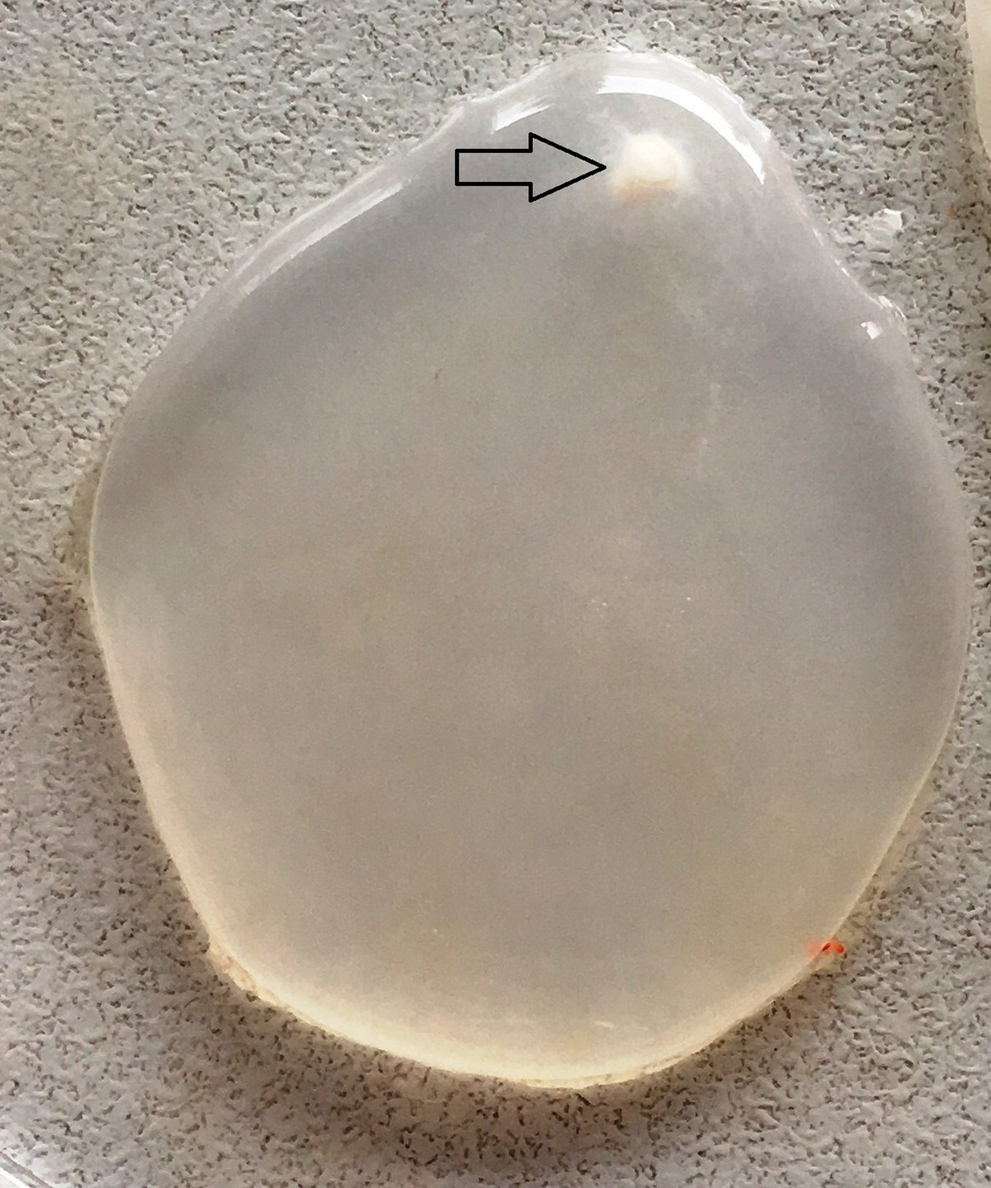
Fig. 1. A Cysticercus tenuicollis cyst collected from mesenterium. Arrow: appearance of the invaginated scolex in the cyst fluid.
In this study, the target 471 bp amplicons were successfully determined in the PCR analyses of all C. tenuicollis isolates from sheep and goats (Fig. 2) and high-quality sequence chromatograms were obtained for each isolate.

Fig. 2. Showing the bands formed by PCR amplification of the mt-ND1 gene region of sheep and goat isolates of Taenia hydatigena (471 bp). M: DNA marker (100 bp), 1: sheep isolate, 2: negative control, 3: goat isolate.
In this study, nucleotide changes obtained through a DNA sequence analysis of the ND1 gene region of 20 sheep with C. tenuicollis samples were determined by the alignment and shown in Fig. 3. Nucleotide changes as a result of the sequence analysis of the sheep samples are also evident in the genetic tree (Fig. 4). It was determined that KY7 is the most distinctive sample, and that KY4 and KY2 are in the same cluster and they are in the same cluster together with the dog reference sequence, with KY17, KY20, KY5, KY1 and KY13. Again, KY14, KY6, KY19, KY18, KY10, KY9 and KY15 were observed to be in the same cluster. KY11 and KY3 were observed to be in close clusters with reference goat and pig sequences, and it was noted that KY12, KY16 and KY8 were close to them, but in different clusters. In haplotype analysis of sheep sequence samples, 16 different haplotypes were determined, which is then demonstrated in Fig. 5.

Fig. 3. Alignment of sequences of mt-ND1 gene region of sheep isolates of T. hydatigena. KY1-KY20: Sequences of sheep isolates. Dog isolate (KC876043), sheep isolates (DQ995654 and KT372542), goat isolate (JN831274) and pig isolate (JN831286) show the reference sequences.

Fig. 4. Phylogenetic tree view of sheep isolates using mt-ND1 gene sequences and reference sequences.

Fig. 5. The appearance of mt-ND1 (471 bp) haplotypes of sheep isolates of T. hydatigena. The size of the circles is related to the haplotype frequency. Small circles indicate additional mutational areas.
The alignment result for the goat samples is demonstrated in Fig. 6 where the genetic tree view is exhibited in Fig. 7. Accordingly, KC4 is located in a different cluster than the other samples. Goat (JN831274) and pig (JN831286) sequences were located in the same cluster, where KC15 and KC12 were noted as sequences closest to them. Again, KC11 and KC14 were observed as the closest sequences to this group. KC1 and reference sheep sequence (DQ995654) were included in the same cluster, KC16, KC17, KC18, KC19, KC20 and KC6 were found to be the most relevant sequences. The sequence KC10 was found to be at an equal distance to the other reference sheep sequence (KT372542); KC5, KC8, KC3 and KC7 were determined to follow them respectively in accordance with their distance. Finally, it was observed that KC13 and KC2 were in the same cluster and KC9 was at the closest sequence. In haplotype analysis regarding the goat sequence samples, 15 different haplotypes were determined and this result is then exhibited in Fig. 8.

Fig. 6. The alignment of the mt-ND1 gene region of T. hydatigena goat isolates. KC1-KC20: Sequences of goat isolates. Dog isolate (KC876043), sheep isolates (DQ995654 and KT372542), goat isolate (JN831274) and pig isolate (JN831286) show the reference sequences.

Fig. 7. Phylogenetic tree constructed using of reference sequences and mt-ND1 gene sequences of goat isolates.

Fig. 8. The appearance of mt-ND1 haplotypes of goat isolates of T. hydatigena. The size of the circles is related to the haplotype frequency. Small circles indicate additional mutational areas.
Discussion
Taenia hydatigena, is a widespread parasite, commonly encountered in Canidae, and that can infect a wide variety of mammals together with its larvae, C. tenuicollis (Murrell et al., Reference Murrell, Dorny, Flisser, Geerts, Kyvsgaard and McManus2005). Mature parasites excrete their eggs through the host's feces, and intermediate hosts intake these eggs during grazing. Factors such as the presence of large pasture areas, shepherd dogs, numerous stray dogs, uncontrolled house slaughtering and improper disposal of carcases facilitate the spread of other important metacestode infections, such as hydatidosis and coenurosis in addition to C. tenuicollis (Scala et al., Reference Scala, Garippa, Varcasia, Tranquillo and Genchi2006).
In many studies conducted in the slaughterhouses in Turkey, the parasite larvae were determined to be quite common. The prevalence of C. tenuicollis is reported as 56.7% for lambs in Samsun (Zeybek, Reference Zeybek1980), 31.8% for sheep, and 28.57% for goats in Ankara (Sarimehmetoglu et al., Reference Sarimehmetoglu, Gonenc, Piskin and Ayaz1993), 2% in Marmara Region (Oncel, Reference Oncel2000), 24.1% in Bursa (Senlik, Reference Senlik2008), 8.18% for cattle, 65.67% regarding the sheep, 61.60% regarding the goats in Tatvan (Deger and Bicek, Reference Deger and Bicek2005), 6.5% for the cattle in the slaughterhouses of Hakkari and Van, 49.2–59.3% regarding the sheep in the same district, 26.1–56.7% regarding the goats there (Aydın, Reference Aydın2003; Deger et al., Reference Deger, Bicek, Gul and Eraslan2001) and 12.3% in Malatya (Kara et al., Reference Kara, Gicik, Sari, Bulut and Arslan2009). However, since all the studies conducted in Turkey are based on carcass follow-up, only one molecular study has been found (Utuk and Piskin, Reference Utuk and Piskin2012), who conducted a sequence analysis, multiplying the mt-CO1 gene region of C. tenuicollis isolate from a goat by PCR, (GenBank accession number: JN827307) and they reported a 99% similarity regarding the above-mentioned sample compared with the priorly published sequences in GenBank. However, since they did not use sufficient amount of samples, they could not perform any haplotype analysis.
In many countries of the world, especially in Europe, the prevalence of C. tenuicollis, its molecular genotyping and the economic losses caused by it have been studied. In a study conducted in Italy, the prevalence of C. tenuicollis in lambs was found to be 14.6% (1135/7781). The related economic loss due to this is calculated as €3.336.577 per year (Scala et al., Reference Scala, Pipia, Dore, Sanna, Tamponi, Marrosu, Bandino, Carmona, Boufana and Varcasia2015). In India, a total of 3199 carcases, including 760 sheep and 2439 goats were examined against C. tenuicollis, and 135 (4.22%) out of 3199 carcases were evaluated as positive. It was observed that most of the cysts were found in the abdominal cavity, and only a small portion was embedded in the liver. The highest prevalence was found out to be in the goats (4.83%), while the sheep exhibited a prevalence of 2.23% (Singh et al., Reference Singh, Sharma, Gill and Sharma2015).
In a study conducted in Palestine, 32 out of 1468 sheep (2.15%) were identified to have been infected with C. tenuicollis. It was observed that the livers regarding 30 of these 32 sheep (93.8%) were affected. The mt-CO1 gene region of these isolates was amplified and sequenced then nine different haplotypes were determined. Ten point mutations were determined among the main haplotypes. In addition to that, one-variable areas were detected in the 6, 72, 102, 141, 207, 231 and 264th nucleotides where three parsimony informative areas were determined in 51, 213 and 219th nucleotides (Adwan et al., Reference Adwan, Jayousi, Abuseir, Abbasi, Adwan and Jarrar2018).
In another study conducted in Italy, Boufana et al. (Reference Boufana, Scala, Lahmar, Pointing, Craig, Dessì, Zidda, Pipia and Varcasia2015) made a molecular analysis on a total of 35 T. hydatigena isolate (27 of them with C. tenuicollis while eight were mature). Twenty-three of the isolates were obtained from sheep where one was obtained from goat and one from a wild boar. The CO1 and ND1 gene regions of these isolates were amplified through PCR and their sequence analysis was conducted. Consequently, 17 haplotypes were determined with CO1 and 17 with ND1.
As a result of this study, and the sequence analysis of the mt-ND1 gene region, 16 different haplotypes were detected regarding 20 sheep isolates where 15 different haplotypes were detected in 20 goat isolates. These results show that gene flow occurs following a cross-fertilization of the mature parasites regarding the T. hydatigena species, and this manifests itself as nucleotide changes. If this situation persists, the intraspecies variation in T. hydatigena will increase, new strains and genotypes will emerge which may affect host adaptation and life cycle of the parasite.
Author ORCIDs
Sami Simsek, 0000-0002-3567-326X
Acknowledgements
The authors would like to thank all veterinarians and workers for their kind help during sample collection.
Financial support
This study has been supported by a grant from Firat University Scientific Research Projects Unit (Project No. VF.17.08).
Conflict of interest
The authors fully declare any financial or other potential conflicts of interest.
Ethical standards
Not applicable.


