Introduction
Thyroid nodules are common: they are detectable in 10 per cent of women and 2 per cent of men.Reference Hegedus1 A prospective study in North America found asymptomatic thyroid nodules in 67 per cent of their study population upon ultrasonography.Reference Ezzat, Sarti, Cain and Braunstein2 A similar study conducted in Germany found thyroid nodules in 20 per cent of the population aged 20–79 years. The prevalence rises with increasing age up to 52 per cent for women and 29 per cent for men aged 70–74 years.Reference Volzke, Ludemann, Robinson, Spieker, Schwahn and Kramer3 The high prevalence of thyroid nodules means that rational evidence-based management strategies are required to identify patients with a significant malignancy risk. These strategies need to be applicable to a wide spectrum of clinical presentations, ranging from small, sub-centimetre nodules to large, growing symptomatic nodules.Reference Hegedus4
The incidence of malignancy in thyroid nodules varies between 5 per cent and 15 per cent, and depends on age, sex, radiation exposure, family history and other factors.Reference Hegedus1, Reference Cooper, Doherty, Haugen, Kloos, Lee and Mandel5, Reference Yeung and Serpell6 In the period 1971–1995, the annual UK incidence was reported as 2.3 per 100 000 women and 0.9 per 100 000 men, with approximately 900 new cases and 250 deaths resulting from thyroid cancer every year recorded in England and Wales.Reference Coleman, Babb and Damiecki7 This has increased to an annual incidence to 6 per 100 000 women and 2 per 100 000 men, with approximately 2654 new cases in the UK in 2010.8 Patients with solitary nodules have a significantly higher incidence of thyroid carcinoma compared with those with multinodular goitre.Reference Abu-Eshy, Khan, Khan, al-Humaidi, al-Shehri and Malatani9 A truly solitary thyroid nodule in a patient under 30 years of age has a 10–20 per cent chance of malignancy, rising to 20–40 per cent in elderly men.Reference Ramsden and Watkinson10
This study aimed to investigate the prevalence of solitary thyroid nodules in patients who underwent thyroid surgery and the clinical significance of these nodules. The value of demographic and sonographic features of solitary nodules in predicting the malignancy risk was also determined and the malignancy rate in our case series was compared with that of other centres.
Materials and methods
A retrospective review of the case notes of all adult patients aged between 18 and 75 years who underwent thyroid surgery from January 2003 until December 2009 was performed. Patients with solitary nodules were identified based on their medical notes and pre-operative ultrasonography. All patients with solitary thyroid nodules who had had pre-operative ultrasonography and fine needle aspiration cytology (FNAC) were included in the study. A nodule was considered solitary if no other nodules in the gland were identified by ultrasonography. The presence of other nodules, irrespective of size, excluded patients from the study. Sonographic features and cytology and histology data were recorded and analysed. All FNAC was performed by experienced radiologists with cytology technicians in attendance to ensure uniform sample preparation. An assessment of cellularity was not made at the time of aspiration.
Statistical analysis
Data was analysed using Prism statistical software version 3.02 (GraphPad, Groningen, the Netherlands). The malignancy risk was stratified according to ultrasonography and FNAC findings. To compare the malignancy risk according to clinical factors, Fisher's exact test was used for discrete variables and the Mann–Whitney U test was used for continuous variables. A p value of less than 0.05 was considered statistically significant.
Ethical considerations
All patient reviews, data collection and analyses were carried out in accordance with appropriate institutional and hospital audit department regulations and approval.
Results
A total of 225 patients underwent thyroid surgery. Of the 76 clinically palpable solitary thyroid nodules, 15 were excluded because more than one nodule was identified by ultrasonography. The prevalence of ‘true’ solitary thyroid nodules was 27.1 per cent (61 out of 225 patients); affected patients had a mean ± standard deviation (SD) age of 52 ± 16 years (women, 52.1 ± 16.5 years; men, 51.4 ± 14.1 years). Forty-four out of 61 patients (72.1 per cent) were women. No patient reported exposure to childhood head and neck irradiation or a family history of thyroid medullary carcinoma. In 30 patients nodules were in the right lobe, in 26 they were in the left node and in the remaining 5 they were in the isthmus. These locations correlated with the ultrasonography locations for the nodules and no other nodules were detected in pathology specimens, thus confirming that the nodules removed were those detected by ultrasonography. The maximum nodule diameter ranged from 0.7 cm to 6.2 cm (median ± SD, 3.5 ± 1.26).
During the study, FNAC findings were not classified according to the British Thyroid Association Thy classification. Instead, results were recorded in a descriptive manner and subsequently classified into five categories: insufficient material for diagnosis, benign or colloid nodule, atypia, neoplasm and malignant (Table I).
Table I Fnac results and pathology findings

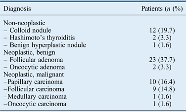
Of the 61 study patients, 5 had non-diagnostic FNAC findings; 3 of these were found to have malignant pathology at surgery. Twenty-eight of the 61 patients had neoplastic FNAC (Table II). Seven of the nine samples showing atypia had neoplastic pathology. Therefore, the atypia category was combined with the neoplastic and malignant categories when calculating FNAC sensitivity and specificity values.
Table II Histological diagnosis in 61 patients with solitary thyroid nodule

The sensitivity and specificity for detecting thyroid neoplasms were 73.9 per cent and 80.0 per cent, respectively, with a positive predictive value of 91.2 per cent for all thyroid neoplasms. The false negative rate for all FNAC for neoplasms was 42.0 per cent (8 out of 19). The sensitivity and specificity of FNAC for detecting thyroid malignancies was 38.1 per cent and 95.0 per cent, respectively, with a positive predictive value of 80.0 per cent. The false negative rate for all FNAC for malignancies was 10.5 per cent (2 out of 19).
The malignancy rate in patients with a truly solitary nodule in our patient population was 34.4 per cent, with a mean age at diagnosis of 53 ± 15 years and a female-to-male ratio of 2:1. There was no association between age or maximum nodule dimension with malignancy (p = 0.68 and p = 0.33, respectively). A thyroid nodule measuring more than 4 cm on its longest axis was seen in a third of patients with malignancy and in 27.5 per cent of patients with benign pathology. Most patients (56.0 per cent) had nodules measuring 2–3.9 cm in length. Hypo-echoism was seen in 6 out of 21 (28.6 per cent) patients with malignancy and 62.0 per cent of the malignant solitary nodules had increased vascularity. Four out of 21 patients (19.0 per cent) with malignant pathology had a nodule with a sonographic solid mass appearance and in 19.0 per cent the nodules were cystic or had undergone cystic degeneration. Hypo-echoism or a solid mass appearance was not associated with malignancy risk (p = 0.76 and p = 0.78, respectively). The malignancy risk for solitary thyroid nodules showing microcalcification was also assessed. Microcalcification was seen in the nodules of only eight (13.1 per cent) patients upon ultrasonography and there was no association with malignancy (p = 0.43).
Discussion
The management of differentiated thyroid cancer varies amongst European and North American countries. This reflects different clinical practices, as well as pathogenesis factors such as obesity and iodine diet supplementation, which influence the presentation and the management strategy.Reference Pacini, Schlumberger, Dralle, Elisei, Smit and Wiersinga11 The British Thyroid Association and American Thyroid Association have therefore set out guidelines based on the best evidence for managing thyroid nodules and differentiated thyroid cancer to minimise bias and differences in opinion.Reference Cooper, Doherty, Haugen, Kloos, Lee and Mandel5, 12, 13 Because thyroid gland nodules are so common (with an incidence of 2.0–13.7 per cent), the major challenge remains assessing which nodules require surgical excision and which can be followed conservatively.Reference Delbridge14
The incidence of malignancy in solitary thyroid nodules is reported to range from 10.2 to 21.0 per cent.Reference Abu-Eshy, Khan, Khan, al-Humaidi, al-Shehri and Malatani9, Reference Wong, Ong, Tan and Rauff15–Reference Khadilkar and Maji20 In our case series, 34.4 per cent of solitary thyroid nodules were confirmed as malignant by histopathological analysis. This figure assumes that all solitary nodules in this series were detected and that the standard departmental practice of operating on all solitary nodules was followed. The authors acknowledge that this figure may be higher than the true percentage of solitary thyroid nodules because cases in which surgery was not performed were not included. The malignancy rate of 34.4 per cent is much higher than in previous reported series. There are no obvious predisposing factors in the Scottish Highland population that might influence the rate of differentiated thyroid cancer, although the area was affected by fall-out from the Chernobyl incident. In this series, case selection was more rigorous than in other series because all nodules were truly solitary, with some less than a centimetre in size. Other case series of solitary thyroid nodules have included patients with sub-centimetre nodules (less than cm in diameter) in addition to the dominant nodule. Thus, these glands could be termed ‘oligo-nodular’. Despite the perceived lack of clinical significance of sub-centimetre nodules, their presence may indicate that the underlying disease process is more likely to be hyperplastic than neoplastic.
In other respects, our case series reports similar findings to many others. Two-thirds of patients with thyroid malignancy were women. A rise in differentiated thyroid malignancy in women was observed in a recent population study in the USA.Reference Enewold, Zhu, Ron, Marrogi, Stojadinovic and Peoples21 Other studies have demonstrated similar findings of rapidly increasing papillary carcinoma rates in women.Reference Abu-Eshy, Khan, Khan, al-Humaidi, al-Shehri and Malatani9, Reference Enewold, Zhu, Ron, Marrogi, Stojadinovic and Peoples21 Most of our patients had a papillary carcinoma (52.0 per cent), with follicular carcinoma being the next most common diagnosis (38.0 per cent).
Published guidelines and current practice are that nodules larger than 1 cm are investigated because they have more potential to be malignant. Nodules less than 1 cm should only be evaluated if there are suspicious ultrasonography findings, a history of head and neck irradiation, or thyroid malignancy in one or more first-degree relatives.12 Polyzos et al. found a higher malignancy risk for thyroid nodules with a maximum diameter of 4.5 cm or greater upon ultrasonography.Reference Polyzos, Kita, Efstathiadou, Goulis, Benos and Flaris22 Mazzaferri and Sipos found that thyroid nodules measuring 5 mm or smaller have a high rate of false positive ultrasonography findings and often yield inadequate FNAC findings.Reference Mazzaferri and Sipos23 However, following cytological analysis of their patients Gul et al. reported the malignancy rate of sub-centimetre nodules to be significantly higher than of nodules greater than 1 cm.Reference Gul, Ersoy, Dirikoc, Korukluoglu, Ersoy and Aydin24 The American Thyroid Association guidelines suggest that nodules that grow by at least 50 per cent in volume or by at least 20 per cent in one dimension should be considered for fine needle aspiration (FNA) or surgical excision, but only if they exceed 5 mm.12 It is argued that the prognosis of malignant tumours of less than 5 mm is so good that attempts to diagnose and treat all small thyroid cancers to prevent rare malignant outcomes may cause more harm than good.12 In our case series, the smallest solitary nodule sampled with papillary carcinoma measured only 8 mm in its longest axis. At our institution, thyroid ultrasonography is performed by experienced radiologists and if any suspicious features are seen, the radiologist will perform FNAC under ultrasound guidance. When aspirating such small nodules, there is a high probability of obtaining insufficient cytology samples for diagnosis, especially for inexperienced ultrasonographers. This may cause unnecessary anxiety in patients.
Various sonographic features are reported to be associated with an increased malignancy risk. These include microcalcification, absence of a peripheral halo, irregular borders, echogenicity and intra-nodular hypervascularity. Irregular margins, intra-nodular vascular spots and microcalcification were shown to be independent risk factors for malignancy.Reference Papini, Guglielmi, Bianchini, Crescenzi, Taccogna and Nardi25 The malignancy incidence for solitary calcified thyroid nodules is reported to be 2.5 times higher than for nodules without calcification; they have a relative malignancy risk of 22.8 compared with multiple non-calcified thyroid nodules.Reference Kakkos, Scopa, Chalmoukis, Karachalios, Spiliotis and Harkoftakis26 A combination of these features might have a high predictive value for local malignancy and metastasis to regional cervical lymph nodes.Reference Mazzaferri and Sipos23, Reference Gul, Ersoy, Dirikoc, Korukluoglu, Ersoy and Aydin24, Reference Popowicz, Klencki, Lewinski and Słowińska-Klencka27 Additionally, ultrasound-guided aspirates of suspicious cervical lymph nodes can be analysed for the presence of thyroglobulin. This was shown to be a highly sensitive method for detecting regional thyroid cancer metastases, even in the presence of anti-thyroglobulin antibodies or undetectable serum thyroglobulin.Reference Cignarelli, Ambrosi, Marino, Lamacchia, Campo and Picca28–Reference Snozek, Chambers, Reading, Sebo, Sistrunk and Singh31 Using the authorised ultrasonography report, we found in our case series that none of the previously described sonographic features had a significant predictive value for differentiating malignant from benign solitary thyroid nodules (p > 0.05), despite a high proportion of malignant solitary nodules having increased vascularity.
The sensitivity of thyroid FNAC for thyroid malignancy ranges from 65 per cent to 98 per cent, with a specificity of 72–100 per cent and reported false negative rates of 1–11 per cent.Reference Gharib, Papini, Paschke, Duick, Valcavi and Hegedüs32–Reference Wu, Jones and Osman35 Although ultrasound-guided FNAC has a low false negative rate, Bashier et al. reported a low positive predictive value for follicular neoplasm with FNAC (44 per cent).Reference Bashier, Abdin, Elhassan, Sanhouri and Ahmed17 In the current study, FNAC accurately detected follicular neoplasms in 91.1 per cent (34 out of 37) of cases. The false negative rate for all malignancy types was 10.5 per cent. The reasons for false negative results (i.e. inadequate smears or missed sampling of the lesion) in our series were similar to those of other reported series.Reference Hall, Layfield, Philippe and Rosenthal36–Reference Ramacciotti, Pretorius, Chu, Barsky, Brennan and Robbins38 In our case series, 8.0 per cent of patients had a non-diagnostic sample, probably related to sample adequacy not being assessed at the time of FNAC. There was also a high proportion of follicular carcinomas relative to papillary carcinomas. FNAC cannot distinguish between benign and malignant follicular lesions; tissue diagnosis is almost always required. Reported false positive results range from less than 1.0 per cent to 7.7 per cent.Reference Caruso and Mazzaferri33, Reference Wu, Jones and Osman35 In our series, false positive results were 20 per cent for neoplasia but 0 per cent for malignancy. Factors leading to over-diagnosis include the presence of benign mimics of neoplasia, degenerative changes in the sample and over-interpretation of scanty material.
• The reported malignancy incidence for solitary thyroid nodules is 10.2–21.0 per cent
• In this series, 34.4 per cent of solitary thyroid nodules were histopathologically malignant
• There was rigorous selection for truly solitary nodules, some less than a centimetre
• True solitary nodules should be managed with great suspicion of malignancy
The indeterminate or atypical category has always been the most challenging diagnostic category for thyroid FNAC. This category includes hyperplastic nodules as well as benign and malignant follicular lesions. Few if any cytological criteria allow a reliable distinction between benign and malignant follicular lesions.Reference DeMay39 Malignancy risk in the indeterminate category is reported to range from 15 per cent to 20 per cent and has been associated with microfollicular cytology, nodules larger than 4 cm, nodule fixation and male sex.Reference Schlinkert, van Heerden, Goellner, Gharib, Smith and Rosales40, Reference Tuttle, Lemar and Burch41 The current study demonstrated that 29.6 per cent (8 out of 27) of indeterminate or atypical cytological diagnoses correlate with malignancy. In view of this, surgery is recommended for this patient group, especially for those with suspicious radiological findings.
A precise correlation between cytology and histology findings was achieved in only 34.0 per cent of patients in this series. Difficulties in distinguishing between benign and malignant follicular lesions accounted for the largest group of diagnostic mismatches. Molecular profiling and several potential immunohistochemical markers of thyroid malignancy have been recently studied for distinguishing malignant from benign follicular neoplasms and therefore avoiding unnecessary surgery.Reference Zeiger, Smallridge, Clark, Liang, Carty and Watson42–Reference Finley, Arora, Zhu, Gallagher and Fahey49 Microarray technology and cluster analysis have also emerged as potential predictors of malignancy.Reference Napoli, de Nigris and Sica50 This technology is still at an experimental phase and is limited by its high cost, low availability and lack of validation. However, a confident cytological diagnosis of malignancy is highly specific. Thus, despite its limitations, thyroid FNAC remains an important screening tool for thyroid nodule management.
Conclusion
Thyroid nodule management remains a clinical challenge despite guidelines. Clinically, solitary thyroid nodules should be investigated thoroughly with a high index of suspicion because there is at least a 10–20 per cent probability of malignancy. We have shown a higher rate of malignancy than in other published series but our cases are truly solitary nodules based upon ultrasonography and histopathological examination, rather than oligo-nodular glands. Therefore, a truly solitary nodule should be viewed with greater suspicion and surgery is strongly recommended. Thyroid lobectomy with isthmusectomy for follicular neoplasms and total thyroidectomy for papillary carcinomas remain the standard treatments. At present, the role of ultrasound-guided FNA for sub-centimetre solitary nodules remains uncertain.


