Introduction
Tonsillectomy carries the risk of arterial injury due to: the variable course of the great arteries and their close proximity to the tonsil; the abundant blood supply of the tonsillar region;Reference Sholevar, Hunsicker and Stools1 and the possibility of an aberrant course of the internal carotid artery.Reference Paulsen, Tillmann, Christofides, Richter and Koebke2–Reference Weibel and Fields4 Blunt or penetrating trauma due to tonsil dissection and/or suturing to achieve haemostasis may result in a periarterial haematoma which expands with time. Ingrowing fibrotic tissue and endothelial cells subsequently encapsulate the haematoma. Liquefaction of the central part of the haematoma results in a potential space, with communication to the injured artery, resulting in a circumscribed dilatation of the blood vessel. Such a lesion is termed a ‘false aneurysm’ or ‘pseudoaneurysm’, since only part of the vessel wall is involved, in contrast with ‘true aneurysms’, which involve the whole vessel wall.
Pseudoaneurysms in the neck have been described in relation to peritonsillar abscess,Reference Blum and McCaffrey5, Reference Conrad, Krokenberger, Hufen and Harder6 radiation therapyReference Girishkumar, Sivakumar, Andaz, Santosh, Solomon and Brown7 and local infection.Reference Pourhassan, Grotemeyer, Fokou, Heinen, Balzer and Ramp8, Reference Maurizi, Almadori, Paludetti, Ottaviani and Loschi9 Considering the frequency of tonsillectomy, a thorough knowledge of this potential post-operative complication is desirable.
This study identified patients with a proven post-tonsillectomy pseudoaneurysm from amongst those with post-tonsillectomy bleeding who had undergone arteriography after surgical attempts to achieve haemostasis had failed.
Patients and methods
The study included a retrospective chart review of all patients scheduled for tonsillectomy between 1 January 1988 and 31 October 2004 at our institution (Malteser Krankenhaus St Anna, Duisburg, Germany). This chart review was restricted to the search criteria ‘arteriography’ and ‘pseudoaneurysm’.
A similar review concerning neurological lesions was previously undertaken by the authors. From the expert's office: localized neural lesions following tonsillectomy.Reference Windfuhr, Schloendorff, Sesterhenn and Kremer10
A comprehensive literature review was also performed using the Medline computerised database (search items: “tonsillectomy” “aneurysm”). Articles, cited in these publications but not identified by the Medline literature review were included in the analysis of this study.
Results
Hospital data
We analysed data of 8837 patients (4515 males (51.1 per cent) and 4322 females (48.9 per cent)) who had undergone tonsillectomy with or without adenoidectomy.
Post-tonsillectomy haemorrhage requiring revision surgery was recorded for 286 patients (3.2 per cent). Primary haemorrhage (i.e. occurring less than 24 hours after surgery) had occurred in 196 patients (68.5 per cent), while the remainder had experienced secondary haemorrhage (i.e. occurring more than 24 hours after surgery).
We could identify only two cases in which arteriography was performed.
The first was a five-year-old girl with a proven post-tonsillectomy pseudoaneurysm (case 19 in Table I; see also Figure 1).

Fig. 1 Post-tonsillectomy pseudoaneurysm of the left lingual artery. Of 8837 tonsillectomy procedures and 286 cases of post-operative haemorrhage requiring revision surgery under general anaesthesia, this was the first case of proven post-tonsillectomy pseudoaneurysm at our institution. (a) Transfemoral digital subtraction angiography in a 5-year-old girl, 36 days following tonsillectomy, establishing the diagnosis of post-tonsillectomy pseudoaneurysm (on that day, bleeding with spontaneous cessation had followed a brief bleeding episode 8 days after surgery). (b) The same imaging modality showing occlusion of the post-tonsillectomy pseudoaneurysm with six platinum coils, producing complete resolution.
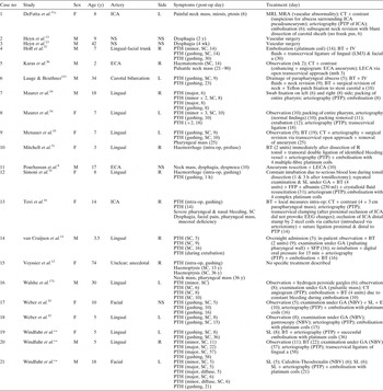
Table I Post-tonsillectomy pseudoaneurysms: published cases

*Ipsilateral peritonsillar abscess, abscess tonsillectomy; †post-tonsillectomy abscess formation; ‡tonsillectomy + uvulopalatoplasty. **Current study. No = number; post-op = post-operative; intra-op = intra-operative; F = female; M = male; ICA = internal carotid artery; ECA = external carotid artery; NS = not specified; L = left; R = right; y = years; mth = months; wk = weeks; h = hours; PTH = post-tonsillectomy haemorrhage; SC = spontaneous cessation; MRI = magnetic resonance imaging; MRA = magnetic resonance angiography; CT = computed tomography; PTP = post-tonsillectomy pseudoaneurysm; BT = blood transfusion; IV = intravenous; a = artery; LECA = ligation of ECA & individual branches; GA = general anaesthetic; SL = transoral suture ligation; FFP = fresh frozen plasma; EEG = electroencephalogram; SFP = suture of faucal pillars; NBV = no bleeding vessel identified; E = transoral electrocautery
The second was a 51-year-old man who had undergone unsuccessful incision and drainage of a peritonsillar abscess, with tonsillectomy the following day under general anaesthesia. Abrupt, brisk bleeding had begun during tonsil dissection, requiring several suture ligations and also swab fixation to the tonsillar fossae. Direct vascular injury (either by tonsil dissection or previous drainage procedures) was excluded by immediate arteriography, with normal findings. However, the following day, recurrent bleeding after swab removal under general anaesthesia prompted a neck exploration, including exploration of the external carotid artery and its individual branches. No injured neck arteries were identified, and the subsequent course was uneventful (this patient is not included in Table II).
Table II Arteriography findings: experts collection

No = number; PTH = post-tonsillectomy haemorrhage; post-op = post-operative; M = male; F = female; SC = spontaneous cessation; E = transoral electrocautery; SL = transoral suture ligation; NBV = no bleeding vessel identified; LECA = ligature of external carotid artery & individual branches; BT = blood transfusion; asc = ascending; SFP = suturing of faucal pillars; a = artery
Data from the experts collection
We identified 128 cases related to post-tonsillectomy haemorrhage. Only 12 reports matched the search criteria ‘arteriography’, including two cases which also matched the search criteria ‘pseudoaneurysm’ (cases 20 and 21; Table I). Both patients had experienced a gushing haemorrhage with spontaneous cessation and an uneventful recovery after treatment.
Of the 12 patients undergoing arteriography, the results were: normal findings (cases one to four, Table II); injuries of the facial, ascending pharyngeal or lingual artery (cases five to eight, Table II); and vascular abnormalities (cases nine and 10, Table II).
Discussion
Early diagnosis and adequate management of post-tonsillectomy pseudoaneurysm is mandatory to prevent rupture with haemorrhagic shock and exsanguination. Post-tonsillectomy pseudoaneurysms may not rupture in every patient, as has been reported by DeFatta et al.,Reference DeFatta, Verret and Bauer11 Veyssier et al. Reference Veyssier, Biou, Langman and Vilain12 and Heyn et al. Reference Heyn, Metz and Olthoff13 In contrast, our study shows that post-tonsillectomy pseudoaneurysms are typically associated with bleeding complications (Table I). Van Cruijsen et al. Reference van Cruijsen, Gravendeel and Dikkers14 have stated that post-tonsillectomy pseudoaneurysm only involves children under the age of 10 years, presumably due to the smaller anatomy and thinner pharyngeal muscles. However, the current and previous studiesReference Pourhassan, Grotemeyer, Fokou, Heinen, Balzer and Ramp8, Reference Veyssier, Biou, Langman and Vilain12, Reference Heyn, Metz and Olthoff13, Reference Laage and Beuthner15–Reference Walshe, Ramos, Low, Thomas, McWilliams and Hone17 indicate that the occurrence of post-tonsillectomy pseudoaneurysm is not restricted by age (Table I). The clinical signs of this lesion may be confusing; a high index of clinical suspicion is thus mandatory in order to include post-tonsillectomy pseudoaneurysm in the differential diagnosis of head and neck masses.Reference Haynes, Schwaber and Netterville18–Reference Liu and Filipp26
Onset
False aneurysms in the head and neck have been reported to occur as early as four hoursReference Ferry and Kempe27 and as late as eight monthsReference Beale28 after trauma or maxillofacial surgery.Reference DiStefano, Maimon and Mandel29 In one case, a post-tonsillectomy pseudoaneurysm, resulting from a postsurgical parapharyngeal abscess, was attributed to bacterial spread caused by infiltration of local anaesthetic agent.Reference Laage and Beuthner15 One of our patients presented a comparable history of repeated peritonsillar abscesses despite repeated incision and drainage. In contrast to the case of delayed post-tonsillectomy haemorrhage reported by Laage and Beuthner,Reference Laage and Beuthner15 our patient suffered an immediate, gushing haemorrhage on tonsil dissection. We suspected that this patient's unusual intensity of intra-operative bleeding resulted from a pseudoaneurysm, since a comparable case was reported in the literature.Reference Tovi, Leiberman, Hertzanu and Golcman16 However, subsequent arteriography and neck exploration was capable to rule out this possible differential diagnosis.
Our patients with proven post-tonsillectomy pseudoaneurysm experienced bleeding 21, 36 and 58 days following tonsillectomy, variously, at a stage when their wounds should have been (almost) healed. This finding is supported by most other relevant reported cases, which describe post-tonsillectomy pseudoaneurysm associated with bleeding as occurring only exceptionally within the first few hours,Reference Simoni, Bello and Kent32 being much more likely to occur 8,Reference Weber, Keerl, Hendus and Kahle33, Reference Maurer, Beck and Mann34 10,Reference Walshe, Ramos, Low, Thomas, McWilliams and Hone17, Reference Maurer, Beck and Mann34, Reference Menauer, Suckfull, Stabler and Grevers35 14,Reference Tovi, Leiberman, Hertzanu and Golcman16, Reference Hertzanu, Hirsch and Tovi30, Reference Karas, Sawin and Sie36 15,Reference Weber, Keerl, Hendus and Kahle33 16Reference van Cruijsen, Gravendeel and Dikkers14, Reference Weber, Keerl, Hendus and Kahle33 or 30Reference Hoff, Graumuller and Pau37 days, or even 36 years,Reference Veyssier, Biou, Langman and Vilain12 after tonsillectomy. Regrettably, the phenomenon of delayed post-tonsillectomy bleeding is not restricted to just those haemorrhages of pseudoaneurysm origin, as our results attest (see Table II).
Site
An analysis of magnetic resonance images in 100 children revealed that the distance from the internal carotid artery to the tonsillar fossa varied from 6.0 to 28.6 mm, depending on age and weight.Reference Deutsch, Kriss and Willging38 The external carotid artery courses more laterally and anterior to the internal carotid artery, suggesting that the latter should be involved in most cases of post-tonsillectomy pseudoaneurysm,Reference Tovi, Leiberman, Hertzanu and Golcman16, Reference Menauer, Suckfull, Stabler and Grevers35, Reference Karas, Sawin and Sie36 due to its closer relationship to the tonsil.Reference Deutsch, Kriss and Willging38 However, the internal carotid artery was not involved in any of our patients (cases 19–21 of table I) and only two authors have reported such involvement.Reference DeFatta, Verret and Bauer11, Reference Tovi, Leiberman, Hertzanu and Golcman16 We consider the inferior tonsillar pole to carry a much greater risk of arterial injury during tonsillectomy, due to the variable course of the facial artery looping over the submandibular gland in this area. The lingual artery may also run close to the inferior tonsillar pole. Both arteries were involved in all of our patients (table I, case 19–21) and also in most other reported cases (Table I).
Simoni et al. Reference Simoni, Bello and Kent32 have stated that pseudoaneurysms of the lingual artery are rare. This directly contrasts with our own findings: we found the lingual artery to be the most commonly involved artery in cases of post-tonsillectomy pseudoaneurysm and other tonsillectomy-related arterial injury (Table I). The facial artery is rarely involved in post-tonsillectomy pseudoaneurysm; there was only one such case in our patient population, and two cases reported in the literature.Reference Weber, Keerl, Hendus and Kahle33, Reference Hoff, Graumuller and Pau37
Diagnosis and treatment
Rapid diagnosis of a post-tonsillectomy pseudoaneurysm is based on arteriography; ultrasound and computed tomography with contrast are also options. Arteriography with simultaneous embolisation has been reported as management for a ruptured lingual artery,Reference Levy, Horowitz and Cahill39 true and false aneurysms involving the lingual,Reference Walshe, Ramos, Low, Thomas, McWilliams and Hone17 external carotidReference Hoff, Graumuller and Pau37 and internal carotid artery.Reference Tovi, Leiberman, Hertzanu and Golcman16, Reference Hertzanu, Hirsch and Tovi30 Steel coils have been used to occlude a pseudoaneurysm of the internal carotid artery;Reference Tovi, Leiberman, Hertzanu and Golcman16 however, platinum coilsReference Simoni, Bello and Kent32, Reference Weber, Keerl, Hendus and Kahle33 are advantageous due to their compatibility with microcatheter systems used in superselective catheterisation. Tovi et al. Reference Tovi, Leiberman, Hertzanu and Golcman16 and Hertzanu et al. Reference Hertzanu, Hirsch and Tovi30 (reporting on the same patient) have suggested a combined surgical-radiological approach for selected cases.
Embolisation was successful in three patients of our study (case 19 and 21 of table I; case 6 of table II), pseudoaneurysm and one with a damaged facial artery. Opatowsky et al. Reference Opatowsky, Browne, McGuirt and Morris40 reported the use of embolisation in two children with repeated post-tonsillectomy bleeding despite previous surgical revision. No definitive source of bleeding was identified by arteriography in either child. Prompted by a suspicious appearance, embolisation of the ascending palatine artery in one child and of the lingual plus ascending pharyngeal artery in the other child prevented further bleeding. Levy et al. Reference Levy, Horowitz and Cahill39 also reported the use of embolisation, in a 10-year-old girl with repeated bleeding episodes with spontaneous cessation. Post-tonsillectomy haemorrhage recurred in the recovery room, after haemostasis had previously been achieved under general anaesthesia. Arteriography was undertaken, including successful embolisation of a damaged left lingual artery.
Embolisation, even if superselective, was not successful in three patients, in our study, case 2, 4 and 5 of table II. Hoff et al. Reference Hoff, Graumuller and Pau37 reported post-tonsillectomy haemorrhage recurrence 16 days after previous embolisation (performed 14 days after tonsillectomy) comparable to the case of Hoff is case 20 of table I. As in one of our patients, immediate ligation of the injured vessel via an open transcervical approach proved successful.
Arteriography revealed normal findings in four of our patients (cases 1–4 of table II). It remains speculative whether repeated arteriography would have subsequently revealed a post-tonsillectomy pseudoaneurysm. Maurer and colleaguesReference Maurer, Beck and Mann34 reported a normal finding 10 days after surgery, yet a confirmed post-tonsillectomy pseudoaneurysm 8 days later, when bleeding recurred. Our study results suggest that normal arteriography does not exclude the risk of later bleeding (Table II).
• A ruptured pseudoaneurysm is an extremely rare cause of post-tonsillectomy haemorrhage
• Whenever episodes of gushing and secondary post-tonsillectomy bleeding with spontaneous cessation occur, arteriography should be considered to rule out vascular injuries, including pseudoaneurysm formation
• Simultaneous, superselective embolisation has been proven to be safe and effective. However, this is impractical in the acute emergency presented by life-threatening bleeding, aspiration or expanding cervical haematoma
In cases of aspiration or life-threatening bleeding, arteriography is not practical. Instead, immediate surgery and intensive care are mandatory for a successful outcome. Whenever ligature of the external carotid artery is considered, the surgeons should exclude an aberrant arterial blood supply by branches of the internal carotid artery, as was shown for two patients in our study (Table II). Moreover, ligature of the external carotid artery may result in post-operative blindness due to atypical collaterals between the internal and external carotid artery.Reference McIntosh, Douglas, Lee, Allen and Mahadevan41
Conclusion
A ruptured pseudoaneurysm is an extremely rare cause of post-tonsillectomy haemorrhage; other causes of episodic post-tonsillectomy bleeding are much commoner. However, whenever episodes of gushing and secondary post-tonsillectomy bleeding with spontaneous cessation occur, arteriography should be considered to rule out vascular injury, including post-tonsillectomy pseudoaneurysm. Embolisation of the injured artery should be performed proximal and distal to the lesion, due to the extensive retrograde blood flow.
Simultaneous, superselective embolisation has been proven to be safe and effective. However, it is impractical in the acute emergency presented by life-threatening bleeding, aspiration or expanding cervical haematoma.