


INTRODUCTION
Gap junctions (GJ) are clusters of intercellular channels that provide cells with means of communicating directly with their neighbours, a process which is essential for the development of tissues and organs in vertebrates and invertebrates. Vertebrate GJs are composed of 2 families of channel trans-membrane proteins: connexins (Goodenough et al. Reference Goodenough, Goliger and Paul1996) and pannexins (Panchin, Reference Panchin2005). Invertebrate organisms have 1 family of topologically and functionally similar proteins, the innexins (Inx) (Phelan, Reference Phelan2005) that are homologous only to vertebrate pannexins (Baranova et al. Reference Baranova, Ivanov, Petrash, Pestota, Skoblov, Kelmanson, Shagin, Nazarenko, Geraymovych, Litvin, Tiunova, Born, Usman, Staroverov, Lukyanov and Panchin2004).
GJs are used to transport small molecules and ions from one cell to another (Simon and Goodenough, Reference Simon and Goodenough1998). Decades of information accumulated in the field of innexins/connexins have shown that the adhesive properties of GJs, as well as the non-junctional properties of Inx (Bao et al. Reference Bao, Samuels, Locovei, Macagno, Muller and Dahl2007) and pannexins (Dahl and Locovei, Reference Dahl and Locovei2006) are as important as the transporting properties of GJ channels in tissues such as the neocortex (Elias et al. Reference Elias, Wang and Kriegstein2007).
Ultrastructural evidence of GJs has been described in parasitic flatworms such is Hymenolepis diminuta and Mesocestoides corti (Lumsden and Hildreth, Reference Lumsden, Hildreth, Arme and Pappas1983; Richards and Arme, Reference Richards and Arme1983; Burns et al. Reference Burns, Howard, Allen, Van Velsen and McKerr1995). Electron microscopical studies also revealed a large number of GJs in the neck and immature proglottids of experimental adult Taenia solium worms (Willms et al. Reference Willms, Robert and Caro2003). In this tapeworm, cytoplasmic sacs of glycogen are connected by numerous discrete GJs to other cells throughout the maturing strobilar tissue of these parasites.
Little is known about the transport of metabolites in the parasitic flatworm T. solium, which is acoelomate and must therefore make use of the tegumentary surface (Smyth and McManus, Reference Smyth, McManus, Smyth and McManus1989) and syncytial organization of its tissues to absorb nutrients and deliver these to distant tissues in the proglottids, possibly by using a GJ channel pathway.
Since the use of degenerate primers corresponding to highly conserved regions of Inx genes have so far been unsuccessful in obtaining coding sequences for T. solium Inx(s) by PCR, in this study we report the use of biochemical methods to prepare enriched membrane fractions containing GJs to relative homogeneity, and the preparation of polyclonal antibodies against a synthetic peptide copied from a highly conserved region of invertebrate GJ proteins, which bind to Western blot (WB) proteins of the enriched membrane fraction as well as to sections of larvae and worm tissues.
MATERIALS AND METHODS
Rabbit polyclonal serum
A peptide sequence corresponding to the conserved regions of innexins/pannexins reported by EMBL/DDBJ/GenBank Database Library was constructed. A 20-amino acid synthetic peptide NH2-NYYQWVPIILALQALLFYFP coupled to keyhole limpet haemocyanin bound to the COOH-terminal (INVITROGEN) was used in order to raise polyclonal antibodies in rabbits (anti-Ts-INX1).
Rabbits (New Zealand) were immunized subcutaneously (s.c.) with 250 μg of the KLH-coupled peptide in 10 μg of saponine (Quillaja Bark, Sigma-Aldrich) as adjuvant and boosted 3 times at 3-week intervals. Rabbits were anaesthetized by s.c. injection with ketamine (15 mg/kg) and xylazine (1 mg/kg) and blood obtained by cardiac puncture. Polyclonal serum was tested in WB. Rabbit pre-immune serum was used as control.
Isolation of GJ-enriched membrane fractions (GJ-EMF)
The procedure described by Ryerse (Reference Ryerse1989) was followed to obtain the membrane fractions. Taenia solium cysticerci (100 g) were dissected from the skeletal muscle of naturally infected pork, rinsed in phosphate-buffered saline (PBS, pH 7·2) and frozen at −70°C. Larvae were thawed and homogenized in a Polytron (Brinkmann Instruments, Inc) at maximal power for 4 min, suspended in 1 litre of ice-cold GJ buffer (1 mm NaHCO3, pH 7·2, containing protease inhibitors: 1 mm PMSF, 50 μg/ml TLCK and 5 μm leupeptin). The homogenate was filtered through gauze and centrifuged. The soft upper pellet was separated from the surrounding rough dark pellet, washed and suspended in GJ buffer and centrifuged. This pellet was resuspended in GJ buffer and 60% sucrose to obtain a 42·5% sucrose-homogenate, overlaid with 30% sucrose and centrifuged. The crude membrane fraction was collected from the 30/42·5% sucrose interface, dissolved in GJ buffer and centrifuged once more. The fraction was suspended in GJ buffer mixed with 8 m urea and 0·4 g of N-lauroyl-sarcosine and spun at 45 000 g in a Beckman Ti 70 rotor for 30 min at room temperature. The pellets were pooled and washed in GJ buffer and stored at −20°C until use. All steps were carried out at 4°C unless otherwise noted.
Ultrastructural and WB analysis of GJ-EMF
An aliquot of the GJ-EMF pellet was used to solubilize the proteins in SDS-PAGE sample buffer (Laemmli, Reference Laemmli1979) and separate them in 10% polyacrylamide gels, stained with Commassie Blue or transferred to nitrocellulose membranes for Western blotting with rabbit sera. Transferred proteins were incubated with either rabbit pre-immune or polyclonal anti-TsINX1 immune sera diluted 1:1000, and further incubated with goat-anti-rabbit IgG bound to horseradish peroxidase (Zymed Laboratories) diluted 1:2000.
A fraction of the GJ-EMF pellet was fixed in Karnosvky (Reference Karnovsky1965) solution for 2 h, rinsed in cacodylate buffer and embedded in Polybed for electron microscopy. Thin sections were stained with uranyl acetate and lead citrate and observed in a JEOL 1200EX electron microscope.
Immunocytochemistry of tapeworm and larvae
Taenia solium tapeworms were obtained from experimentally infected hamsters as described by Willms et al. (Reference Willms, Robert and Caro2003, Reference Willms, Fernández Presas, Jiménez, Landa, Zurabian, Juárez Ugarte and Robert2005) and larval parasites were dissected from infected pork meat. Worms were washed in PBS, adult parasites were cut into segments and frozen at −70°C. Frozen sections, 5–6 μm thick, were deposited on poly-L-lysine treated slides, hydrated 3×10 min in PBS and covered with PBS containing 3% BSA and 0·05% Tween 20 for 30 min. Blocked parasite sections were incubated with rabbit anti-TsINX1 or pre-immune sera diluted 1:500 in PBS with 3% BSA, 0·05% Tween 20, rinsed 3 times in PBS 0·05% Tween 20 and incubated with fluorescent-labelled goat-anti-rabbit IgG (Zymed Laboratories) diluted 1:200 in 1% Evans Blue-PBS for 2 h. All incubations were carried out at 4°C. Sections were rinsed in PBS and photographed in a NIKON Eclipse E600 epi-fluorescence microscope.
Amino acid sequence alignment
Comparison of amino acid sequences of conserved trans-membrane regions of TsINX-peptide and T. solium glucose transporters TGTP1 and TGTP2, was carried out using Biology Workbench 3.2 (SDSC; http://workbench.sdsc.edu).
RESULTS
PAGE gels of the GJ-EMF contained at least 8 protein bands. WB analysis of PAGE gels incubated with anti-TsINX1 and goat-anti-rabbit IgG-peroxidase, revealed 2 positive bands of 55 and 67 kDa (Fig. 1). Electron microscopy of the semi-purified fraction (Fig. 2) showed membranes with GJ's, which are also seen in the GJ-EMF as increased density zones (Fig. 3). Clear GJs measured 10–14 nm across.

Fig. 1. PAGE and WB image of purified gap junction fraction. Lane A: PAGE gel; lane B: Western blot of GJ-EMF incubated with antiserum to INX peptide; lane C: GJ-EMF incubated with rabbit pre-immune serum.

Fig. 2. Electron micrograph section of semi-purified GJ-EMF fraction obtained from the pellet of the sucrose gradient, illustrating a number of GJs (arrows). gly, Glycogen.

Fig. 3. Electron micrograph section of purified GJ-EMF obtained from the sucrose gradient interface illustrating a number of gap junctions (arrows).
Light micrographs of cysticerci and tapeworm tissues incubated with anti-TsINX1 and anti-rabbit IgG bound to fluorescein, showed intense green fluorescence over the tegumentary surface with patchy fluorescent areas in the parenchyma (Fig. 4B and D). Sections incubated with rabbit pre-immune serum were negative (Fig. 4A and C).

Fig. 4. Immunohistochemistry of frozen sections from (A and B) Taenia solium strobilae and (C and D) cysticerci. (A and C) Control sections incubated with pre-immune rabbit serum, and anti-rabbit IgG-fluorescein. (B and D) Sections incubated with rabbit anti-TsINX1 serum and anti-rabbit IgG-fluorescein. Binding of antibody to tegument is clearly seen as an intense fluorescent band in the tegument (B and D, arrow), and also as patchy fluorescent areas in the parenchyma (double-headed arrows).
Comparison of trans-membrane amino acid sequences of Inx and T. solium glucose transporters TGTP1 and TGTP2, revealed very low homology between these proteins (Fig. 5), revealing only a minor discontinuous sequence shared between the synthetic peptide and the reported TGTP2 sequence.
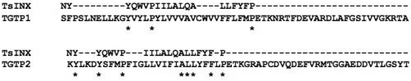
Fig. 5. Comparison of amino acid alignment of conserved trans-membrane regions of TsINX-peptide and Taenia solium glucose transporters TGTP1 and TGTP2. *, Identical amino acids.
DISCUSSION
The results described here constitute the first evidence that larvae and adult T. solium tissues contain a large number of innexin/pannexin epitopes in the tegumentary surface and in the parenchyma. Although the GJ-EMF contained at least 8 protein fractions, only 2 of these were recognized by the antibody prepared against the synthetic peptide aligned to a highly conserved sequence of innexins reported in GenBank; furthermore, these proteins had molecular weights in the range of those reported for other gap junction hexamer subunits (Potenza et al. Reference Potenza, del Gaudio, Rivieccio, Russo and Geraci2002).
Taeniids exhibit continuously differentiating tissues, so that it may be inferred that GJs and their constituent innexin proteins, probably represent an important pathway for the transport of small molecules and ions through cell-to-cell junctions, which may be functional throughout the strobilar/proglottid tissues.
Numerous members of Inx have been described in Ecdysozoans and Lophotrochozoans (for review see Panchin, Reference Panchin2005) and are found to be stage-specific in the morphogenesis of invertebrates (Bauer et al. Reference Bauer, Lehmann, Martini, Eckardt and Hoch2004; Dykes and Macagno, Reference Dykes and Macagno2006). In the phylum platyhelminthes, the Inx genes have been found in Girardia tigrina (Panchin et al. Reference Panchin, Kelmanson, Matz, Lukyanov, Usman and Lukyanov2000), Dugesia japonica (Nogi and Levin, Reference Nogi and Levin2005) and Schmidtea mediterranea (Oviedo and Levin, Reference Oviedo and Levin2007) and are associated with expression patterns in the intestine, nervous system or parenchyma of these planarian flatworms.
Innexins comprise a large family of proteins with identity/homology to pannexins found in vertebrates (Panchin, Reference Panchin2005; Shestopalov and Panchin, Reference Shestopalov and Panchin2008), that show little homology with mammalian connexins (Goodenough et al. Reference Goodenough, Goliger and Paul1996). It is now evident, that a number of species have several isoforms of innexins/pannexins in their tissues (Phelan, Reference Phelan2005). The present results demonstrate that Taeniids have innexins, but by using antibodies to a highly conserved trans-membrane sequence of innexins, it cannot be ruled out that there is more than one isoform of these proteins; for example, the nematode Caenorhabditis elegans is reported to have 25 different innexins (Starich et al. Reference Starich, Sheehan, Jadrich and Shaw2001) The particular highly conserved sequence we used for raising antibodies is most closely related to the planarian G. tigrina, which is also a platyhelminth (Panchin et al. Reference Panchin, Kelmanson, Matz, Lukyanov, Usman and Lukyanov2000).
Glucose, which is stored as glycogen in cestodes, has been shown to decrease in concentration from the scolex to the distal strobilae in Hymenolepis diminuta (Cornford, Reference Cornford1990) and by ultrastructural analysis of T. solium strobilae, it was found that cytoplasmic glycogen decreases significantly from the scolices to the mature proglottids, suggesting that glycogen stores are used up as the proglottids mature to the gravid stage (Willms et al. Reference Willms, Robert and Caro2003). In T. solium strobilae, cytoplasmic glycogen is also found linked by GJs to other cell types, and is particularly abundant in immature proglottids. In vitro studies of T. solium strobilae exposed to increasing concentrations of glucose and the GJ specific LY fluorochrome, showed a significant increase of LY in the tegument of strobilae incubated in 20 mm glucose as opposed to strobilae incubated in 5 mm glucose (Willms et al. Reference Willms, Fernández Presas, Jiménez, Landa, Zurabian, Juárez Ugarte and Robert2005). Taken in conjunction with our present observations, which revealed significant tegumentary recognition sites for an Inx peptide sequence, it can be suggested that the tegumentary surface of these cestodes also has innexins that may be involved in the transport of glucose and other small molecules from the external surface to the parenchymal tissues of the worm.
The delivery of glucose in the strobilar tissues, which would appear to be an indispensable function in these flatworms, may be carried out by one form of innexin, whereas other functions such as the movement of ions in nervous function may be performed by a different innexin isomer. In vertebrate tissues, Sato et al. (Reference Sato, Haimovici, Kao, Li and Roy2002) showed that exposure to high glucose concentrations significantly reduced the number of GJ's in microvascular endothelium. However, in T. solium strobilae, high glucose concentrations significantly increase LY uptake in the tegument, an observation, which in the absence if typical ultrastructural GJs, also suggests the possibility that the epitopes recognized by the anti-TsINX1 may correspond to hemi-channel structures.
It should be noted that the typical GJ structures seen in the parenchyma of T. solium strobila are not evident in the tegumentary tissue, in which we have previously described what appear to be closely apposed membrane structures (Willms et al. Reference Willms, Fernández Presas, Jiménez, Landa, Zurabian, Juárez Ugarte and Robert2005) which tend to fuse, forming what Richards and Arme (Reference Richards and Arme1983) described as heptalaminar junctions. These observations suggest that the innexin epitopes recognized by the anti-TsINX1 in the tegumentary surface may belong to isomers of hemi-channel forming non-junctional innexins, similar to what has been described in other invertebrates (Bao et al. Reference Bao, Samuels, Locovei, Macagno, Muller and Dahl2007). Additionally, junction proteins have been found to interact with a wide variety of proteins (Hervé et al. Reference Hervé, Bourmeyster, Sarrouilhe and Duffy2007).
It has previously been shown that T. solium has at least 2 glucose transporters (TGTP1 and TGTP2) (Rodríguez et al. Reference Rodríguez-Contreras, Skelly, Landa, Shoemaker and Laclette1998; Willms et al. Reference Willms, Fernández Presas, Jiménez, Landa, Zurabian, Juárez Ugarte and Robert2005). TGTP1 has been found in larval and adult tegument tissue; however, TGTP2 has only been found in the tegument of larvae and not in the worm (Rodríguez et al. Reference Rodríguez-Contreras, Skelly, Landa, Shoemaker and Laclette1998), so that cross-reactions between the discontinuous ALL—F-P motif found in the synthetic peptide sequence and the TGTP2 transporter are unlikely, as evidenced by the strong fluorescence in the adult tegument after incubation with anti-TsINX1. The possibility that other tegumentary membrane proteins may be functionally coupled to Inx hemi-channels should be explored in the future.
The GJ-enriched fraction will be used for further isolation of T. solium innexins. It is expected that isolation of these protein fractions will allow for purification and sequencing of the Inx proteins and eventual isolation of the corresponding cDNA sequences.
The authors thank J. A. Jiménez Rodríguez for his help in obtaining live taeniid strobilae and preparation of frozen sections. This work was supported by Papiit-UNAM Grant numbers IN238602 and IN210407.


