


Introduction
Studies of brain morphology in schizophrenia provide overwhelming evidence of abnormal brain structure. The brain regions most consistently reported as abnormal in established schizophrenia include the medial temporal lobes (particularly the hippocampus), the prefrontal lobes and the thalamus. Meta-analyses have reported tissue loss in these brain regions when people with schizophrenia are compared to controls using both region of interest (ROI) and fully automated methods of image analysis (Lawrie & Abukmeil, Reference Lawrie and Abukmeil1998; Nelson et al. Reference Nelson, Saykin, Flashman and Riordan1998; Wright et al. Reference Wright, Rabe-Hesketh, Woodruff, David, Murray and Bullmore2000; Konick & Friedman, Reference Konick and Friedman2001; Honea et al. Reference Honea, Crow, Passingham and Mackay2005; Ellison-Wright et al. Reference Ellison-Wright, Glahn, Laird, Thelen and Bullmore2008).
The point in illness development at which these abnormalities arise has been more difficult to ascertain. Two meta-analyses of first-episode studies using ROI techniques reported that hippocampal volume reduction was detectable in individuals experiencing their first episode of illness (Steen et al. Reference Steen, Mull, McClure, Hamer and Lieberman2006; Vita et al. Reference Vita, De Peri, Silenzi and Dieci2006); meta-analysis of voxel-based morphometry (VBM) studies suggested that abnormalities were also present in the thalami and regions of the frontal lobes (Ellison-Wright et al. Reference Ellison-Wright, Glahn, Laird, Thelen and Bullmore2008). Several studies have looked even earlier than first episode of psychosis, evaluating brain structure in people who are well but at elevated risk of schizophrenia. The Edinburgh High Risk Study (EHRS) has previously reported that abnormalities of the amygdala–hippocampal complex (AHC), prefrontal lobes and thalami are detectable in those who are clinically well but at genetic risk of schizophrenia (Lawrie et al. Reference Lawrie, McIntosh, Hall, Owens and Johnstone2008). Brain structural abnormalities in this population were generally an attenuated form of that seen in the first episode of illness (Lawrie et al. Reference Lawrie, Whalley, Kestelman, Abukmeil, Byrne, Hodges, Rimmington, Best, Owens and Johnstone1999; Job et al. Reference Job, Whalley, McConnell, Glabus, Johnstone and Lawrie2003). Independent studies of individuals at risk of schizophrenia for familial reasons report similar findings (Diwadkar et al. Reference Diwadkar, Montrose, Dworakowski, Sweeney and Keshavan2006; Boos et al. Reference Boos, Aleman, Cahn, Hulshoff Pol and Kahn2007). Studies of people identified as being at elevated risk of schizophrenia on the basis of expressed symptoms (rather than genetic risk) report generally comparable results (Wood et al. Reference Wood, Pantelis, Velakoulis, Yücel, Fornito and McGorry2008; Fusar-Poli et al. Reference Fusar-Poli, Borgwardt, Crescini, D'Este, Kempton, Lawrie, Guire and Sacchetti2011). Taken together, these findings suggest that, whereas subtle brain structural abnormalities are detectable in people at elevated risk of schizophrenia (whether for genetic or symptomatic reasons), further changes occur during transition from the at-risk state to frank illness. This has been confirmed in ROI and automated analyses of longitudinal data for the prefrontal and temporal lobes in both genetic and clinical high-risk populations (Pantelis et al. Reference Pantelis, Velakoulis, McGorry, Wood, Suckling, Phillips, Yung, Bullmore, Brewer and Soulsby2003; Job et al. Reference Job, Whalley, Johnstone and Lawrie2005; McIntosh et al. Reference McIntosh, Owens, Moorhead, Whalley, Stanfield, Hall, Johnstone and Lawrie2011).
Ascertaining what drives the brain structural changes that characterize the transition from at-risk to illness state is the next step in advancing our understanding of schizophrenia. In this regard, longitudinal imaging studies of individuals at elevated risk are likely to be crucial. Through such studies we can establish whether exposure to putative environmental risk factors is associated with the development of brain structural changes, without the confounding effects of antipsychotic drugs and other aspects of the management and effects of illness. If demonstrated, this also provides further evidence for their role in the aetiology of schizophrenia and might provide important justification for some preventive interventions.
Cannabis is arguably the environmental factor with greatest evidence for increasing risk of schizophrenia (Arseneault et al. Reference Arseneault, Cannon, Witton and Murray2004; Semple et al. Reference Semple, McIntosh and Lawrie2005; Zammit et al. Reference Zammit, Moore, Lingford-Hughes, Barnes, Jones, Burke and Lewis2008). However, only a minority of people who use cannabis will develop schizophrenia, suggesting that additional factors are present in some individuals that make them particularly susceptible to the risk-modifying effects of exposure. This postulate is very much in keeping with the stress–diatheses model of schizophrenia, which posits that genetic vulnerability interacts with environmental factors to influence an individual's risk of developing the condition (van Os et al. Reference van Os, Kenis and Rutten2010). There is evidence that such an interaction exists with cannabis. For example, relatives of people with schizophrenia have been reported to be particularly susceptible to the psychotomimetic effects of the drug (Kahn et al. Reference Kahn, Linszen, van Os, Wiersma, Bruggeman, Cahn, de Haan, Krabbendam and Myin-Germeys2011). A genetic vulnerability to psychosis has also been reported to increase the risk that cannabis users will develop psychosis (Hall & Degenhardt, Reference Hall and Degenhardt2006).
If schizophrenia is associated with brain structural abnormalities and cannabis use is a risk factor for schizophrenia, the question naturally arises is cannabis itself associated with abnormalities of brain structure? This has not been demonstrated consistently in the cannabis-using population without major psychiatric disorders (Quickfall & Crockford, Reference Quickfall and Crockford2006; Martín-Santos et al. Reference Martín-Santos, Fagundo, Crippa, Atakan, Bhattacharyya, Allen, Fusar-Poli, Borgwardt, Seal and Busatto2010). By contrast, accelerated grey matter (GM) loss is reported in those who use the drug following onset of schizophreniform illness (Rais et al. Reference Rais, Cahn, van Haren, Schnack, Caspers, Hulshoff Pol and Kahn2008). A particular susceptibility to the effects of cannabis use conferred by a genetic propensity to schizophrenia may be central to understanding this apparent contradiction. It may be that, in the ‘well’ population, brain structural change in association with cannabis use is only detectable in those already at elevated risk of the condition for genetic (or potentially other) reasons. In short, susceptibility to brain structural consequences of cannabis use may be a feature of vulnerability to schizophrenia, present prior to the development of illness. Through such an interaction, cannabis may contribute to the brain structural changes associated with transition from at-risk to illness state.
In keeping with this, a cross-sectional analysis of baseline data from the EHRS found that people who were clinically well but at genetically high risk of schizophrenia did exhibit brain structural abnormalities in association with cannabis use. Such an association was not observed in controls (Welch et al. Reference Welch, McIntosh, Job, Whalley, Moorhead, Hall, Owens, Lawrie and Johnstone2011a). Differential sensitivity to the brain structural consequences of cannabis use in those with a family history of schizophrenia has subsequently been reported in an independent cohort (Habets et al. Reference Habets, Marcelis, Gronenschild, Drukker and van Os2011). Furthermore, a longitudinal ROI study of the EHRS cohort demonstrated progressive thalamic volume loss with use of the drug (Welch et al. Reference Welch, Stanfield, McIntosh, Whalley, Job, Moorhead, Hall, Owens, Lawrie and Johnstone2011b).
In the current study we sought to extend these findings, using a fully automated image analysis technique to compare longitudinal brain structural changes in individuals from this high-risk cohort who did and did not use cannabis during the time between the scans. The focus of this study was those brain regions known to be abnormal in schizophrenia; namely, the hippocampi, the prefrontal lobes and the thalami. If an association was seen between cannabis use and progressive structural abnormalities in these regions, it would provide further evidence that people at elevated genetic risk of schizophrenia are particularly vulnerable to the effects of cannabis.
Method
Any brain structural changes associated with cannabis use would be expected to be subtle and consequently difficult to detect with standard automated techniques of image analysis. Conventional VBM techniques, for example, are unable to separate effects due to imperfect image realignment from changes in tissue density. This can result in increased variance in analyses and consequently make the detection of changes such as any cannabis-associated effect difficult. A refinement to VBM is tensor-based morphometry (TBM), which uses the deformation field created when warping subjects' follow-up brain scan to their baseline scan. Unlike VBM, this technique is able to distinguish intrinsic changes in brain anatomy from translational shifts caused by imperfect image registration (Moorhead et al. Reference Moorhead, McKirdy, Sussmann, Hall, Lawrie, Johnstone and McIntosh2007). In essence, TBM applies a high-dimensional (HD) warp to correct for slice misalignment between time-points, thus ensuring that the same voxels are compared in successive scans from the same subject. This more sensitive technique of longitudinal image analysis was therefore used in this study.
Participants
Data were collected from people at elevated risk of schizophrenia as part of the EHRS. Details of the recruitment process have been described previously (Hodges et al. Reference Hodges, Byrne, Grant and Johnstone1999). In brief, individuals with schizophrenia, with a family history of schizophrenia and with adolescent relatives, were identified from hospital case records. We then approached their relatives, and high-risk subjects aged 16–25 years who agreed to participate were given a detailed clinical, neuropsychological and brain imaging assessment. Assessments were repeated after approximately 2 years in consenting participants who had enrolled in the first 2 years of the study and had not made the transition to schizophrenia. As part of this repeat assessment, use of alcohol, tobacco and illicit drugs (including cannabis) in the interim period was ascertained by self-report. Exposures in this period were dichotomized as follows: cannabis use during this period or not; alcohol use exceeding UK government recommendations during this period (>14 units/week for women and >21 units/week for men) or not; ecstasy use during this period or not; amphetamine use during this period or not; and tobacco smoker during this period or not. When dichotomizing use in this manner, even a single episode of substance use (aside from alcohol) resulted in inclusion in the ‘use’ group. The choice of a dichotomous rather than a continuous measure of drug and alcohol use reflected the manner in which drug/alcohol use was recorded at the data collection stage.
Magnetic resonance imaging (MRI) scanning and analysis
Each participant underwent MRI scanning on a 1-T Siemens (Erlangen, Germany) Magnetom scanner at both baseline (T1) and follow-up (T2). Details of image acquisition and processing have been given elsewhere (Whalley et al. Reference Whalley, Kestelman, Rimmington, Kelso, Abukmeil, Best, Johnstone and Lawrie1999). When running TBM analyses, similarly to VBM, corrections are made for multiple comparisons at whole-brain level. In the context of TBM it is common practice to restrict the analysis to regions determined a priori to be of particular interest. This is achieved by using the small volume correction (SVC) function in SPM (http://www.fil.ion.ucl.ac.uk/spm/), significance being correcting for the voxels included in this restricted analysis rather than the whole brain. As discussed in the Introduction, the three brain regions for which there is considerable evidence of structural change during transition from at-risk state to frank schizophrenia are the hippocampi, the frontal lobes and the thalami. Thus, in the following analyses SVCs for these regions were applied. The actual contrasts applied in these analyses were the same as those applied in our previously published volumetric analysis of this dataset (Welch et al. Reference Welch, Stanfield, McIntosh, Whalley, Job, Moorhead, Hall, Owens, Lawrie and Johnstone2011b).
Image preprocessing
We implemented the TBM protocol released for the Statistical Parametric Mapping (SPM2) application by J. Ashburner (http://www.fil.ion.ucl.ac.uk/spm/). This protocol was implemented by and discussed in Kipps et al. (Reference Kipps, Duggins, Mahant, Gomes, Ashburner and McCusker2005) and implemented by Whitford et al. (Reference Whitford, Grieve, Farrow, Gomes, Brennan, Harris, Gordon and Williams2006). We implemented this staged protocol as described below and in the publication of Moorhead et al. (Reference Moorhead, McKirdy, Sussmann, Hall, Lawrie, Johnstone and McIntosh2007).
(1) The SPM brain extract function was used to recover the first- and second-round native space brains from the participants' T1 scans and the SPM2 default T1-weighted single subject using raw space segmentations. These extractions were used to exclude non-brain tissue from the analysis.
(2) The SPM co-register function was used to register the first- and second-round extracted brains with the extracted brain from the SPM single subject, without rescaling. This provided a co-registration mapping of the brain tissue for each subject and with alignment along the Montreal Neurological Institute (MNI) template anterior–posterior (AP) commissure axis. The mappings to obtain these AP commissure axis registrations were then applied to the T1 raw images (non-brain-extracted) to obtain co-registered native space T1 images. The SPM segment function was then used to extract GM segments for the native space co-registered first- and second-round scans.
(3) The SPM deformations toolbox was used to implement an HD warp between the second- and first-round co-registered brains given by step 2. The resultant warp was then used to implement an HD registration of the second-round GM segment with the first-round GM segment. This HD warp is used to minimize local registration differences between the first- and second-round tissue segments. We subtracted the first-round GM segment from the second-round HD warped GM segment to give a native space GM difference image. The Jacobian determinants for the HD warp were then evaluated.
(4) In a procedure analogous to modulated VBM, localized tissue change is recovered in the form of GM and white matter (WM) modulated difference images. In this, the Jacobian determinants from HD deformation between the first and second rounds are used to ensure that the assessments of tissue changes over time are corrected for MRI sampling noise.
(5) To obtain subject-to-subject co-registration, we applied non-linear warping to normalize the first-round extracted brain from step 2 with the SPM2 single-subject T1-extracted brain acquired from step 1. The normalization warp was then applied to the modulated difference image from step 4. Steps 1–5 were also repeated for WM.
Unfortunately, scans from two individuals in the cannabis-consuming group could not be preprocessed successfully for the TBM analysis. This resulted in 23 subjects who consumed cannabis between scan points being contrasted with 32 subjects who did not.
Statistical analysis
Differences in the modulated difference images (from step 5) between high-risk subjects who did and did not consume cannabis between T1 and T2 were compared in SPM5 using the general linear model. A GM mask was included. Age, inter-scan interval, sex and use of alcohol, cigarettes, ecstasy and amphetamines were included in the model as covariates. These are the same covariates that were included in our earlier volumetric analysis of the same dataset (Welch et al. Reference Welch, Stanfield, McIntosh, Whalley, Job, Moorhead, Hall, Owens, Lawrie and Johnstone2011b). T contrasts were thresholded at p = 0.001 (uncorrected). Whole-brain analysis was supplemented with an SVC for the AHC, the frontal lobes and the thalami, all corrected for multiple comparisons [family-wise error (FWE) <0.05]. The TBM whole-brain and SVC analyses were implemented in MNI space. The voxel coordinates of the results were extracted from MNI standard space. We also report the Talairach coordinates, obtained using the Matlab function mni2tal (Brett, Reference Brett1999). The anatomic location of significant results was checked manually using the Talairach atlas (Talairach & Tournoux, Reference Talairach and Tournoux1988), supplemented by use of Talairach Daemon (http://ric.uthscsa.edu/projects/talairachdaemon.html).
Additional steps were taken to confirm that the results were genuine and to exclude possibilities that, for example, they were due to the presence of an artefact. First, statistical parametric maps were superimposed on scans from the study to ensure that regions of maximal difference were indeed arising in anatomically feasible locations. Second, an identical analysis was run on expansion images to investigate volume gain in association with cannabis consumption. This would only be expected to be seen in the presence of an artefact. Third, it is notable that none of the subjects who did not consume cannabis in the inter-scan period consumed ecstasy or amphetamine, whereas substantial numbers of the cannabis-consuming subjects did (see Table 1). This raises concerns that structural abnormalities associated with use of these substances could potentially confound the results outlined above. This emphasizes the importance of having included these variables as covariates. Additionally, however, given that this imbalance of variables was so marked across the comparator groups, we considered it important to take further steps to confirm that this imbalance was not resulting in spurious findings. The above analyses were thus repeated excluding the 11 subjects who consumed either amphetamine or ecstasy or both drugs in the inter-scan period. The analysis included all the other covariates from the previous analyses.
Table 1. Demographic and clinical characteristics of individuals at high familial risk of schizophrenia who did and did not consume cannabis between T1 and T2

RISC, Rust Inventory of Schizotypal Cognitions; s.d., standard deviation.
a Independent t test.
b χ2 test.
c Fisher's exact test.
Results
Demographic and relevant clinical details for the 55 subjects included in the TBM analysis are detailed in Table 1. Participants who used and did not use cannabis between the scan points did not differ significantly in terms of gender, age at time of initial scan, rating on the Rust Inventory of Schizotypal Cognitions (RISC) at T1, or the proportion exceeding government recommended maximum weekly alcohol consumption between scanning points. Significantly more of the cannabis-using group did, however, smoke cigarettes and consume ecstasy and amphetamines in the inter-scan period. All the aforementioned variables, including those not significantly different between the groups, were included as covariates in the TBM analysis.
The initial analysis, conducted at whole-brain level, compared GM loss in cannabis consumers versus non-consumers between T1 and T2. Although they did not reach statistical significance at the level of whole-brain analysis, three regions of prominent GM loss were observed in the former compared to the latter group. These were located in the left prefrontal lobe, left caudate and right anterior hippocampus. The caudate was not part of our initial hypothesis, and thus this area of GM loss was not investigated any further. The analysis was, however, rerun with bilateral SVCs for the AHC, the frontal lobes and the thalami.
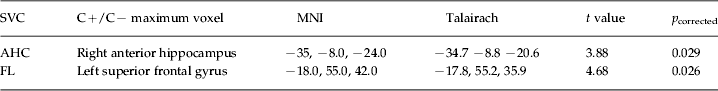
On applying the bilateral AHC SVC, significantly greater GM tissue loss was found in the cannabis-using group compared to the non-using group. The group differences in tissue change over time were estimated using t contrasts; the number of degrees of freedom (df) in these contrasts was 46. A single significant maximal voxel location at p corrected = 0.029 (t contrast value = 3.88) was found at MNI coordinates [−35, −8.0, −24]. This was converted into Talairach coordinates, detailed in Table 2, and is seen on sagittal, coronal and transverse views in Fig. 1 b.

Fig. 1. (a) SPM superimposed on a ‘glass brain’ showing voxels of reduced density in those exposed to cannabis in the interim period when compared to those not exposed. (b, c) SPM overlay on a structural image demonstrating the region of density loss in (b) the right anterior hippocampus and (c) the left superior frontal gyrus.
Table 2. Maximum voxel results for contrast of cannabis consumers (C+) versus cannabis non-consumers (C–) between T1 and T2 after application of the amygdala–hippocampal complex (AHC) and frontal lobe (FL) small volume corrections (SVCs)
MNI, Montreal Neurological Institute.
When a bilateral prefrontal lobe SVC was applied, greater GM tissue loss was again found in the cannabis-using group compared to the cannabis non-using group. As before, the group differences in tissue change over time were estimated using t contrasts, the df in these contrasts being 46. A single significant maximal voxel location at p corrected = 0.026 (t contrast value = 4.68) was found at MNI coordinates [−18.0, 55.0, 42.0]. This was again converted into Talairach coordinates (detailed in Table 2) and is marked by cross-hairs on sagittal, coronal and transverse views in Fig. 1 c. There were no significant findings on applying the bilateral thalamus SVC.
As discussed previously, the analyses outlined here were repeated excluding the 11 subjects who consumed either amphetamine, ecstasy or both drugs in the inter-scan period. The analyses included all the other covariates discussed earlier. On running these analyses, an area of significantly greater GM loss was again seen in the right anterior hippocampus. This was again statistically significant on applying the AHC SVC (p corrected = 0.039, t contrast value = 3.90, Talairach coordinates 31.7, −10.7, −19.7). The region of GM loss observed in the left prefrontal lobe did not remain significant after removal of these subjects (p corrected > 0.1, t contrast value = 2.69). Given the persistence of the hippocampal result, however, this seems most likely to be due to loss of power rather than the initial finding being spurious.
Discussion
This is the first longitudinal study to use automated methodology to examine the effects of cannabis use on brain structure in a genetically high-risk population. Within this population, cannabis use was associated with right anterior hippocampal and left superior frontal lobe GM loss. The former finding remained significant even after the exclusion of individuals who had used other drugs during the inter-scan interval.
Tissue loss in those at high risk of schizophrenia has been reported in several independent populations, including those identified on the basis of both genetic and symptomatic risk (Pantelis et al. Reference Pantelis, Velakoulis, McGorry, Wood, Suckling, Phillips, Yung, Bullmore, Brewer and Soulsby2003; Job et al. Reference Job, Whalley, Johnstone and Lawrie2005; Borgwardt et al. Reference Borgwardt, McGuire, Aston, Gschwandtner, Pflüger, Stieglitz, Radue and Riecher-Rössler2008; Sun et al. Reference Sun, Phillips, Velakoulis, Yung, McGorry, Wood, van Erp, Thompson, Toga and Cannon2009). Of most direct relevance to the current findings, a recent study from the EHRS reported that accelerated tissue loss in the prefrontal lobes differentiated those who developed schizophrenia from those who remained well (McIntosh et al. Reference McIntosh, Owens, Moorhead, Whalley, Stanfield, Hall, Johnstone and Lawrie2011). This finding was significant when people making the transition to psychosis were compared to either all those who remained well or just those high-risk subjects who had partial psychotic symptoms but did not develop frank illness. That accelerated prefrontal lobe tissue loss distinguishes high-risk individuals who do and do not make the transition to psychosis is also suggested by longitudinal studies of people identified on the basis of expressed symptoms (rather than genetic risk). The Melbourne group reported significantly greater brain contraction in the right prefrontal region in the 12 ultra-high-risk (UHR) individuals from a cohort of 35 who went on to develop psychosis; five of these 12 were diagnosed with schizophrenia spectrum disorders (Sun et al. Reference Sun, Phillips, Velakoulis, Yung, McGorry, Wood, van Erp, Thompson, Toga and Cannon2009). A Swiss study of 20 UHR individuals, 10 of whom had developed psychosis, reported comparable findings of longitudinal tissue loss in the orbitofrontal and right superior frontal cortices in those who developed psychosis, with no longitudinal changes in those who remained well (Borgwardt et al. Reference Borgwardt, McGuire, Aston, Gschwandtner, Pflüger, Stieglitz, Radue and Riecher-Rössler2008). Similarly, a recent report about a symptomatic UHR population suggested that the (left) hippocampal volume is also falling in these individuals presenting with symptoms (Wood et al. Reference Wood, Kennedy, Phillips, Seal, Yücel, Nelson, Yung, Jackson, McGorry and Velakoulis2010). In the case of the thalamus, previous reports from the EHRS have reported that people who are well but at increased genetic risk of schizophrenia have reduced thalamus volume compared with controls, and studies have established thalamus reduction as a measure of genetic liability to psychosis (McDonald et al. Reference McDonald, Bullmore, Sham, Chitnis, Suckling, MacCabe, Walshe and Murray2005; McIntosh et al. Reference McIntosh, Job, Moorhead, Harrison, Whalley, Johnstone and Lawrie2006). Further thalamus reductions are possible between the vulnerability state and frank psychosis (Chan et al. Reference Chan, Di, McAlonan and Gong2011). Overall, therefore, the data suggest that prefrontal, hippocampal and thalamus volume loss is present to some degree in those at increased risk of schizophrenia for both genetic and symptomatic reasons, but further loss is associated with the transition from at-risk state to illness. Of direct relevance to the current study, a previous analysis of the current dataset using an ROI approach did not show any changes over time for the prefrontal lobe, AHC and thalamus in either group (Lawrie et al. Reference Lawrie, Whalley, Abukmeil, Kestelman, Miller, Best, Owens and Johnstone2002). It may therefore be crucial to consider cannabis use as a moderator or mediator of volumetric changes in the prodrome to psychosis.
The question of what determines these structural changes has previously been little addressed. The data provided here suggest that cannabis is at least a potential cause. As discussed in the Introduction, it may be that people with schizophrenia have a particular sensitivity to the effects of cannabis on brain structure (Rais et al. Reference Rais, Cahn, van Haren, Schnack, Caspers, Hulshoff Pol and Kahn2008), and that this sensitivity extends to those who are at elevated genetic risk of the condition (Habets et al. Reference Habets, Marcelis, Gronenschild, Drukker and van Os2011; Welch et al. Reference Welch, McIntosh, Job, Whalley, Moorhead, Hall, Owens, Lawrie and Johnstone2011a, Reference Welch, Stanfield, McIntosh, Whalley, Job, Moorhead, Hall, Owens, Lawrie and Johnstoneb). Furthermore, Bangalore et al. (Reference Bangalore, Prasad, Montrose, Goradia, Diwadkar and Keshavan2008) have reported that, within a group of people experiencing their first episode of psychosis, those who used cannabis exhibited decreased GM volume of the right posterior cingulate and left hippocampus when compared to those who had not used the drug. Of interest, no differences were noted in the regions investigated on comparing cannabis-unexposed, first-episode psychosis subjects to controls (Bangalore et al. Reference Bangalore, Prasad, Montrose, Goradia, Diwadkar and Keshavan2008).
However, it is unlikely that all the brain structural abnormalities that develop during the transition from the at-risk state to the first episode of psychosis are attributable to cannabis use. Some would probably occur even in the absence of cannabis use, as we have shown previously, for example, in those at high genetic risk with or without psychotic symptoms (McIntosh et al. Reference McIntosh, Owens, Moorhead, Whalley, Stanfield, Hall, Johnstone and Lawrie2011), perhaps as a consequence of genetic risk interacting with synaptic remodelling and the other processes that characterize brain development in late adolescence and early adulthood. As endogenous cannabinoid receptors are known to be important in synaptic plasticity, this is also one possible explanation for how cannabis exposure may lead to brain structural abnormalities (El Khoury et al. Reference El Khoury, Gorgievski, Moutsimilli, Giros and Tzavara2012). Furthermore, other environmental risk factors (such as the experience of life stressors) may also lead to derangement of these processes and increase the likelihood of brain structural abnormalities arising. There is certainly an increase in independent stressful life events in the 3 months preceding onset of psychosis (Bebbington et al. Reference Bebbington, Wilkins, Jones, Foerster, Murray, Toone and Lewis1993), although imaging data examining brain structural manifestations of this effect are lacking. While acknowledging this possibility, however, it does seem likely that, at least in a subset of vulnerable individuals, cannabis does play an important role in the development or progression of brain structural abnormalities in schizophrenia. Indeed, the proposal that genetic variables are important determinants of whether an individual is susceptible to the psychotomimetic effects of cannabis is gaining increasing support (van Winkel et al. Reference van Winkel, Kahn, Linszen, van Os, Wiersma, Bruggeman, Cahn, de Haan, Krabbendam and Myin-Germeys2011 ).
Pinpointing when, in the course of transition from at-risk state to illness, cannabis exerts its maximal effects is crucial to the development of effective preventive interventions. It may be, for example, that the effects of cannabis on brain structure are of greatest magnitude some months or years prior to the actual transition to psychosis, limiting the potential efficacy of interventions aimed at addressing cannabis use in those presenting with an at-risk mental state (ARMS). Longitudinal studies examining individuals identified on the basis of symptomatic (rather than genetic) risk will only identify changes occurring at the point of transition rather than earlier in the course of development. This may explain the finding that, in contrast to findings from population studies, cannabis use does not seem to influence the rate of transition in individuals identified on the basis of symptomatic risk (Phillips et al. Reference Phillips, Curry, Yung, Yuen, Adlard and McGorry2002).
To our knowledge, whether or not longitudinal cannabis-associated effects on brain structure are detectable in symptomatic high-risk groups has not yet been addressed. One cross-sectional study of symptomatic high-risk subjects has been published. Of note, this reported that cannabis use was inversely correlated with prefrontal GM volume in both at-risk individuals and controls (Stone et al. Reference Stone, Bhattacharyya, Barker and McGuire2012). The cross-sectional nature of this study means, of course, that causality is impossible to ascertain.
Limitations
Primary among the limitations of this study is that we did not examine the effects of cannabis use in normal controls. This, particularly when the findings of Stone et al. (Reference Stone, Bhattacharyya, Barker and McGuire2012) are considered, raises the possibility that the effects observed are a general consequence of cannabis use rather than being specific to those at elevated genetic risk of schizophrenia. Only four of the control subjects in the EHRS consumed cannabis in the interim period; too few for such an analysis to be statistically meaningful.
As discussed in the Introduction, however, there is little evidence of cannabis-associated brain structural abnormalities in the normal population. Specifically, two systematic reviews have reported no consistent effects (Quickfall & Crockford, Reference Quickfall and Crockford2006; Martín-Santos et al. Reference Martín-Santos, Fagundo, Crippa, Atakan, Bhattacharyya, Allen, Fusar-Poli, Borgwardt, Seal and Busatto2010). Those cross-sectional studies that precede the report of Stone et al. (Reference Stone, Bhattacharyya, Barker and McGuire2012) and do report positive findings may therefore merit particular consideration. Demirakca et al. (Reference Demirakca, Sartorius, Ende, Meyer, Welzel, Skopp, Mann and Hermann2011) reported reduced GM volume in the right anterior hippocampus and Yücel et al. (Reference Yücel, Solowij, Respondek, Whittle, Fornito, Pantelis and Lubman2008) bilateral hippocampal volume loss in healthy cannabis smokers. In keeping with Stone et al. (Reference Stone, Bhattacharyya, Barker and McGuire2012), Lopez-Larson et al. (Reference Lopez-Larson, Bogorodzki, Rogowska, McGlade, King, Terry and Yurgelun-Todd2011) reported reduced cortical thickness bilaterally in the superior frontal region in healthy cannabis users. In each of these studies, however, the mean quantities of cannabis consumed by participants greatly exceeded that of even the heaviest cannabis-using individuals in the current study. Consequently, even these relatively isolated positive findings are still consistent with individuals in the current study having an enhanced sensitivity to the effects of the drug on brain structure.
A further limitation of the study is that we were unable to examine the association between these cannabis-associated effects and the development of schizophrenia. The study was inadequately powered to enable this, as only seven of the subjects for whom longitudinal data were available did in fact develop the condition. We do know, however, from previous analyses of longitudinal data from the EHRS, that greater volume loss is indeed observed in the prefrontal lobes of those who become unwell compared to those who do not (McIntosh et al. Reference McIntosh, Owens, Moorhead, Whalley, Stanfield, Hall, Johnstone and Lawrie2011), this also being seen in some parts of the temporal lobe (Job et al. Reference Job, Whalley, Johnstone and Lawrie2005). This study suggests that cannabis use may be one factor contributing to this differential effect. The present study was also inadequately powered to explore dose–response effects. Additionally, we cannot exclude the possibility that some of the changes we report here could potentially reflect the delayed effects of other factors (associated with subsequent cannabis use) before the first scan.
Lastly, findings from a previous volumetric analysis of these scans should be discussed (Welch et al. Reference Welch, Stanfield, McIntosh, Whalley, Job, Moorhead, Hall, Owens, Lawrie and Johnstone2011b). Application of this methodology did not detect hippocampal volume loss in cannabis users at increased familial risk of schizophrenia, but did report thalamic volume loss. The apparent inconsistency between these two methodologies can probably explained by the differing sensitivities of the two image analysis techniques. Volumetric approaches have greater sensitivity for the detection of diffuse change whereas automated voxel-based approaches are more likely to detect localized effects (Giuliani et al. Reference Giuliani, Calhoun, Pearlson, Francis and Buchanan2005; Emerton et al. Reference Emerton, Jerram, Deckersbach, Dougherty, Fulwiler and Gansler2009). When this is considered, it is not entirely surprising that findings from the two approaches do not exactly tally. The prefrontal lobe and hippocampal volume loss seems to be highly localized (to the superior frontal gyrus and anterior hippocampus respectively), and consequently a voxel-based approach will be more sensitive to detection of these effects. By contrast, the thalamic effects may be more diffuse and more likely to be picked up by a volumetric methodology.
In summary, we report for the first time that cannabis use by individuals at elevated risk for schizophrenia for familial reasons is associated with tissue loss in the right anterior hippocampus and left superior frontal lobe. These are two brain regions in which it is likely that tissue loss is associated with transition from at-risk state to psychosis. Consequently, this study provides further evidence that, in genetically vulnerable individuals at least, cannabis use is an important environmental factor influencing regional brain volumes. This observation may be important in understanding the link between cannabis exposure and the subsequent development of schizophrenia. Although they will be difficult to realize, particular studies of whether cannabis mediates the brain structural changes associated with transition to psychosis are now required.
Acknowledgements
This work was funded by the UK Medical Research Council and the Dr Mortimer and Theresa Sackler Foundation.
Declaration of Interest
None.


