


Introduction
A mucocele, which may contain mucoid or mucopurulent fluid, is a benign cystic disorder. Post-Caldwell–Luc maxillary mucoceles originate from within the maxillary sinus. If they expand because of an increase in their fluid content, they may affect the orbit through erosion, and bone destruction may eventually occur. Clinical symptoms associated with maxillary mucoceles include cheek bulging, cheek pain, dental problems, diplopia, orbital pain and vision loss.Reference Simoes, Nogueira-Neto, Gregorio, Caparroz Fde and Kosugi1
Surgery is considered necessary to effectively treat a post-Caldwell–Luc maxillary mucocele. Post-Caldwell–Luc maxillary mucoceles were previously treated by performing the Caldwell–Luc operation; recently, though, transnasal endoscopic surgery through a middle and/or inferior meatal approach has been used.Reference Devars du Mayne, Moya-Plana, Malinvaud, Laccourreye and Bonfils2–Reference Albu and Dutu7 The inferior meatal approach is recommended as the surgical technique of choice.Reference Lee, Baek, Byun and Shin6 However, the recurrence of sinus mucoceles has been reported.Reference Devars du Mayne, Moya-Plana, Malinvaud, Laccourreye and Bonfils2–Reference Makeieff, Gardiner, Mondain and Crampette4 For example, Devars du Mayne et al.Reference Devars du Mayne, Moya-Plana, Malinvaud, Laccourreye and Bonfils2 reported that 23.5 per cent of the patients who underwent surgery for sinus mucoceles developed recurrence after a mean interval of four years.
Mucosal flaps have been used in dacryocystorhinostomy to prevent the opening from closing and the recurrence of obstruction.Reference Elwan8–Reference Mueller, Freitag, Lefebvre and Bleier11 Durr and GoldbergReference Durr and Goldberg12 also used endoscopic partial medial maxillectomy with a mucosal flap to treat five maxillary sinus mucoceles in four patients. Nevertheless, a detailed survey of the usefulness of the mucosal flap, and particularly of inferior meatal antrostomy with a mucosal flap, in preventing recurrence has not been performed for maxillary mucoceles. Therefore, we examined the post-operative outcomes in patients who underwent transnasal endoscopic inferior meatal antrostomy with or without a mucosal flap, and compared the efficacy and safety of the two procedures.
Materials and methods
Surgical approach
Inferior meatal antrostomy was performed using the transnasal endoscopic approach, with preservation of the inferior turbinate (Figure 1). To this end, the inferior turbinate was pushed medially, to allow better access to the inferior meatus and its lateral wall in all patients.

Fig. 1. Preparation of the mucosal flap for the left maxillary mucocele during the surgery and view after the surgery. (a) Incision into the lateral wall of the inferior meatus. (b) Exfoliation of the mucosal flap off the lateral nasal wall of the inferior meatus. (c) Cutting of the bottom of the mucosal flap. (d) Cutting of the top of the mucosal flap. (e) Mucosal flap. (f) Rotation of the mucosal flap. (g) Mucosal flap, which covers the bone of the posterior edge of the opening. (h) Endoscopic view of the opening into the maxillary sinus after the surgery. IT = inferior turbinate; F = flap; C = cyst
An incision was made into the lateral wall of the inferior meatus (Figure 1a), and the lateral nasal mucosa was exfoliated with a suction elevator in patients with a mucosal flap (Figure 1b). The top and bottom of the mucosal flap were cut (Figure 1c and d), and the mucosal flap was prepared.
In patients without a mucosal flap, the lateral wall mucosa of the inferior meatus was removed without preparation of the mucosal flap.
Osteotomy of the medial maxillary wall was performed. Following the confirmation of a mucocele, an incision was made into the wall of the cyst to first drain the contents before the opening was enlarged. Any damage to the nasolacrimal duct was carefully avoided during this process.
In the last step of the procedure, the bone at the posterior edge of the opening was covered with the mucosal flap in patients with a mucosal flap (Figure 1e–g).
Surgery with the mucosal flap was performed only in patients who consented to undergo such a procedure. Patients who did not consent to undergo surgery with the mucosal flap underwent the endoscopic inferior meatal antrostomy without the mucosal flap.
Patients
This retrospective study comprised patients who underwent endoscopic inferior meatal antrostomy for the treatment of recurrent post-Caldwell–Luc mucoceles in the maxillary sinus from 1 January 2001 to 31 May 2015.
Patients were included if the existence of the opening to the maxillary sinus mucoceles could be evaluated with a fibrescope or computed tomography (CT) within three to five years after inferior meatal antrostomy (Figures 1h and 2). In all cases, the fibrescope procedure was performed for the first time. A CT scan was performed in all cases when the opening was not identified with the fibrescope.
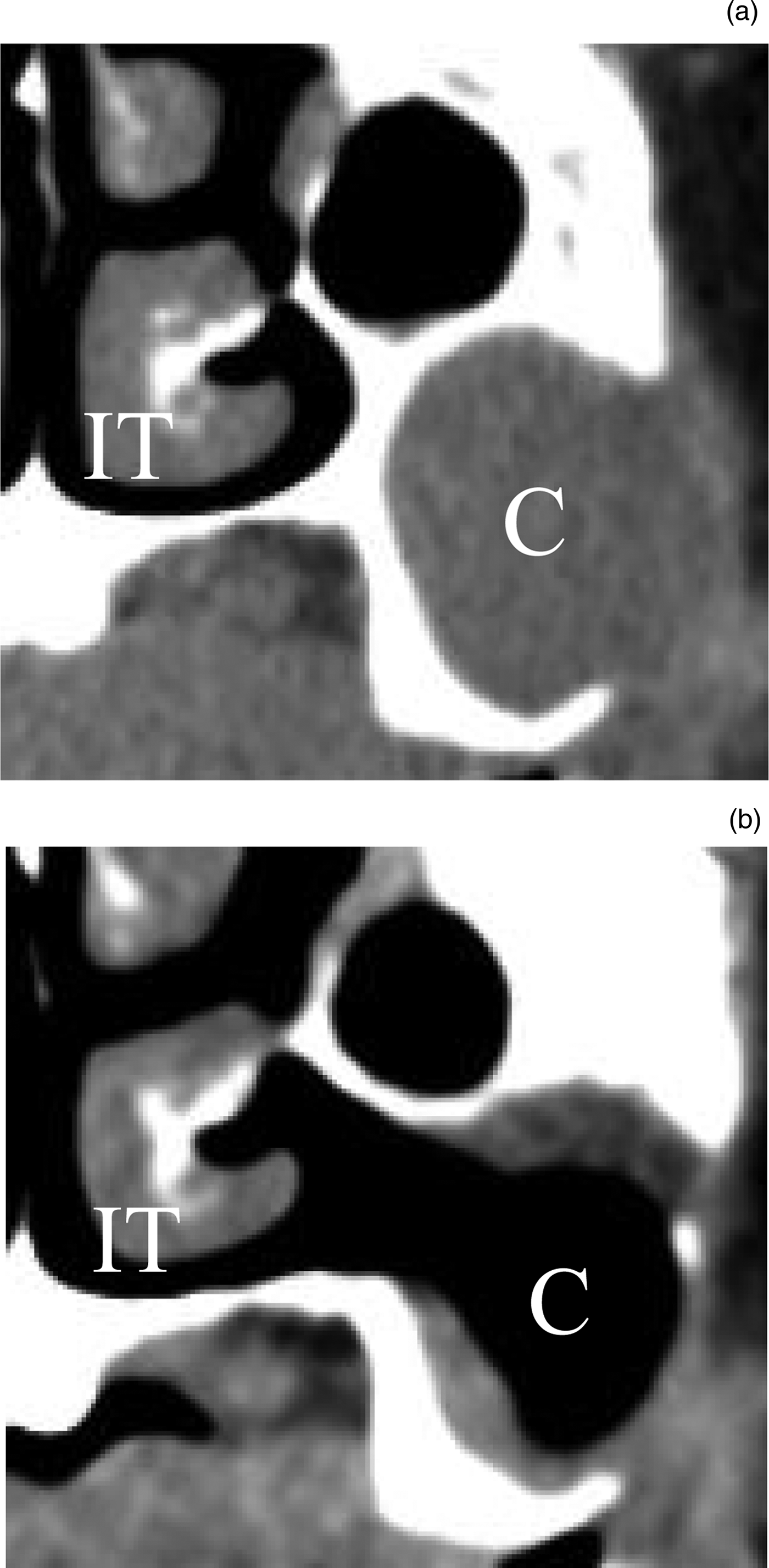
Fig. 2. Computed tomography scan (a) before and (b) after inferior meatal antrostomy with a mucosal flap, in patients with a maxillary mucocele. IT = inferior turbinate; C = cyst
This study was approved by the Nagoya City University Ethical Committee.
Statistical analysis
A statistical comparison of the outcomes obtained for surgery with or without the mucosal flap and sex was performed using chi-square tests. A statistical comparison of results between the two groups was performed using the Mann–Whitney U test for age, diameter and follow-up duration. A statistically significant difference was defined as p < 0.05.
Results
Patients’ demographics
Seventy patients who underwent inferior meatal antrostomy for the treatment of recurrent post-Caldwell–Luc maxillary mucoceles were included in this study. All patients had undergone a Caldwell–Luc operation more than 15 years previously for the treatment of sinusitis.
Transnasal endoscopic inferior meatal antrostomy without a mucosal flap was performed in 49 patients (26 males and 23 females); the mean age at surgery was 64.5 years (range of 55–82 years). Twenty-one patients (10 males and 11 females) underwent transnasal endoscopic inferior meatal antrostomy with a mucosal flap; the mean age at surgery was 64.3 years (range of 56–80 years). There was no statistically significant difference between the two groups with respect to age and sex.
Comparison of surgery with or without mucosal flap
The presence of an opening to the maxillary sinus mucoceles was examined with either a fibrescope or CT within three to five years after surgery.
The mean follow-up duration was 47.2 months (range of 37–59 months) for patients who had undergone surgery without a mucosal flap, and 48.4 months (range of 38–56 months) for patients who had undergone surgery with a mucosal flap. There was no statistically significant difference in the follow-up duration between the two groups.
The antero-posterior, lateral and longitudinal diameters of the maxillary cysts were measured using CT. The maximum antero-posterior diameters of patients who underwent surgery with or without a mucosal flap were 3.3 ± 0.2 cm and 3.2 ± 0.1 cm, respectively. The maximum lateral diameters of patients with or without a mucosal flap were 3.7 ± 0.3 cm and 3.4 ± 0.1 cm, respectively. The maximum longitudinal diameters of patients with or without a mucosal flap were 3.4 ± 0.2 cm and 3.2 ± 0.1 cm, respectively. There were no significant differences between the two groups with respect to the antero-posterior, lateral or longitudinal diameters of cysts.
A closing of the inferior meatal antrostomy was observed in 9 (18.4 per cent) of the 49 patients who had undergone transnasal endoscopic inferior meatal antrostomy without a mucosal flap. Patency of the inferior meatal antrostomy was confirmed in all 21 patients who had undergone the procedure with a mucosal flap. There was a statistically significant difference in the rate of closing between the group with and the group without a mucosal flap (p < 0.05; Table 1).
Table 1. Relationship between mucosal flap and recurrence

Data represent numbers of patients
Complications associated with surgery
No intra-operative or post-operative complications were observed in the 70 patients with recurrent maxillary sinus mucoceles. The nasolacrimal duct and inferior turbinate were preserved in all patients.
Discussion
In this study, we followed up 70 patients who had undergone inferior meatal antrostomy with or without a mucosal flap for recurrent maxillary sinus mucoceles after a Caldwell–Luc operation. No intra-operative or post-operative complications were observed. This suggests that inferior meatal antrostomy with or without a mucosal flap is a safe procedure.
No closings of the antrostomy were observed in the 21 patients who had undergone inferior meatal antrostomy with a mucosal flap. The rate of closing in patients who underwent the procedure with the mucosal flap was significantly lower than that in patients who underwent the procedure without the mucosal flap. These results suggest that a mucosal flap prevents the opening from closing, thereby reducing the recurrence rate of maxillary sinus mucoceles. Thus, inferior meatal antrostomy with a mucosal flap may be an effective strategy for the treatment of maxillary sinus mucoceles.
Several surgical techniques have been developed to prevent the recurrence of maxillary mucoceles. For example, Ono et al.Reference Ono, Ito, Homma, Okada, Murata and Ikeda13 used a T-tube stent to prevent recurrence. However, a stent might need to be removed; as it is a foreign body, it may cause problems such as infection. In contrast, a mucosal flap is made of autologous material and removal is not needed, which is an additional advantage of using mucosal flaps.
Devars du Mayne et al.Reference Devars du Mayne, Moya-Plana, Malinvaud, Laccourreye and Bonfils2 reported that 16 (23.5 per cent) of 68 patients developed recurrence after surgery for sinus mucoceles. Makeieff et al.Reference Makeieff, Gardiner, Mondain and Crampette4 reported that one (12.5 per cent) out of eight maxillary sinuses with mucoceles that were treated endoscopically developed a recurrence, and one (50 per cent) of two sinuses that were treated by a classical external approach (Caldwell–Luc operation) developed a recurrence. Huang et al.Reference Huang, Chen, Lee, Chang, Chen and Chen3 observed recurrence in 5 (14.9 per cent) out of 34 maxillary mucoceles after transnasal endoscopic surgery. In contrast, other studies have reported no recurrences after surgery for maxillary sinus mucoceles.Reference Caylakli, Yavuz, Cagici and Ozluoglu5–Reference Albu and Dutu7
To our knowledge, no studies have, to date, reported on the recurrence rate after the second surgery in only patients with recurrent maxillary sinus mucoceles. The present study showed that the rate of closing of antrostomy was 18.4 per cent after inferior meatal antrostomy without a mucosal flap in patients with recurrent maxillary mucoceles.
Durr and GoldbergReference Durr and Goldberg12 performed partial medial maxillectomy with a mucosal flap for maxillary sinus mucoceles, and conducted partial resection of the anterior inferior turbinate to obtain adequate access to the lateral nasal wall of the inferior meatus. Our method, as described here, does not involve resection of the inferior turbinate, but forceps were used to push the inferior turbinate medially. Our results indicate that inferior meatal antrostomy without resection of the inferior turbinate may be used to successfully treat maxillary sinus mucoceles. Further, while Durr and GoldbergReference Durr and Goldberg12 covered the floor of the nose and the inferior bony border during maxillectomy with the mucosal flap, we used a technique in which only the bone at the posterior edge of the opening into the mucocele was covered with the mucosal flap. Thus, there is a difference between these techniques. However, it is not known whether this accounts for the difference in outcomes. Further studies are needed to address this question.
• Recently, transnasal inferior meatal antrostomy has been used for treating post-Caldwell–Luc mucoceles in the maxillary sinus
• Maxillary sinus mucocele recurrence has been reported after surgery
• Transnasal inferior meatal antrostomy, with or without a mucosal flap, is a safe method
• Transnasal inferior meatal antrostomy with a mucosal flap is effective at preventing antrostomy closing
Competing interests
None declared


