


Case
A male patient was given a diagnosis of univentricular heart and explorative sternotomy was performed while in elementary school; however, no surgical intervention was attempted because of a lack of clinical symptoms. As he reached adulthood, he had developed slightly reduced exercise tolerance and ceased to undergo regular check-ups. At 42 years of age, he was admitted to hospital complaining of dyspnoea on exertion and leg oedema. He was diagnosed with congestive heart failure and tricuspid atresia by transthoracic echocardiography. His symptoms improved with diuretic treatment, and he was referred to our hospital for further testing. Physical examination showed moderate cyanosis with an oxygen saturation of 85%. Detailed transthoracic echocardiography showed tricuspid atresia (muscular type), transposition of the great arteries, atrial septal defect, large and unobstructive ventricular septal defect in subaortic position, hypoplastic right ventricle, and dilated left ventricle. Left ventricular ejection fraction was estimated at 41%. Pulmonary valve was bicuspid and moderate stenosis (peak velocity, 3.3 m/second). Mild aortic valve regurgitation was observed. Sinus of Valsalva dilated to 51 mm in diameter. A catheterisation study revealed elevated pulmonary arterial pressure and impaired ventricular function (Table 1). Serial examinations confirmed a diagnosis of tricuspid atresia with discordant arterial ventricular connection, associated ventricular septal defect and pulmonary stenosis, and medical treatment was chosen as an appropriate option because the patient was not considered to be a candidate for Fontan circulation due to his elevated pulmonary pressure and impaired ventricular function, and sufficient medication was not administered at this time.
Table 1. Results of catheterisation studies of first presentation and 2 years later
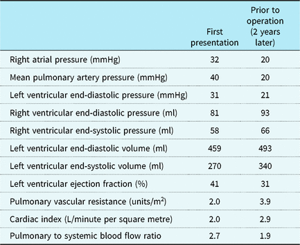
Despite receiving intensive medical treatment, our patient required repeat hospitalisations for congestive heart failure. Transthoracic echocardiography performed 2 years after his first presentation showed more decreased left ventricular function (left ventricular ejection fraction, 37%), mild mitral valve regurgitation, and moderate-to-severe aortic valve regurgitation. A catheterisation study also revealed a further increase in his ventricular volume (Table 1). Contrast-enhanced CT showed further enlargement of sinus of Valsalva (59 mm, Supplementary Fig S1). Considering his well-balanced pulmonary-to-systemic flow ratio and atrioventricular valve function, we speculated that the chief cause of our patient’s heart failure was the progressive aortic valve regurgitation and his ventricular function improved by controlling aortic valve regurgitation. Furthermore, he was not suitable for Fontan operation because of his elevated pulmonary arterial pressure and decreased left ventricular function. Although valve sparing root replacement was considered, we chose aortic root replacement with mechanical valve because of shorter ischaemic time and secure control of the regurgitation. We performed aortic root replacement (modified Bentall procedure) with a 23-mm Regent (St. Jude Medical, Saint Paul, Minnesota, United States of America) and a 26-mm Triplex (Terumo, Shibuya, Tokyo) under general anaesthesia (Fig 1). The patient was weaned from cardiopulmonary bypass without difficulty, and there were no major complications post-operatively. He was discharged on post-operative day 30. Transthoracic echocardiography performed 2 weeks after the operation revealed improved left ventricular function (left ventricular end-diastolic volume, 354 ml; left ventricular end-systolic volume, 201 ml; left ventricular ejection fraction, 43%). His aortic saturation was almost unchanged post-operatively and his cardiopulmonary exercise testing 1 year after the operation showed somewhat reduced exercise tolerance functions (peak oxygen uptake: 13.8 ml/minute per kilogram, anaerobic threshold: 7.8 ml/minute per kilogram). However, considering the fact that he required repeat hospitalisations before the operation and was back to full-time work post-operatively, we guessed his exercise tolerance function was improved. Histopathological examination of the ascending aortic wall which was removed in this operation showed cystic medial necrosis (Supplementary Fig S2).

Figure 1. Intraoperative picture after resternotomy, showing transposition of the great arteries, dilated aortic root and hypoplastic right ventricle. Ao: aorta, LV: left ventricle, RA: right atrium.
Discussion
Univentricular heart is a rare congenital heart malformation that occurs in 1–2% of all cases of congenital heart disease in newborns.Reference Samanek and Voriskova 1 Without surgical intervention, the prognosis of univentricular heart is quite poor.Reference Moodie, Ritter, Tajik and O′Fallon 2 However, certain patients with univentricular heart can reach adulthood without surgical treatment, and some have reportedly survived beyond 60 years of age.Reference Ammash and Warnes 3 Hager et al revealed that patients with univentricular heart who could reach adulthood had a single ventricle of left ventricular morphology, transposition of the great arteries without systemic outflow obstruction, adequately functioning atrioventricular valve, and moderate pulmonary outflow obstruction.Reference Hager, Kaemmerer, Eicken, Fratz and Hess 4 As our patient was diagnosed with tricuspid atresia with discordant arterial connection, unobstructive and large ventricular septal defect in subaortic position, and moderate pulmonary stenosis, he was inferred to have these characteristics as well.
Progressive aortic dilatation and regurgitation in various types of congenital heart diseases such as tetralogy of Follot and single ventricle with pulmonary atresia or stenosis were reported.Reference Niwa, Perloff and Bhuta 5 Aortopathy in congenital heart disease in adult has been getting more attention recently.Reference Niwa 6 Aortic root dilatation, decreased aortic elasticity, and secondary aortic regurgitation impair systemic ventricular function. Although some cases about surgical interventions for aortic valve regurgitation with a Fontan circulation were reported,Reference Murakami, Mori, Inoue, Kaneko and Nakashima 7 to our knowledge, our case was the first report describing a surgical procedure for aortic valve regurgitation with unoperated univentricular heart. Since it was difficult to estimate what degree the pulmonary blood flow would be relevant to heart failure in this patient, concomitant pulmonary artery banding was planned when recovery of cardiac function was inadequate. Consequently, ventricular overload was reduced and then ventricular function was improved even in the unoperated univentricular heart by controlling aortic valve regurgitation. If his pulmonary arterial pressure and left ventricular function improved sufficiently in the future, he could be a candidate for a Fontan circulation.
As the prognosis of patients with a Fontan circulation has been improving,Reference Pundi, Johnson and Dearani 8 it is predicted that more patients with univentricular heart suffer from aortopathy and aortic valve regurgitation in the future and surgical intervention for them may improve their ventricular functions. Therefore, we should follow adult patients with univentricular heart with careful attention to aortopathy and aortic valve regurgitation, and a specialised heart team should carefully discuss their treatment strategies and the timing of surgical intervention.
Acknowledgements
We thank Dr. Koichiro Niwa for his advice about our manuscript. Mr. Fujimura supported us to reconstruct 3D-CT image.
Financial Support
This research received no specific grant from any funding agency, commercial or not-for-profit sectors.
Conflicts of Interest
None We declare we have no conflicts of Interest.
Supplementary material
To view supplementary material for this article, please visit https://doi.org/10.1017/S1047951119001719


