


Introduction
Three-dimensional (3D) printing within healthcare has become increasingly popular and the future of healthcare is being challenged in dramatically new ways as many aspects of healthcare have become outdated.Reference Dunham 1 3D printing allows the production of any given design specification to be three dimensionally printed directly from computer software.Reference Dunham 1 Traditional use of fibreglass wraps for patient fractures can directly impact upon patient’s lives due to the bulky design and the detrimental impact of water upon the integrity of the material used is a major limitation. They are time consuming to construct and often require more than one staff member to make. The emergence of creating 3D printed braces have not only resulted in decreased construction time, but their slim breathable design allows the patient to engage in day to day activities including; showering, performing household duties and does not inhibit clothing options as the cast is no longer cumbersome.Reference Dunham 1 In addition, many surgical departments have begun to explore the use of 3D printing over traditional methods to create medical devices such as stents, skin grafts and artificial limbs.Reference Dunham 1 This demonstrates the revolutionary benefit 3D printing can achieve over traditional practice, treating patients more efficiently and providing enhanced treatment options to patients that were previously deemed unfeasible.Reference Dunham 1 – Reference Su, Moaran and Robar 4 It has been argued that the practical use of 3D printing in radiation therapy (RT) can improve patient outcomes and reduce practitioner resources enabling increased patient throughput without compromising practice standards.Reference Dunham 1 – Reference Su, Moaran and Robar 4
Within RT, challenges may arise when treating superficial lesions due to the phenomenon known as the ‘skin-sparing effect’. This effect occurs when using high energy photons as the maximum dose is deposited below then skin surface, negating the requirement of the intended treatment coverage at the skin’s surface.Reference Zou, Fisher and Zhang 2 – Reference Su, Moaran and Robar 4 In order to minimise this effect and achieve the desired dose at the skin surface for superficial lesions, tissue equivalent material known as bolus can be placed on the patient’s skin and over the treated volume [planning target volume (PTV)] to enable sufficient dose coverage.Reference Zou, Fisher and Zhang 2 – 5 International Commission on Radiation Units and Measurements (ICRU62) recommends the treated tumour volume is covered by 95% of the prescribed dose when radiation treatment is delivered. Many superficial tumours do not adhere to the ICRU62 guidelines of 95% coverage largely due to body contour irregularities, tissue inhomogeneity and variations in PTV.Reference Zou, Fisher and Zhang 2 , 5 Therefore bolus placement over superficial lesions is imperative to adhere to these international guidelines and to achieve desirable outcomes for patients.
Method
A literature review of the journal databases; Science direct, Medline, CINAHL, Ovid and Google Scholar was conducted. To ensure appropriate literature was sought, the search terms utilised Boolean terminology and included keywords; (‘3d’ OR ‘3-dimentional’ OR ‘three dimensional’) ‘bolus’ OR ‘boli’ conform*, (‘Radiation therapy’ OR ‘radiotherapy’) Printing. The initial search returned 40 articles. To minimise this number and maintain relevance, further criteria was set to exclude research older than 5 years and limited to work published in the English language. Remaining articles were then reviewed and selected based upon satisfying criteria of evaluating ‘3D bolus printing’ and ‘3D bolus printing within radiotherapy AND conformity’. Key papers identified in the references of these articles were then reviewed, known as ‘citation pearl growing’ or ‘snowballing’, in order to ascertain relevant literature has been included.
A total of four journal articles were identified and analysed for this paper. Articles included in this evaluation were critically analysed to ensure that the findings were feasible in accordance with the SIGN critical appraisal tool. 6
Why 3D print within radiation therapy?
The benefits of 3D printing within the wider healthcare sector have prompted the use of 3D printing to be explored within RT. On a local level, the disadvantages attributed to the production of bolus within our department mirrors those seen in other departments. It is evident that bolus production can be limited by several factors; the accuracy of bolus fabrication is limited to the expertise of the practitioner, time and resources for both patients and staff required to construct bolus and the capacity of prefabricated bolus to conform to irregular surfaces to minimise error in dose distribution.Reference Dunham 1 , Reference Su, Moaran and Robar 4
Butson et al.Reference Butson, Martin and Cheung 7 measured the effects of air gaps of 2, 4 and 10 mm underneath 10 mm bolus and increasing beam angels of incidence upon dose distribution for 8×8 cm and 10×20 cm field sizes on the skin surface using a 6 MV photon beam. Measurements were recorded using both radiochromic film and an Attix ionisation chamber (Attix; RMI, Middleton, WI, USA). Each method produced similar results, finding a 2 mm air gap in both 8×8 cm and 10×20 cm field sizes to have negligible effect upon dose distribution at the skin surface. The study also found a gap of 4 mm for both field sizes to have little effect upon dose distribution, producing dose reductions of 2–6% when measured with varying angles of incidence between 0 and 60°. The greatest reduction in dose was evident for a gap of 10 mm in the smaller 8×8 field size at the largest angle of incidence of 60°, resulting in a dose reduction of >10%.
Khan et al.Reference Khan, Villarreal-Bearajas and Udowics 8 evaluated the effect of air gaps of between 0 and 50 mm on skin surface dose (Dsurf) using 10 mm of SUPERFLAB bolus (Radiation Products Design Inc., Albertville, MN, USA) and measuring depth of maximum dose (Dmax) in solid water and RANDO phantoms (Radiology Support Devices, Long Beach, CA, USA) The study produced similar findings to Buston et al.Reference Butson, Martin and Cheung 7 indicating smaller field sizes have the greatest variation in Dsurf. Although the study shows similar effects upon field size, the study does not explore the effect of beam obliquity upon Dsurf. The effect of beam obliquity is of critical importance due to the advancement of volumetric arc therapy (VMAT) and intensity modulated radiation therapy (IMRT), becoming the principle choice of treatment technique for many body sites. Both VMAT and IMRT techniques incorporate small field sizes and can utilise up to 360° of differing treatment angles, therefore a 4 mm gap or greater between skin and bolus may significantly produce a bigger dose variation than those shown within these studies.
A major limitation of both of these studies is measurements were obtained from rigid phantoms and not patients undergoing active RT. Patient contours are not as stable and defined as a solid rigid phantoms and this may influence the level of bolus conformity, therefore possibly resulting in different measurement outcomes. Furthermore, it could be claimed that variations in patient positioning could further impact upon Dsurf and result in greater inaccuracies on dose delivered to the skin surface. However, the studies do provide a baseline to draw conclusions from and highlight the effect air gaps can have upon Dsurf, contributing to a decrease of >10% of the prescribed dose. This reinforces the need to explore the use of custom 3D printing to create conformal bolus and its need is of even greater importance when considering the increased use of VMAT and IMRT technology.
Feasibility of 3D printed bolus, fabrication device and materials
Despite a wealth of literature existing regarding the emergence of 3D printing in medicine, there is a paucity of research into its application within RT. RT has specific needs that must be achieved before the implementation of personalised 3D bolus. First, fabrication material that closely resembles tissue density must be established. Second, selected materials should be durable and contain no air gaps within the material itself. Finally, assessment of conformity of the printed material to a patient’s skin surface can then be considered.Reference Zou, Fisher and Zhang 2 – Reference Su, Moaran and Robar 4 , Reference Burleson, Baker and Hsia 9 Few studies have investigated fabrication material and devices to determine those suitable for RT.
Burleson et al.Reference Burleson, Baker and Hsia 9 aimed to demonstrate that 3D printers have the ability to create 3D printed bolus for RT treatments. They explored the use of a generic fused deposition modelling (FDM) fabrication device and focussed on two printing materials; acrylonitrile butadiene styrene (red-ABS) and clear polylactic acid (clear-PLA) to create 3D printed bolus. The rationale for selecting the two chosen printing materials are clearly explained, showing the physical properties of both materials compared with water, as bolus should resemble these properties. The tissue maximum ratio of each material compared with measurements in solid water phantoms using a 6 MV beam were examined. The dose difference between the two materials and compared with the standard bolus materials SUPERFLAB and wax was less than 4%, shown in Figure 1. The findings show red-ABS followed the trend of water more closely than that of the clear-PLA. However, the study cited red-ABS became compromised once it reached a certain height, edges started to curve and layers began to separate and the authors chose not to continue testing the material for the remainder of the study, the height in which it started to fail was not stipulated by the authors. It is unsure from the study whether this is due to the material properties or the limitations of the fabrication device. Such ambiguity makes it difficult to draw valid conclusions and further investigations should be carried out to determine the effect of bolus size in clinical situations, for example its application for chestwall treatments for patients with breast cancer. The percentage depth dose (PDD) was also measured and shown in Figure 2. These highlight that clear-PLA builds up quicker with a steeper fall off gradient compared with water. It is essential that this is acknowledged because if clear-PLA was to be used in a clinical setting there is a potential to use a lower thickness than that of standard bolus material to achieve the same bolus effect.

Figure 1 Tissue maximum ratio (TMR) curves of clear-PLA and red-ABS compared with standardly used bolus.Reference Burleson, Baker and Hsia 9 Abbreviation: PLA, polylactic acid.

Figure 2 Percentage depth dose (PDD) curves of clear-PLA compared with water.Reference Burleson, Baker and Hsia 9 Abbreviation: PLA, polylactic acid.
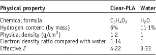
Table 1 indicates that clear-PLA does not fully resemble water and the PDD measured by the treatment planning system demonstrates the Hounsfield unit (HU) of 0 cannot be standardly applied to bolus material as current practice permits. Burleson et al.Reference Burleson, Baker and Hsia 9 examined both the electron density and mass density of clear-PLA and assigned the value of HU 260 to the material within the treatment planning system as shown in Figure 3. They found a difference of 0.25 mm in depth dose at Dmax and just a 0.5% variation in PDD, suggesting clear-PLA is an adequate fabrication material when created using FDM technology.

Figure 3 Electron density to determine Hounsfield unit value.Reference Burleson, Baker and Hsia 9
Table 1 Comparison of physical property’s of clear polylactic acid (PLA) and waterReference Burleson, Baker and Hsia 9
Similarly, Yeo et al.Reference Yeo 10 and Su et al.Reference Su, Moaran and Robar 4 explored the use of clear-PLA as a 3D printing material to improve bolus conformity. Both studies mirror Burleson et al.Reference Burleson, Baker and Hsia 9 findings, citing differences of 0.5% in PDD between clear-PLA and solid water. These outcomes further reinforce the belief that clear-PLA is a sufficient fabrication material when constructed by FDM technology. Su et al.Reference Su, Moaran and Robar 4 further explored fabrication of 3D bolus using RANDO phantoms of varying anatomical locations, finding clear-PLA to be durable, capable of conforming to the skin’s surface and dosimetrically suitable. The main focus of Su et al.Reference Su, Moaran and Robar 4 study was to investigate the feasibility of using 3D bolus printing to be applied for modulated electron therapy (MERT) treatments. However, exploration of this is above the scope of this study, but does further promote clear-PLA as a suitable material.
Kim et al.Reference Kim, Shin and Kay 11 recognised the lack of clarity within the literature to determine the use of fabricated 3D printed bolus within the field of RT and attempted to evaluate how effectively 3D printed materials could be incorporated into Varian’s Eclipse planning system (Varian Medical Inc., Palo Alto, CA, USA). The study discovered 3D printed bolus must be converted into a stereolithography (STL) format that enables the printing of 3D computer-aided design (3D CAD) to ensure data integrity as it is not possible to directly convert the data from Eclipse planning system. This clearly highlights a major limitation of using 3D printed bolus and the additional costs and resources required to guarantee accurate dosimetric calculation may discourage departments to invest in such technical advancements. However, Kim et al.Reference Kim, Shin and Kay 11 endeavours to explain the necessary steps required to create 3D printed bolus and convert it to the STL format.
OsiriX MD ver. 2.8.x (OsiriX, Geneva, Switzerland) was used for 3D rendering of the designed bolus structure in DICOMRT format. In order to convert the file into the STL format, 3Ds Max 2013 (Autodesk, San Rafael, CA) was used. Insight ver. 9.1 (Stratasys, Eden Prairie, MN) was used to print out the STL file of this designed bolus on a Fortus 400 mc 3D printer (Stratasys, Eden Prairie, MN). The Fortus 400 mc is a fused deposition modelling technique. (p. 2)
The extra steps required to convert the data increase the potential for systematic error to occur within the printing process. To minimise this, detailed quality assurance (QA) must be established. The authors found the printed bolus to conform well to irregular surfaces and dosimetric results favoured the production of 3D printed bolus material, ABS-M30.Reference Kim, Shin and Kay 11 The use of ABS-M30 to create 3D bolus is unique to this study and the rationale for its use is unclear, with the author stating ‘ABS-M30 is commonly used by the FDM fabrication device’ (p. 2). While this may be true, no other data exists within the literature to support its use within the RT setting.
It is worth noting that the studies discussed received partial funding from 3D printing manufacturers.Reference Zou, Fisher and Zhang 2 – Reference Su, Moaran and Robar 4 , Reference Burleson, Baker and Hsia 9 , Reference Kim, Shin and Kay 11 . Although the literature advocates the FDM printing device and the clear-PLA material to be the more superior products, other fabrication devices and material are available on the market that have not been researched in this review. It is possible that other products may exist of superior quality and before implementing 3D printing it would be prudent to consider all options available.
Implementation
This review has highlighted several key advantages of using 3D printed bolus, including; a reduction in production time when compared with the creation of traditional bolus, decrease in cost and an improved patient experience as bolus can be printed directly from computed tomography/magnetic resonance images thus limiting patient time spent at the planning stage. However, in order to successfully implement 3D printing within RT a number of factors must be considered. Rigorous QA procedures must be established to ensure patient safety and the size of the bolus must be taken into account as 3D printers have a limited size capability.Reference Burleson, Baker and Hsia 9 Such barriers are not insurmountable to ensure the patients benefit by experiencing a much more pleasurable experience and shorter times through the creation of 3D bolus.
The impact upon workload for medical physicists should be considered as the majority of the initial QA processes will be performed by that staff group. The emergence of more sophisticated technology may include bio-printing which enables tissue equivalent material to be printed within a structure.Reference Harris 12 This type of printing could potentially be introduced in the fabrication of a beam direction shell and incorporate another solid material to be used within the shell to act as bolus. This has the potential to reduce uncertainties of air gaps which are known to have a large dosimetric impact upon VMAT techniques.Reference Butson, Martin and Cheung 7 , Reference Khan, Villarreal-Bearajas and Udowics 8 This of great significance as VMAT techniques are internationally becoming the preferred treatment option for head and neck cancer, therefore the introduction of bio-printing could prove to be revolutionary. Su et al.Reference Su, Moaran and Robar 4 have paved the way by exploring the use of 3D bolus for MERT, creating a solid foundation for future research to be based on.
One study identified clear-PLA to be structurally sound after being exposed to radiation 20 times, but often a course of radiation treatment may be in excess of that number.Reference Burleson, Baker and Hsia 9 Therefore, further studies should obtain dosimetric measurements to determine the effect of radiation on the material itself. In view of this, robustness tests must be conducted before implementation which is of critical importance considering current 3D printers take ~4–6 hours to produce a small block.Reference Burleson, Baker and Hsia 9 If during a course of treatment the 3D bolus happened to break, it would take a considerable period to create another and may result in delays to a patient’s treatment.Reference Burleson, Baker and Hsia 9
3D printing has the potential to dramatically change practice not just for bolus production but also for individual patient immobilisation and overall experience. While current printers allow 3D bolus to be printed in any shape they are limited by the size by the 3D printer. However, due to the velocity 3D printing is evolving it is unlikely that such a problem will continue to remain.
Conclusion
This critique has aimed to highlight the possibility to produce a superior custom 3D printed bolus. Customised 3D printed bolus has been shown to potentially replace and improve upon commercially available boluses, potentially reduce bolus placement errors and overcome some of the disadvantages in traditionally made boluses. This is of increasing importance due to the emergence of VMAT and IMRT techniques where dose escalation and precision are imperative to positive treatment outcomes. As with any new technique it is prudent to develop QA procedures to safeguard both patients and health professionals.Reference Burleson, Baker and Hsia 9 Given the paucity of literature and the potential bias from the vendors producing 3D printers, caution must be used when deciding what fabrication device and fabrication material are to be used, as there may be invested interest in the new technology and products explored.
Acknowledgements
The authors would like to thank the staff of the Wellington Blood and Cancer Centre for their financial support.
Conflicts of Interest
None.


