Introduction
Persistence of otitis media with effusion (OME) beyond childhood and into adolescence or adulthood is associated with ongoing hearing impairment, primarily attributable to the presence of fluid in the middle-ear cleft. While this may be managed by attention to the underlying condition (e.g. adenoidectomy or treatment of chronic rhinosinusitis), the middle-ear condition may persist. Placement of a ventilation tube will restore middle-ear aeration but is not without consequence. Common complications are infection, wax accumulation around a tube, tympanic membrane atrophy and perforation;Reference McLelland1 ventilation tube extrusion is often associated with reoccurrence of OME. The placement of long-term tubes will allow for longer periods of improved hearing, but at the expense of a higher complication rate, and the duration of tube retention is variable.Reference Klingensmith, Strauss and Connor2, Reference Kim, Moore and Rockley3 Perforation following ventilation tube extrusion is more common when wide flanged tubes are used and is usually associated with some degree of hearing loss, which is often the symptom that is being treated.Reference Kay, Nelson and Rosenfeld4–Reference Vlastarakos, Nikolopoulos, Korres, Tavoulari, Tzagaroulakis and Ferekidis6
Positioning a ventilation tube outside the margin of the tympanic membrane is an alternative for securing long-term ventilation, and is referred to as either ‘extra-tympanic’ or ‘subannular’ ventilation tube placement. Often a bony groove is drilled across the tympanic sulcus, the aim being to allow better positioning of the tube and to provide a bony ridge that may hinder tube extrusion.Reference Simonton7–Reference Jahn9 This is more complex than transmyringeal placement, and involves the elevation of a tympanomeatal flap as well as drilling of the groove; the procedure is usually performed under general anaesthesia. It is not uncommon for the ventilation tube flange to require modification so that it sits securely in the groove.Reference Duadia, Yelavich and Dawes10 Whether placement of subannular ventilation tubes is the optimum procedure for managing adolescent and adult recurrent or persistent OME remains controversial.Reference Duadia, Yelavich and Dawes10–Reference Saliba, Boutin, Arcand, Freohlich and Abela15
Subannular ventilation tubes are associated with the complications that accompany long-term intubation, although these occur less frequently than with transmyringeal long-term ventilation tubes.Reference Duadia, Yelavich and Dawes10, Reference Elluru, Dhanda, Neely and Goebel12, Reference Jassar, Coatesworth and Strachan14, Reference Saliba, Boutin, Arcand, Freohlich and Abela15 One additional potential complication is injury to the chorda tympani nerve. This exits the facial nerve approximately 3.2 mm above the stylomastoid foramen and courses anterolaterally, within a bony canal, to enter the middle ear at about the medial end of the tympanomastoid suture.Reference McManus, Dawes and Stringer16, Reference Ott, Tebben, Losenhause and Issing17 The bone between the chorda tympani nerve and annulus forms the lateral boundary of the facial recess; when this is removed, there is access to the posterior tympanum.
When undertaking placement of a subannular tube, it is essential to have a good understanding of the relationship between the drilled groove and the chorda tympani nerve, and of the space available in the hypotympanum and retrotympanum for the tube flange. With the aim of maximising surgical safety and procedure success, we used microslice computed tomography (CT) to determine the average depth of the hypotympanum and retrotympanum, and determined the risk to the chorda tympani nerve by imaging sets of bones before and after drilling the groove for a subannular ventilation tube in both the conventional and recommended positions.
Materials and methods
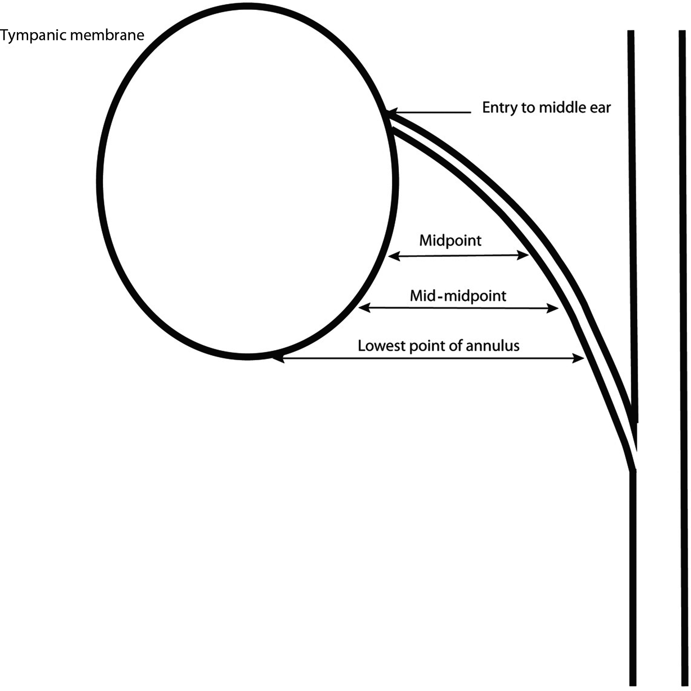
Forty cadaver temporal bones were imaged using microslice CT as described by McManus et al.Reference McManus, Dawes and Stringer16 Using a mathematical algorithm, the image data were recalculated so that axial images were viewed in Reid's plane and coronal views were perpendicular to this; this was used to define the course of the chorda tympani nerve.Reference McManus, Dawes and Stringer16 Measurements relating the chorda tympani nerve to the annular groove used the chorda tympani nerve exit from its posterior canaliculus and the lowest point of the annulus as identifiable reference points. Two additional points were defined: the midpoint, that point midway between chorda tympani nerve middle-ear entry and the lowest point of the annulus; and the point midway between the midpoint and the lowest point of the annulus, referred to as the mid-midpoint (Figure 1). At these points, the distance between the annular groove and chorda tympani nerve was recorded. The position of the midpoint and the mid-midpoint in relation to the annulus were measured for each specimen (Figure 2).

Fig. 1 Measurements made between tympanic sulcus and chorda tympani nerve.

Fig. 2 Measurements of the distance between the tympanic sulcus and chorda tympani nerve (x axis = distance in millimetres from the centre, y axis = height of the tympanic membrane as a percentage of the total height).
Ten of the dataset temporal bones subsequently had a groove drilled across the posterior annulus at the position usually employed when placing a subannular tube.Reference Duadia, Yelavich and Dawes10 Using the described microslice CT protocol and mathematical algorithm to convert the images to Reid planes,Reference McManus, Dawes and Stringer16 the depth and width of the groove and the distance between the chorda tympani nerve and the groove were measured. These were then assessed. A further 10 bones of the original dataset had a groove drilled as inferiorly as possible, followed by reimaging and measurement of the depth and width of the groove and its distance from the chorda tympani nerve. All drilling procedures were performed by the senior author.
Twenty of the original temporal bone dataset images were viewed separately. Measurements of the depth of the hypotympanum were taken at the lowest point of the annulus (measurement ‘a’), and at a point midway between this and the posterior annulus at the level of the umbo (measurement ‘b’). The depth of the facial recess was measured at the point midway between the lowest point of the annulus and umbo (measurement ‘c’), and at the level of the umbo (measurement ‘d’) (Figure 3).

Fig. 3 Measurements made of the hypotympanum and retrotympanum (facial recess). ‘a’ = depth of hypotympanum at the lowest point of the annulus; ‘b’ = depth of the hypotympanum at a point halfway between the lowest point of the annulus and the umbo; ‘c’ = depth of the retrotympanum at a point halfway between the lowest point of the annulus and the umbo; ‘d’ = depth of the retrotympanum at the umbo
Results
The full dataset comprised 17 right side and 23 left side temporal bones. For presentation of the results, the figures are drawn showing data from both sides presented as the left ear viewed from a lateral perspective.
The mean entry point of the chorda tympani nerve into the middle ear was 62 per cent (33–81 per cent) of the height of the tympanic membrane (Figure 2); in two specimens, the chorda tympani nerve exited the canaliculus below the level of the umbo. The mean distance from the annulus to the chorda tympani nerve was: 1.4 mm (standard deviation (SD) = 0.6) at the midpoint, 3.4 mm (SD = 0.7) at the mid-midpoint and 7.5 mm (SD = 1.3) at the lowest point of the annulus.
Nineteen sets of drilled bone images were suitable for image analysis, 10 from the initial position and 9 from the second revised position. In two bones of the initially drilled set, the groove came in contact with the posterior canaliculus (Figure 4). The mean dimensions of the two sets of bones are given in Table I. The second set of drilled grooves was significantly more shallow and narrow than the first set. The senior author found drilling the lower placed groove more technically challenging, resulting in greater variability in its position. The dimensions of the grooves were superimposed on the full dataset results (Figure 5) to demonstrate the risk of each position to a larger set of bones.

Fig. 4 Sagittal microslice computed tomography image showing proximity of groove to the chorda tympani nerve. S = superior; A = anterior; P = posterior; I = inferior

Fig. 5 Relationship of the two grooves drilled across the tympanic sulcus to the chorda tympani nerve. (x axis = distance in millimetres from the centre, y axis = height of the tympanic membrane as a percentage of the total height). Black areas = mean groove depth, grey areas = two standard deviations
Table I Comparison of groove dimensions and proximity to chorda tympani nerve between drilled bone sets

Data represent means ± standard deviations. *p < 0.05 (t-test)
Table II and Figure 6 show the depth of the hypotympanum and facial recess at the points defined. There is variability in the space available for placement of a ventilation tube flange; the bony groove depth may be up to 1.2 mm and at times this will be dictated by individual anatomy.

Fig. 6 Depth of the hypotympanum and retrotympanum (x axis = distance in millimetres from the centre, y axis = height of the tympanic membrane as a percentage of the total height). ‘a’ = depth of hypotympanum at the lowest point of the annulus; ‘b’ = depth of the hypotympanum at a point halfway between the lowest point of the annulus and the umbo; ‘c’ = depth of the retrotympanum at a point halfway between the lowest point of the annulus and the umbo; ‘d’ = depth of the retrotympanum at the umbo
Table II Depth of hypotympanum and facial recess at points shown in figure 3

SD = standard deviation
Recommendation
The bony groove for posterior placement of a subannular ventilation tube should be positioned at 20 per cent of the height of the tympanic sulcus above the lowest point of the annulus (Figure 7). Placing the groove higher than this carries a greater risk of encountering the chorda tympani nerve in its posterior canaliculus. When modifying the flange of a long-term ventilation tube, this should initially be trimmed to between 1 and 2 mm in its posterior and inferior part. If it fails to sit well in the bony groove, further trimming may be required.

Fig. 7 Recommended position for a groove drilled across the tympanic sulcus and its relationship to the chorda tympani nerve (x axis = distance in millimetres from the centre, y axis = height of the tympanic membrane as a percentage of the total height). Black area = mean groove depth, grey area = two standard deviations
Discussion
The maintenance of middle-ear aeration in patients with chronic Eustachian tube dysfunction is an ongoing problem for otologists. A variety of ventilation tubes have been designed to resist extrusion. However, meta-analysis has shown the risk of perforation, otorrhoea and cholesteatoma to be at least double with long-term ventilation tubes compared with short-term ventilation tubes.Reference Kay, Nelson and Rosenfeld4 Subannular ventilation tubes have been described for use in both chronic otitis media and atelectatic ears, and may provide long-term ventilation with fewer complications.Reference Duadia, Yelavich and Dawes10, Reference Elluru, Dhanda, Neely and Goebel12, Reference Jassar, Coatesworth and Strachan14, Reference Saliba, Boutin, Arcand, Freohlich and Abela15
The duration of ventilation varies between clinical reports, but overall it appears to be no longer than that for T-tubes. Thus, the primary benefit of subannular ventilation tube placement appears to be from the lower complication rate, particularly with respect to infection and perforation. Subannular placement was initially aimed at either protecting an atelectatic tympanic membrane from further damage or providing longer duration of hearing improvement (ventilation), with the placement of part of the tube flange behind bone being considered to delay extrusion.Reference Silverstein8, Reference Jahn9
Injury to the chorda tympani nerve has not been described, other than by Haberkamp and SilversteinReference Haberkamp and Silverstein18 who reported a 13 per cent prevalence. For some techniques employed, this will be because either there is no bone removal or any bone removal is conducted well away from the chorda tympani nerve. However, the more traditional positioning for subannular tube placement is across the posterior half of the tympanic sulcus as this is more easily accessible via a permeatal approach.
SilversteinReference Silverstein8 and JahnReference Jahn9 created either a bony tunnel or a groove to allow placement of the subannular ventilation tube with less elevation of the annulus; they did not report injury to the chorda tympani nerve. However, our findings indicate that there is variability in the course of the chorda tympani nerve, and when the nerve runs closer to the annulus as it ascends to enter the middle ear there is a greater risk of injury. Ott et al.Reference Ott, Tebben, Losenhause and Issing17 describe variability of chorda tympani nerve entry to the middle ear, and the distribution of height at middle-ear entry was similar to that shown in Figure 2. Gopalan et al.,Reference Goplan, Kumar, Gupta and Phillips19 and Michael and Raut,Reference Michael and Raut20 have recorded the prevalence of chorda tympani nerve injury and dysgeusia following middle-ear surgery; approximately 15 per cent of patients will report altered taste in the early post-operative period, but for the majority this will resolve completely. Tympanotomy, as opposed to either mastoidectomy or myringoplasty, was associated with a higher rate of symptom reporting. The procedures associated with tympanotomy alone are either ossiculoplasty or stapedectomy, and, in contrast to myringoplasty, these often require mobilisation of the chorda tympani nerve. With subannular ventilation tube placement, there is little need to mobilise the chorda tympani nerve. It is the creation of a bony groove across the tympanic sulcus that places the chorda tympani nerve at risk.
• Drilling across the tympanic sulcus places the chorda tympani nerve at risk
• Drilling at a point below 20 per cent of the height of the tympanic membrane minimises this risk
• The hypotympanum and retrotympanum are shallow (1–2 mm); subannular ventilation tube design should accommodate this
We have mapped the course of the chorda tympani nerve in relation to the tympanic sulcus, and assessed the risk of injury to the chorda tympani nerve following drilling a groove across the tympanic sulcus. We have demonstrated that positioning this closer to the lowest point of the annulus can effectively protect the chorda tympani nerve from physical injury.
Further understanding of the mechanism by which subannular ventilation tubes extrude may be of use in improving their design. Flange design may contribute to both duration of ventilation and rates of perforation after extrusion: larger flanged tubes are retained longer, but these are associated with a greater rate of perforation following either extrusion or removal.Reference Klingensmith, Strauss and Connor2–Reference Vlastarakos, Nikolopoulos, Korres, Tavoulari, Tzagaroulakis and Ferekidis6 It is not known whether ventilation tubes extrude because of inflammation, migration of the tympanic membrane epithelium or erosion through the fibrous annulus. The use of biocompatible materials such as hydroxylapatite and titanium may reduce any effect that inflammation has on extrusion and improve longevity. When considering the design of a subannular ventilation tube, placement in the postero-inferior mesotympanum and hypotympanum is most easily achieved; the tube will ideally have an eccentrically placed flange, the shorter part fitting into the facial recess and hypotympanum and the longer part accommodated within the mesotympanum. It is likely that the creation of a trans-sulcus groove will necessitate trimming of the posterior part of the flange; it will be the more anterior flange which provides for longer tube retention, and which following either elevation of an atelectatic segment or tympanoplasty may prevent adhesion formation.
Conclusion
This microslice CT study of the anatomy of the chorda tympani nerve, and posterior mesotympanum and hypotympanum, provides information that will both minimise chorda tympani nerve trauma and inform future ventilation tube design.
A clear recommendation is that a groove drilled across the posterior tympanic sulcus should be placed in relation to the lower 20 per cent of the tympanic membrane.
Acknowledgements
Lauren McManus' work was funded by a University of Otago Faculty of Medicine scholarship. We wish to thank Andrew McNaughton, Otago Centre for Confocal Microscopy, for his expertise with microslice CT scanning.


