Introduction
Radiotherapy (RT), either as a definitive or post-operative adjuvant treatment, is crucial for the management of head and neck cancer.Reference Yeh 1 Intensity-modulated RT is a conformal technique used to reduce the adverse effects of RT and control the irradiation doses received by the target. Well-designed, randomised, controlled clinical trials have shown that the use of intensity-modulated RT to spare the parotid glands significantly reduces the incidence of xerostomia and improves patients’ quality of life.Reference Nutting, Morden, Harrington, Urbano, Bhide and Clark 2 Therefore, intensity-modulated RT has increasingly been used to treat head and neck cancer.
Otitis media with effusion (OME) is a known clinical outcome of RT, and has been largely attributed to the deterioration of Eustachian tube function.Reference Bluestone 3 – Reference Wakisaka, Yamada, Motoyoshi, Ugumori, Takahashi and Hyodo 10 Currently, Eustachian tube function is often assessed using sonotubometry,Reference van Heerbeek, van der Avoort, Zielhuis and Cremers 11 – Reference Ohta, Sakagami, Suzuki and Mishiro 14 a method based on the arrival of sound pressure in the auditory canal from the nostril via the Eustachian tube. Swallowing, yawning and the Valsalva manoeuvre provoke Eustachian tube opening, which increases the sound pressure level (SPL) in the auditory canal.
Some previous studies have examined the relationship between radiation doses and OME;Reference Wang, Wang, Zhang, Guo, Hoffman and Jiang 15 – Reference Wang, Li, Miyamoto, Chen, Zhou and Zhang 17 however, these included patients treated with intensity-modulated RT for nasopharyngeal cancer, which itself could cause Eustachian tube dysfunction and OME. Accordingly, the present study was conducted to examine the relationship between Eustachian tube dysfunction and RT-induced OME in patients with non-nasopharyngeal head and neck cancer, and to identify the threshold radiation dose via intensity-modulated RT required to induce OME.
Materials and methods
This study was approved by the Ethics Committee of Hyogo College of Medicine Hospital (approval number: 1898). All patients provided written informed consent to participate in this prospective study.
Eighteen patients (16 male, 2 female) with head and neck cancer who underwent intensity-modulated RT at the Hyogo College of Medicine Hospital, between March 2015 and July 2016, were enrolled in this study. The patients’ ages ranged from 31 to 81 years, with a mean of 69.2 years. Other characteristics of the patients are shown in Table I. Disease staging was performed according to the UICC TNM Classification of Malignant Tumours, seventh edition.Reference Sobin, Gospodarowicz and Wittekind 18
Table I Patients’ characteristics

TNM = tumour–node–metastasis; RT = radiotherapy; M = male; F = female
Regarding treatment, six patients received cisplatin as an intravenous (IV) infusion (100 mg/m2 two times on weeks one and four) and three patients received cisplatin as an intra-arterial injection (100 mg/m2 four times on weeks one, three, five and seven) during intensity-modulated RT. Three other patients received docetaxel IV (20 mg/m2 three times on weeks one, three and five) and five received cetuximab IV (400 mg/m2 loading dose, followed by 250 mg/m2 weekly) during intensity-modulated RT. Three patients underwent surgical treatments before RT, including neck dissection alone (case number 1) and neck dissection with primary resection (numbers 10 and 18) (Table I).
No patient exhibited a soft tissue shadow in the middle ear on computed tomography (CT) scans, or had a tumour that reached the Eustachian tubes or middle ears. Accordingly, all ears were considered to be without otitis media with effusion (OME) before RT.
Radiotherapy and irradiation doses
For treatment planning, all patients underwent CT scanning in the supine position using an Aquilion LB CT unit (Toshiba, Otawara, Japan). The CT datasets were transferred to a XiO treatment planning system (Elekta, Stockholm, Sweden) or MIM Maestro planning software (MIM Software, Cleveland, Ohio, USA) to outline the volumes of interest, including the Eustachian tubes. The datasets were subsequently transferred to a commercial Monte Carlo based treatment planning system (Monaco 5.0; Elekta, St Louis, Missouri, USA), where the RT plans were designed for volumetric modulated arc therapy.
Regarding treatment, a simultaneous integrated boost intensity-modulated RT technique was used, with a photon energy of 6 MV and dose calculation grid size of 2 mm. The high-risk clinical target volumes (e.g. gross tumours, post-operative tumour beds) received total radiation doses of 60–70 Gy in 30–35 once-daily fractions of 2–2.12 Gy. The planned RT was delivered using an Elekta Synergy device (Elekta, Crawley, UK). Daily image guidance was also performed.
Regarding irradiation dose determination, on the CT datasets, the contoured Eustachian tube was defined as the area between the pharyngeal ostium and tympanic ostium of the Eustachian tube. The irradiation dose to the Eustachian tube was evaluated using a primary dose–volume histogram, to calculate and record the maximum dose, mean dose and percentage volume covering 100 per cent of the prescribed dose to the Eustachian tube.
Sonotubometry
Sonotubometry was performed (using a JK-05A D type Eustachian tube function meter; Rion, Tokyo, Japan) on 36 ears before, after 20 or 21 intensity-modulated RT fractions, and at 1, 2 and 3 months after the completion of intensity-modulated RT. Specifically, a sound-producing probe was placed in contact with the nostril, and the increased SPL in the ipsilateral auditory canal during swallowing was examined. Participants who had difficulty swallowing because of a dry mouth were allowed to drink water before the test. In accordance with previous reports, normal Eustachian tube function was defined as an ear that exhibited an increase of more than 5 dB in the SPL during swallowing.Reference Beleskiene, Lesinskas, Januskiene, Daunoraviciene, Rauba and Ivaska 12 , Reference Di Martino, Thaden, Antweiler, Reineke, Westhofen and Beckschebe 13 All analysed ears were stratified as having normal Eustachian tube function (group A) or Eustachian tube dysfunction (increase of 5 dB SPL or less, group B). Additionally, OME was set as the endpoint, as fluid in the middle ear might obstruct the sound pressure.
Pure tone audiometry, tympanometry and otoscopy were performed simultaneously. All patients with suspected OME (ears with an air–bone gap of more than 10 dB at an average of four frequencies (0.5, 1, 2 and 3 kHz), with type B tympanometry or with a change in the colour of the tympanic membrane) underwent myringotomy to confirm the presence of fluid in the middle ear and thus diagnose OME.
Statistical analysis
All data are expressed as medians (and ranges) unless otherwise indicated. Regarding Eustachian tube function and OME, incidence rates were assessed using Fisher's exact test and the Mann–Whitney U test, respectively. The optimal cut-off value for the irradiation dose was determined using receiver operating characteristic curve analysis. All statistical analyses were performed using BellCurve for Excel software (Social Survey Research Information, Tokyo, Japan). A p-value of less than 0.01 was considered significant.
Results
Sonotubometric evaluation led to the classification of 25 ears (69.4 per cent) as in group A and 11 (30.6 per cent) as in group B before RT. During and after RT, 22 of the 25 ears (88.0 per cent) in group A shifted to group B (Figure 1), although 4 of these 22 ears (19.0 per cent) returned to group A at 3 months after RT. In a comparison of group sizes from before to three months after RT, group B significantly increased in size following RT (Fisher's exact test, p < 0.01).

Fig. 1 The timing of changes from initially normal Eustachian tube (ET) function during and after the course of intensity-modulated radiotherapy (RT) (25 ears).
Seven ears were found to exhibit otitis media with effusion (OME) after intensity-modulated RT; these included bilateral OME in three participants (numbers 4, 6 and 12) and unilateral OME in one participant (number 10) (Figure 2). All cases of OME occurred in group B; of these, two ears were originally in group B, whereas five ears shifted from group A to group B (Figure 3).

Fig. 2 The overall incidence of otitis media with effusion (OME) during and after the course of intensity-modulated radiotherapy (RT) (36 ears).

Fig. 3 Changes in Eustachian tube function in ears with otitis media with effusion (OME) during and after the course of intensity-modulated radiotherapy (RT) (seven ears). Ear numbers 1 and 2 correspond to case number 4 in Table I, 3 and 4 correspond to number 6, 5 and 6 correspond to number 12, and 7 corresponds to number 10. The occurrence of OME was set as a study endpoint. All cases of OME occurred in group B (Eustachian tube dysfunction).
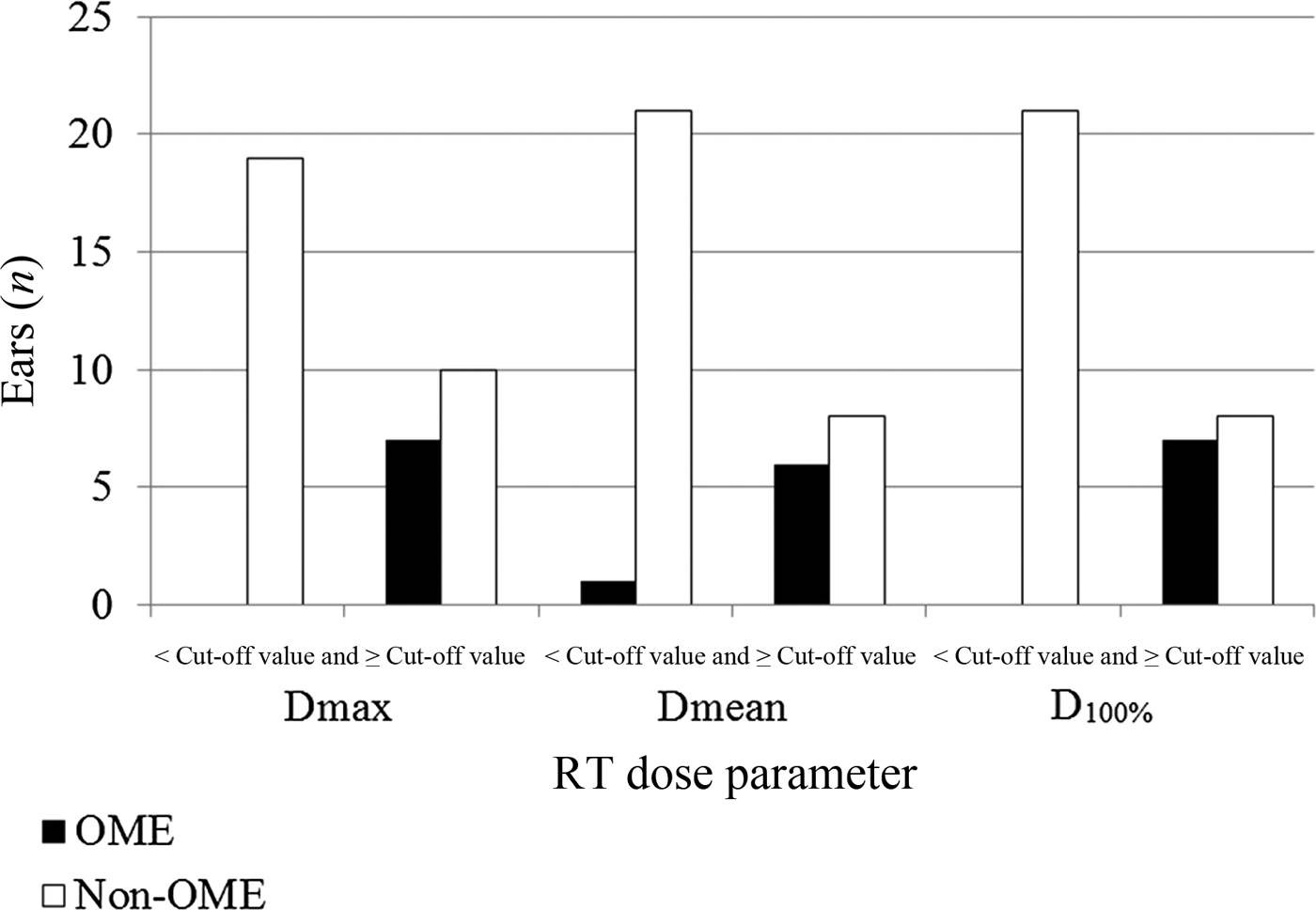
The median (range) maximum dose, mean dose and percentage volume covering 100 per cent of the prescribed dose for all Eustachian tubes were 55.7 Gy (13.6–71.7 Gy), 47.4 Gy (4.4–69.0 Gy) and 33.6 Gy (1.8–61.1 Gy), respectively. Significant differences in the radiation doses to the Eustachian tubes were observed between the ears with and without OME (Mann–Whitney U test, p < 0.01) (Table II). Cut-off values of 59.0 Gy, 54.5 Gy and 38.0 Gy were calculated for the maximum dose, mean dose and percentage volume covering 100 per cent of the prescribed dose, respectively. Notably, all but one ear with OME received a mean dose equal to or above the cut-off value (Figure 4).

Fig. 4 The otitis media with effusion (OME) and non-OME ears (36 ears) were divided into two groups according to the cut-off values of maximum dose (Dmax), mean dose (Dmean) and percentage volume covering 100 per cent of the prescribed dose (D100%). The x-axis indicates ears that received doses of less than, and equal to or greater than, these cut-off values (59.0 Gy, 54.5 Gy and 38.0 Gy, respectively). RT = radiotherapy
Table II Radiotherapy doses in ome and non-ome ears

Differences in the radiotherapy doses administered to the ears with and without otitis media with effusion (OME) were compared using the Mann–Whitney U test. *n = 7; † n = 29
Discussion
Otitis media with effusion (OME) has been linked to Eustachian tube dysfunction. This dysfunction is the result of a decrease in the gas exchange between the middle-ear cavity and mucous membrane microcirculation, which creates negative pressure in the middle-ear cavity, followed by the transudation of effusion.Reference Bluestone 3 Young et al. reported that the Eustachian tube function (based on passive opening pressure) deteriorated in 75 per cent and 83 per cent of ears at six months and five years after RT, respectively; however, these authors did not examine the radiation doses.Reference Young, Lin and Ko 9 In the present study, therefore, the first aim was to determine the radiation doses that cause Eustachian tube dysfunction. However, this aim was not achievable because Eustachian tube function deteriorated in almost all cases (88 per cent). Accordingly, the radiation doses for head and neck cancer were considered too high to allow a comparison between the normal and abnormal Eustachian tube function groups.
-
• Eustachian tube function deterioration is a main cause of otitis media with effusion (OME) after radiotherapy (RT)
-
• Sonotubometry is a simple, less invasive method for detecting physiological changes in Eustachian tube function
-
• Intensity-modulated RT can focus radiation on tumours, thus reducing adverse effects and controlling irradiation dose
-
• The threshold radiation dose for OME occurrence was defined based on an analysis of radiation doses to OME and non-OME ears
Sonotubometry is an objective method used to evaluate Eustachian tube function according to the SPL, and is based on the ventilation system in the middle ear, which is considered a function of the Eustachian tube. For this evaluation, the patient is instructed to insert the ear probe into the ear canal, place the nasal probe to the nostril and swallow. Of note, the Eustachian tube also mediates ciliary clearance, which is also damaged by RT, and can be measured by the saccharin test.Reference Ikehata, Ohta, Mishiro, Katsura, Miuchi and Tsuzuki 19 However, the saccharin test requires an ear perforation, which is not a characteristic of OME. Accordingly, sonotubometry, which can detect physiological changes in Eustachian tube function independently of tympanic membrane perforation, is more useful than other Eustachian tube function tests because of its simplicity and non-invasiveness.Reference Ohta, Sakagami, Suzuki and Mishiro 14
In the present study, the mean dose, maximum dose and percentage volume covering 100 per cent of the prescribed dose were used to evaluate the doses to the Eustachian tubes. To date, radiation doses to the Eustachian tube have been poorly understood. Previously, Emami et al. reported that doses of 30 Gy and 40 Gy were associated with 5 per cent and 50 per cent probabilities of acute serous otitis, respectively; they also suggested that doses between 30 Gy and 40 Gy might be safe for two-dimensional RT.Reference Emami, Lyman, Brown, Coia, Goitein and Munzenrider 20 More recently, Bhandare et al. reviewed the relationship between the cochlear dose volume and hearing loss,Reference Bhandare, Jackson, Eisbruch, Pan, Flickinger and Antonelli 21 but were unable to determine the doses to the Eustachian tubes.
Recently, intensity-modulated RT has played a significant role as the primary curative treatment for head and neck cancers because of better tumour target coverage and the sparing of normal tissue. Although Wang et al. reported that a radiation dose to the Eustachian tube isthmus, under an intensity-modulated RT protocol, with a total dose of 53 Gy, might decrease radiation-induced OME,Reference Wang, Li, Miyamoto, Chen, Zhou and Zhang 17 their study focused on nasopharyngeal cancer, which itself could obstruct the Eustachian tube. In contrast, a study by Upadhya et al., which demonstrated the ototoxicity (including OME) caused by radiation for head and neck cancers excluding nasopharyngeal cancer,Reference Upadhya, Jariwala and Datar 22 did not use intensity-modulated RT and the radiation dose to the Eustachian tube was not thought to be correct. Therefore, the present prospective study has two advantages: a focus on intensity-modulated RT and the exclusion of nasopharyngeal cancer from the head and neck cancer cases.
In our study, OME occurred in seven ears that were classified as in group B. Of these, five and two ears were classified as in groups A and B, respectively, before intensity-modulated RT, with five ears shifting to group B after intensity-modulated RT. The occurrence of OME in seven ears, all in group B, indicated Eustachian tube dysfunction just before the development of OME (Figure 4). This suggests that radiation-induced Eustachian tube dysfunction is closely related to the occurrence of OME.
Given the small size of the Eustachian tube and the risk of human error during contouring, even when using thin-slice CT, we evaluated the mean dose, maximum dose and percentage volume covering 100 per cent of the prescribed dose for the Eustachian tubes, to reduce inter-observer error. Notably, we observed significant differences in the radiation doses administered to OME and non-OME ears. The present data warrant the performance of further prospective studies to clarify the clinical benefit of a dose reduction to the Eustachian tube during intensity-modulated RT.
Our study had some limitations. For example, only a small number of cases were included, and the observation period was short. Furthermore, we did not analyse clinical stage, tumour size, chemotherapy or surgical treatment before intensity-modulated RT. However, to the best of our knowledge, this was the first prospective study to evaluate the relationship between Eustachian tube dysfunction and OME using sonotubometry, and the link between intensity-modulated RT radiation doses and OME, in patients with non-nasopharyngeal head and neck cancer.
Conclusion
We observed a positive relationship between radiation-induced Eustachian tube dysfunction and the occurrence of otitis media with effusion (OME). Furthermore, we used three dose–volume histogram parameters to determine the thresholds of radiation doses to the Eustachian tube that were predictive of the occurrence of OME in a population of patients treated with intensity-modulated RT for head and neck cancer. Further prospective studies are needed to clarify the clinical benefit of reducing the radiation dose to the Eustachian tube.


