


Introduction
Otological surgical practice may require drilling and exploration of the medial wall of the attic. Understanding the anatomical relationships and temporal bone pneumatisation in this region provides the basis for safe and thorough surgical exploration.
Categorisation of temporal bone pneumatisation has classically described five groups of air cell tracts, namely mastoid, perilabyrinthine, apical, peritubal and accessory.Reference Allam1–Reference Nadol and McKenna3
The perilabyrinthine air cell tract is encountered during surgical exploration of the medial attic wall. This group is bisected into separate areas of supralabyrinthine and infralabyrinthine tracts by the bony labyrinth. The supralabyrinthine area (see Figure 1) is further subdivided into posterosuperior, posteromedial and subarcuate tracts. The former two of these extend parallel to each other immediately posterior to the bony labyrinth adjacent to the junction of the middle and posterior fossa dura. The subarcuate tract runs through the arch of the superior semicircular canal.

Fig. 1 Diagram of the supralabyrinthine group of air cell tracts (left ear). Lateral, superior and posterior semicircular canals are labelled. The subarcuate tract (S) passes through the arch of the superior semicircular canal. The posteromedial (PM) and posterosuperior (PS) tracts run parallel to each other. The anterior supralabyrinthine tract (A) exists anterior to the ampullated end of the superior semicircular canal, and is bisected by the labyrinthine segment of the facial nerve (crossed dashed lines).
However, a fourth subset of the supralabyrinthine tract exists – the anterior supralabyrinthine air cells – located immediately anterior to the labyrinth in its superior aspect, and comprising much of the medial attic wall.
This anterior supralabyrinthine tract is less often mentioned in classical descriptions of temporal bone pneumatisation. However, in the clinical experience of the authors, this is an area that commonly requires surgical dissection when managing disease processes such as expansile attic cholesteatoma or irreversible mucosa disease.Reference Fisch4–Reference Sheahan and Walsh6 In addition, an intimate understanding of this region is required during surgical management of the facial nerve in cases of trauma or neurotological skull base surgery. Safe negotiation of this tract requires a working knowledge of the relationships between the ampullated end of the superior semicircular canal, the labyrinthine segment of the facial nerve, the geniculate ganglion and the adjacent middle fossa dura.
In this report, we aim to better clarify the surgically relevant relationships that comprise the limits of the anterior supralabyrinthine air cell tract.
Materials and methods
Surgical dissection of 10 fresh (non-preserved) cadaveric temporal bones was undertaken at the Clinical Training and Education Centre of the University of Western Australia in Perth, Western Australia, Australia. Needlepoint callipers were used to manually measure the distances between the anterior supralabyrinthine anatomical landmarks depicted in Figure 2, with the specific intent to define the triangle of bone between the tympanic facial nerve, labyrinthine facial nerve and superior semicircular canal. Distances were measured utilising microscopic viewing and a ruler calibrated with markings up to 0.5 mm. Points deemed to fall between 0.5 mm markings were recorded using 0.25 mm increments.

Fig. 2 Diagram of anterior supralabyrinthine space landmarks (left ear), with measurements in millimetres: point A (square), point B (star), point C (circle), measurement 1 (tympanic segment from anterior oval window to geniculate ganglion (GG)), measurement 2 (labyrinthine segment from GG to anterior superior semicircular canal (SSC)), measurement 3 (superior fallopian canal at anterior oval window to anterior SSC), measurement 4 (labyrinthine segment at anterior SSC to tegmen) and measurement 5 (superior fallopian canal at anterior oval window to tegmen). # = number; LSC = lateral semicircular canal
Measurement one (the tympanic facial nerve to the geniculate ganglion) was defined as the distance between the tympanic facial nerve at point A (defined by a line passing between the anterior margin of the vestibule and the anterior margin of the otic capsule of the superior semicircular canal) and point B (where the tympanic and labyrinthine aspects of the fallopian canal merge at the geniculate ganglion; this is a palpable, well defined bony landmark).
Measurement two (the geniculate ganglion to the visualised extent of the labyrinthine facial nerve) was defined as the distance between point B (the geniculate ganglion) and point C (where the labyrinthine segment of the facial nerve passes medial to the otic capsule of the superior semicircular canal, as viewed from the lateral vantage point defined by the orientation of the bony external auditory canal).
Measurement three (the distance between the tympanic and labyrinthine facial nerve segments) was defined as the distance between points A and C.
Measurement four (the tympanic facial nerve to the middle fossa dura) was defined as the distance from point A to a line extending perpendicular to the middle fossa dura.
Measurement five (the labyrinthine facial nerve to the middle fossa dura) was defined as the distance from point C to a line extending perpendicular to the middle fossa dura.
We calculated the bony triangle area and angles (including the angle formed by the junction of the labyrinthine and tympanic segments of the facial nerve as they meet at the geniculate ganglion) based on measurements one to three.
Results
Mean measurements are given in Table I.
Table I Supralabyrinthine anatomical dimensions*

* In 10 fresh cadaveric temporal bones (see Figure 2).
The triangle of bone defined by measurements one to three had angles of 38.5° (at point A), 68.4° (at point B) and 73.1° (at point C), and an area of 3.87 mm2.
Discussion
Superior semicircular canal
In our practice, the first step in drilling the medial wall of the attic involves establishing the posterior boundary of the anterior supralabyrinthine air cell tract, by identifying the anterior margin of the otic capsule of the ampullated end of the superior semicircular canal. We have identified a simple ‘rule of thumb’ which acts as a rough guide in undertaking this step: follow a line parallel to the proximal aspect of the vertical segment of the facial nerve, which passes through the tip of the pyramidal process (see Figure 3). Although confirmation of this guideline was not a formal part of the present study, we did nevertheless find it to be fairly consistent in all 10 specimens dissected.

Fig. 3 Simple method to identify the anterior border of the ampullated end of the superior semicircular canal (SSC) otic capsure (left ear): extend a line (dotted line) parallel to the proximal aspect of the vertical segment of the facial nerve (CNVII; dashed line), which passes through the tip of the pyramidal eminence (triangle).
However, simple rules such as this should never replace a surgeon's skilled, careful identification of the superior semicircular canal bone in each individual case, as anatomical variants may exist.
Geniculate ganglion
The next step involves identification of the geniculate ganglion, which is located medial to the anterior attic wall immediately adjacent to the cog. The geniculate ganglion forms the anterior boundary of the anterior supralabyrinthine air cell tract. The cog (comprising the medial aspect of the petro-squamous lamina) is encountered as a variably sized shelf of bone protruding inferiorly from the tegmen tympani, which when prominent may form a marker of separation between the anterior attic wall and the more anteriorly situated epitympanic sinus or cell. The tensor tympani tendon and cochleariform process are also helpful as landmarks, situated just posterior and inferior to the geniculate ganglion, to be used when tracing the tympanic segment of the facial nerve to the geniculate ganglion.
According to the findings of this study, the tympanic–geniculate junction is encountered when proceeding an average of 3.58 mm anterior along the tympanic segment of the facial nerve, measured from the anterior margin of the oval window niche.
Facial nerve
As with most tympanomastoid surgical dissections, an awareness of the facial nerve is critical in order to safely explore the anterior supralabyrinthine air cell tract. The anatomy of the tympanic segment of the facial nerve should be intimately familiar to any competent otological surgeon.
However, dissection and identification of the distal labyrinthine segment of the facial nerve is not a frequent undertaking for most otological surgeons. The labyrinthine segment of the facial nerve is defined as the nerve segment running between the exit point from the internal auditory canal, via the meatal foramen, into the fallopian canal and extending to the geniculate ganglion. Its average total length has been described as approximately 4 mm. However, in our anterior supralabyrinthine air cell tract dissection, the average visible length of this segment, between the geniculate ganglion and the anterior aspect of the bony superior semicircular canal, was only 2.33 mm.
In the course of drilling the anterior supralabyrinthine air cell tract and dissecting the labyrinthine segment of the facial nerve, a well circumscribed ‘anterior supralabyrinthine triangle’ of bone comes into view between the tympanic and labyrinthine segments (see Figure 4). One of the primary purposes of the present study was to assess the average dimensions of this triangle. Although not visualised in this study, it is important to note that Bill's bar exists as a posteromedial projection from the above-defined triangle, lying between the superior ampullary nerve and the facial nerve.

Fig. 4 Diagram of the anterior supralabyrinthine triangle (left ear): a distinct, triangular segment of bone identifiable between the tympanic and labyrinthine segments of the facial nerve. The sharply defined apex of this triangle lies adjacent to the geniculate ganglion (asterisk).
Our data depict a triangle with a mean area of 3.87 mm2, with several clinically relevant elements that can aid identification of the labyrinthine segment.
First, the mean distance between the medial aspect of the tympanic fallopian canal (at the level of the anterior oval window niche, point A) and the labyrinthine facial nerve adjacent to the superior semicircular canal (measurement three) was 3.48 mm. Thus, from point A, the distances to the geniculate ganglion and to the labyrinthine segment at the superior canal are roughly equal (3.48 versus 3.58 mm, respectively).
Second, the mean angle between the course of the tympanic segment and the labyrinthine segment, as they join the geniculate ganglion, was 68°. Because the usual surgical viewpoint is from the oblique lateral, which is not perpendicular to the plane of the measured bony triangle, this angle will appear more acute when viewed intra-operatively.
Tegmen tympani
The tegmen tympani forms the superior boundary of the anterior supralabyrinthine air cell tract. We found the mean distance between the tympanic and the labyrinthine facial nerve segments and the middle fossa dura to be quite small (2.94 and 5.25 mm, respectively). This has clinical relevance in understanding the potential for communication of the anterior supralabyrinthine air cell tract with the petrous apex in cases of infection or cholesteatoma. When compared with the infracochlear and infralabyrinthine approaches to the petrous apex, a transmastoid anterior supralabyrinthine approach between the labyrinthine facial nerve and tegmen tympani would afford limited petrous apex access.
General discussion
These study findings are presented in order to define more clearly the surgical anatomy of the anterior supralabyrinthine air cell tract, and to provide some general guides for surgical dissection.
In doing so, we acknowledge the limitations and margin for error inherent in an anatomical exercise of this nature, involving direct manual measurement of fresh cadaveric temporal bone specimens. Specifically, the absence of a strictly defined lateral axis to more exactly define point C is acknowledged as a potential source of slight error, as the amount of labyrinthine facial nerve on view at this point will vary depending on the anterior-posterior orientation and rotation of the specimen. We also acknowledge that our study findings are based on a limited sample of 10 temporal bones.
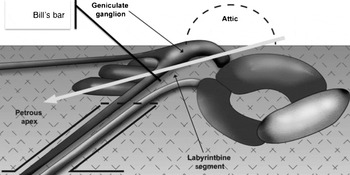
Fig. 5 Diagram of anterior supralabyrinthine air cell tract communication with petrous apex (middle fossa view, right ear). White arrow indicates the route of communication between the anterior supralabyrinthine air cell tract and the petrous apex.
If this effort was intended strictly for the purpose of defining human anatomy, then these limitations would be of concern. However, when viewed within the context of providing basic surgical anatomical guidelines, we believe that such limitations are unlikely to have any major influence on the clinical utility of our study findings.
• In addition to the subarcuate, posteromedial and posterosuperior supralabyrinthine air cell tracts, an important anterior supralabyrinthine air cell tract exists
• The mean distances from the geniculate ganglion to the facial nerve tympanic segment (adjacent to the anterior margin of the oval window niche) and to the facial nerve labyrinthine segment (adjacent to the superior semicircular canal) are both approximately 3.5 mm
• A distinct anterior supralabyrinthine bony triangle exists between the tympanic and labyrinthine segments of the facial nerve, the geniculate ganglion, and the superior semicircular canal, knowledge of which is helpful when drilling the medial attic wall
As noted previously, the anterior supralabyrinthine air cell tract is often omitted from classical descriptions of temporal bone pneumatisation. We find this surprising, given this structure's high degree of clinical relevance with respect to surgical management of chronic otitis media and cholesteatoma. Furthermore, understanding the anatomical relationships of the anterior supralabyrinthine region may be important in other surgical scenarios, such as traumatic facial nerve injury and neurotological skull base surgery, which involve posterior re-routing of the facial nerve.
Conclusion
The surgical anatomy of the anterior supralabyrinthine air cell tract can be defined in terms of a triangle of bone located between the ampullated end of the superior semicircular canal, the labyrinthine and tympanic segments of the facial nerve, and the geniculate ganglion. Understanding the dimensions of this triangle may assist the otological surgeon in safely dissecting this region.
Acknowledgements
The authors would like to acknowledge the staff of the Clinical Training and Education Centre at the University of Western Australia for their help in facilitating the execution of this study.


