


Introduction
Micarea Fr. is a genus of well in excess of 100 crustose lichen species (Czarnota Reference Czarnota2007; International Mycological Association 2019) found across a wide range of habitats and substrata in boreal, austral, temperate and tropical regions of the world. Characters that define the genus include its green photobiont comprising small, often-paired cells, commonly referred to as ‘micareoid’, the often-reduced apothecial excipulum, the 8-spored, Pilocarpaceae-type asci where the well-developed, amyloid tholus is penetrated by a narrow channel, with or without a darker staining tube structure, and simple to branched paraphyses; the hyaline ascospores vary from filiform to fusiform to ovoid-ellipsoid, and from simple to transversely multiseptate. The taxonomic history of Micarea has been summarized by several authors, for example Czarnota (Reference Czarnota2007) and Ekman & Svensson (Reference Ekman and Svensson2014). The modern framework for the species-rank taxonomy of Micarea was essentially erected by Coppins (Reference Coppins1983) in his review of European taxa, and was based largely on thallus chemistry, apothecial anatomy and pigmentation, and ascospore and conidial characters. This general architecture has been maintained by subsequent authors, both in regional revisions (e.g. Czarnota Reference Czarnota2007; Fryday & Coppins Reference Fryday, Coppins, Nash III, Gries and Bungartz2007; Brand et al. Reference Brand, van den Boom and Sérusiaux2014) and in investigations of particular groups within Micarea (e.g. Czarnota & Guzow-Krzemińska Reference Czarnota and Guzow-Krzemińska2010; Guzow-Krzemińska et al. Reference Guzow-Krzemińska, Czarnota, Łubek and Kukwa2016; Launis et al. Reference Launis, Pykälä, van den Boom, Sérusiaux and Myllys2019a). Coppins (Reference Coppins1983) had already recognized that Micarea included several, well-defined species groups, some of which, such as Psilolechia (Coppins & Purvis Reference Coppins and Purvis1987), were worthy of generic rank. Supported by anatomical, morphological and, more recently, DNA-sequence data, this has seen the segregation out of Micarea of Szczawinskia (Funk Reference Funk1983), Brianaria (Ekman & Svensson Reference Ekman and Svensson2014) and Leimonis (Harris Reference Harris2009). However, the genus remains a challenge. New species continue to be recognized from Europe, where most of the detailed research on Micarea has been undertaken (e.g. Coppins Reference Coppins1988, Reference Coppins1995; Coppins & Tønsberg Reference Coppins and Tønsberg2001; Czarnota & Coppins 2005; Coppins & Aptroot Reference Coppins and Aptroot2008; Svensson & Thor Reference Svensson and Thor2011; van den Boom et al. Reference van den Boom, Brand, Coppins and Sérusiaux2018; Launis et al. Reference Launis, Pykälä, van den Boom, Sérusiaux and Myllys2019a), as well as from poorly studied, remote regions (e.g. Coppins Reference Coppins1999; Fryday Reference Fryday2004; Sérusiaux & Coppins Reference Coppins, Smith, Aptroot, Coppins, Fletcher, Gilbert, James and Wolseley2009; Cáceres et al. Reference Cáceres, Mota, de Jesus and Aptroot2013; Brand et al. Reference Brand, van den Boom and Sérusiaux2014).
In Australia, Micarea remains one of the largest, poorly known groups of lichens. Herbaria in all jurisdictions tend to hold extensive, unidentified collections, frequently filed under old ‘Zahlbruckner form genera’ such as Lecidea, Bacidia and Catillaria. At the same time, Micarea itself is often treated as a ‘dustbin’ for small crustose lichens that belong in other genera. Novel species have been published by Stirton (Reference Stirton1876), Jatta (Reference Jatta1911), Coppins & Kantvilas (Reference Coppins and Kantvilas1990), McCarthy & Elix (Reference McCarthy and Elix2016a, Reference McCarthy and Elixb), Elix & McCarthy (Reference McCarthy2018) and Kantvilas (2018b). In addition, numerous chiefly Northern Hemisphere taxa have been recorded for the region (e.g. Rambold Reference Rambold1989; Coppins Reference Coppins, Smith, Aptroot, Coppins, Fletcher, Gilbert, James and Wolseley2009; Elix et al. Reference Elix, McCarthy and Kantvilas2009; Elix Reference Elix2012; Kantvilas et al. 2014; McCarthy Reference McCarthy2015). McCarthy (Reference McCarthy2018) lists 26 species for Australia (including Tasmania), but this is hardly representative of what is clearly a very species-rich genus.
The origins of the present study stem from the 1990s when the authors became aware of the diversity of Micarea in Tasmania's moist, cool areas of wet forest, moorland and alpine vegetation (Coppins & Kantvilas Reference Coppins and Kantvilas1990). Subsequent, mainly floristic and ecological research in these communities (Jarman & Kantvilas Reference Jarman and Kantvilas1995, Reference Jarman and Kantvilas1997; Kantvilas & Jarman Reference Kantvilas and Jarman2012) revealed numerous clearly novel species. The present paper summarizes the current state of knowledge of Micarea in Tasmania, dealing with 35 species, ten of them new to science, as well as taxa from the genera Brianaria, Leimonis and Psilolechia which have been, or may potentially be, confused with Micarea.
Material and Methods
The study is based mainly on specimens held in the Tasmanian Herbarium (HO), supplemented by collections from BM, BG, E, UPS and the private herbaria of K. Kalb and A. Aptroot. Anatomical observations were made on hand-cut sections mounted in water, 10% KOH (K), 50% HNO3 (N), commercial bleach solution (C) or Lugol's Iodine (I). However, in cases where additional critical observations were required to observe paraphyses, excipular hyphae or conidia, lactophenol cotton blue, ammoniacal erythrosin and Powell's ‘vinegar ink’ (Powell Reference Powell2018) mounting media were also employed. Thin-layer chromatography (TLC) was undertaken using standard methods; solvent A was the preferred medium for routine analyses (Orange et al. Reference Orange, James and White2010). High-performance liquid chromatography (HPLC) of a small number of critical specimens was undertaken by Prof J. Elix, Canberra (Elix et al. Reference Elix, Giralt and Wardlaw2003). Nomenclature and interpretation of ascus structure, determined by staining apothecial sections with Lugol's Iodine after pretreatment with dilute KOH, essentially follows Hafellner (Reference Hafellner1984) and Ekman et al. (Reference Ekman, Andersen and Wedin2008). Nomenclature of pigments follows Meyer & Printzen (Reference Meyer and Printzen2000). Ascospore measurements in the descriptions of new species are given in the format: 5th percentile–average–95th percentile, with extreme values given in brackets and n being the number of observations. For taxa where published descriptions already exist, ascospore measurements are derived solely from Tasmanian specimens studied. Most photographs were prepared by Dr J. Jarman, Hobart, and are attributed in the captions. Remaining photographs and drawings (unattributed) were prepared by GK.
Ecology
With the exception of living leaves and man-made substrata such as concrete, glass, rubber and metal, species of Micarea in Tasmania are capable of colonizing almost any surface. These include solid or partially rotting dead wood, bark of all types, including flaky, smooth, papery or fibrous, consolidated soil or peat, and most rock types but essentially excluding limestone. Some species overgrow bryophytes or loose plant litter. Micarea can occur in locally moist and exposed habitats, or in very dry and sheltered sites such as rocky underhangs or the undersides of leaning trees. It ranges from lowland to alpine elevations, although no species are known from littoral rocks. Distinct ecological patterns can be seen in Tasmania's Micarea biota (cf. Brand et al. (Reference Brand, van den Boom and Sérusiaux2014) for Réunion). The following discussion classifies the better-known species where consistent distribution trends can be discerned.
Wet forest species
These are species occurring in old forests as well as species confined to old (or physically massive) trees within such forests (Kantvilas & Jarman Reference Kantvilas and Jarman2004). The climax forest type for much of the high rainfall parts of Tasmania is cool temperate rainforest (Jarman et al. Reference Jarman, Kantvilas and Brown1994) but many wet eucalypt forests, where old-growth characteristics are evident, are also included here. This vegetation is the stronghold of Micarea in Tasmania. Species whose occurrence is intrinsically linked to old trees (or large logs and stumps of old trees) in such forests include M. byssacea, M. ceracea, M. cinereopallida, M. mutabilis, M. pallida, M. prasinastra and M. rubiginosa. Micarea alabastrites, M. cinerea and M. tubaeformis are also included here, although they are generally restricted to younger hosts with smooth bark in such forests. Micarea saxicola also occurs in wet forests but colonizes rocks and is seemingly restricted to (or best developed in) disturbed sites beneath a broken canopy.
Alpine vegetation and wet moorland or heathland
Peaty soil in such vegetation supports M. isabellina and M. magellanica, both of which are important in soil stabilization. Micarea micromelaena occurs directly on rocks and small pebbles of Precambrian metamorphosed sediments such as quartzite, whereas M. oreina is found mostly on Jurassic dolerite. Soil, rocks, small herbs and bryophytes, and plant detritus are often colonized by M. flagellispora and M. melaena. The interface between wet, scrubby or heathy vegetation and wet forests can be dynamic and determined by fire and topographic and edaphic factors. Thus, some essentially moorland taxa such as M. isabellina may be found in rainforests, and typical forest taxa such as M. tubaeformis may occur in heathland.
Dry open eucalypt woodland and grassland
Although disturbance, particularly from fire but also from low-level human activities such as grazing and fire-wood cutting, is frequent in dry, open eucalypt-dominated woodlands, they may nevertheless display some ‘old-growth’ characters and support a significant complement of Micarea species. Consolidated soil in gaps amongst grasses and forbs may be colonized by M. humilis, M. incrassata, M. aff. lapillicola and M. melaenida, whereas M. almbornii and M. argopsinosa appear to prefer elevated soil around the root mounds of upturned trees. The wood of Eucalyptus is long lasting, and the forest floor of dry sclerophyll forests tends to be littered with logs, stumps and fallen limbs, all of which provide potentially good Micarea habitat. Species in such habitats include M. contexta, M. denigrata, M. intersociella and M. peliocarpa.
Ecological changes and Micarea
A study of lichens in wet eucalypt-dominated forests (Jarman & Kantvilas Reference Jarman and Kantvilas2001a, Reference Jarman and Kantvilasb), and the recovery of species following logging and regeneration under different silvicultural treatments (Kantvilas & Jarman Reference Kantvilas and Jarman2006; Kantvilas et al. Reference Kantvilas, Jarman and Minchin2015), offered an informative case study of Micarea ecology. These forests proved to be particularly rich in Micarea, with a total of 18 species recorded in the course of a 13-year study (Kantvilas & Jarman Reference Kantvilas and Jarman2012). The following eleven species were recorded in unlogged forest: M. alabastrites, M. byssacea, M. ceracea, M. cinerea, M. cinereopallida, M. mutabilis, M. prasinastra, M. rubiginosa, M. tubaeformis, M. viridileprosa (all corticolous on trees, stumps or logs) and M. myriocarpa (on soil in sheltered underhangs). Within 3–5 years of timber harvesting and regeneration (usually involving burning), the following eight species were recorded: M. denigrata, M. intersociella and M. nowakii (on stumps and logs), with some surviving M. byssacea (stumps and logs); M. deminuta and M. melaenida (on bare soil or wood fragments on the ground); M. farinosa (on soil in underhangs) and M. peliocarpa (on wood and soil). Thus, with the exception of M. byssacea, which appears to be a relatively ecologically robust species, all the species of Micarea from unlogged forest were replaced by others, suggesting that Micarea presents an excellent subject for ecological studies.
Pigments
Apothecial pigments are critical in characterizing Micarea species. Their importance was already recognized by Coppins (Reference Coppins1983) who categorized and discussed the major pigments in European species, based on their reactions in dilute KOH (K) and HNO3 (N). This approach was extended and applied in later regional revisions of Micarea (e.g. Czarnota Reference Czarnota2007; Brand et al. Reference Brand, van den Boom and Sérusiaux2014). Ekman (Reference Ekman1996) and Meyer & Printzen (Reference Meyer and Printzen2000) sought to formalize the nomenclature of the pigments and to standardize their reactions, and their scheme has proved very useful in taxonomic studies of many groups of lichens. For example, in Australia they have helped to clarify genera as diverse as Mycoblastus (Kantvilas Reference Kantvilas2009), Megalaria (Kantvilas Reference Kantvilas2016) and Bacidia (Kantvilas Reference Kantvilas2018a).
Interpretation and identification of pigments can be difficult. Colour reactions are influenced by concentration and, more importantly, by whether other pigments are present. The outcome of the blending of different pigments is akin to the blending of primary colours to produce a range of tones in, for example, water-colour painting. Even more complex are the personal interpretations of colour that different individuals may have, or the nuances of their microscope (e.g. whether any filters are installed). The colours are rarely pure, such as ‘red’, ‘purple’ or ‘green’ but are a mixture of shades. Terms such as ‘purple-brown’, ‘dull brownish’, ‘crimson-red’ or ‘bluish green’ tend to be far more realistic, but at the same time they can also be highly subjective, even though published species descriptions frequently tend to present them as absolute character states.
Through the careful use of standard reagents, some pigments can be easy to detect. For example, greenish blue pigments rarely pose a challenge because, even in minute concentrations, they will react a crimson reddish in N, whether or not other pigments are also present. However, greater problems are posed by the many brownish pigments in particular, where the colour reactions may be manifest as an intensification of a particular hue rather than a distinct change of colour.
A relatively simplified scheme as applied in the present study of Tasmanian Micarea species is summarized in Table 1, based chiefly on the nomenclature of Meyer & Printzen (Reference Meyer and Printzen2000). Epithecial pigments rarely form a distinct layer but tend to diffuse down between the asci and paraphyses to varying degrees. Likewise, hypothecial pigments may diffuse upwards into the lower hymenium or laterally into the excipulum. Intensity of pigmentation can vary in some species, often in response to the degree of exposure to sunlight, whereas in other species it varies little, regardless of habitat or age. The main pigments seen are listed below.
Atra-red: pinkish or crimson-red in water, intensifying in both K and N.
Cinereorufa-green: ranging from intense greenish blue to greyish green or greyish depending on concentration. In K it may barely change or intensify greenish, but it reacts N+ crimson-red, even in minute concentrations.
Laurocerasi-brown: brown in water but with subtle purplish hues that intensify in K; it turns an orange-brown in N. This pigment can be difficult to interpret when present in low concentrations or occurring together with other pigments.
Melaena-red: purplish or purplish brown in water and could be confused with Laurocerasi-brown, except that it reacts greenish, greenish grey or greenish black in K, depending on concentration; it reacts orange-brown in N.
Melaenida-red: reddish to orange-brown, intensifying reddish or orange in both K and N.
Rubella-orange: orange-red, intensifying orange in K and C (Ekman Reference Ekman1996).
Sedifolia-grey: grey or greyish green, readily identified by the unequivocal K+ violet, C+ violet reaction.
Table 1. Disposition of the main apothecial pigments in Tasmanian species of Micarea.

Key to Tasmanian species of Micarea and other micareoid lichens
1 Ascospores narrowly ellipsoid to fusiform, filiform or acicular, mostly 3- or more septate when mature.……… 2
Ascospores ovoid to oblong or ellipsoid, simple or 1-septate when mature. ………16
2(1) Ascospores acicular to filiform. ………3
Ascospores ellipsoid to fusiform. ………6
3(2) Hypothecium opaque blackish green in section; ascospores filiform. ………4
Hypothecium hyaline or dilutely variously pigmented but never dark and opaque; ascospores acicular. ………5
4(3) Thallus areoles 100–400 µm diam., containing perlatolic acid; pycnidia immersed. ……… Micarea flagellispora
Thallus areoles granular, mostly 50–140 µm wide, containing 2´-O-superlatolic acid; pycnidia conspicuous, trumpet-shaped and resembling the fruiting body of a Calicium. ……… Micarea tubaeformis
5(3) Thallus and apothecia C−; apothecia dark grey to black, with the epithecium C+ violet, K+ violet, N+ violaceous grey (Sedifolia-grey); ascospores curved, indistinctly 3-septate, 10–25 µm long. ……… Micarea intersociella
Thallus and apothecia C+ red (containing gyrophoric acid); apothecia pallid to grey to grey-brown or blackish, often piebald; epithecium C−, K± greenish, N+ crimson-red; ascospores ±sigmoid, mostly 5–7-septate, 21–50 µm long.……… Micarea mutabilis
6(2) Thallus and apothecia in section C+ red (containing gyrophoric acid). ……… 7
Thallus and apothecia C− or C+ pink or orange (alectorialic acid or xanthones)………10
7(6) Thallus sorediate. ……… Micarea pseudocoppinsii
Thallus not sorediate. ……… 8
8(7) Ascospores (3–)7-septate, 24–38(–40) μm long. ……… Micarea cinerea
Ascospores 3(–5)-septate, to 24 µm long. ……… 9
9(8) Apothecia entirely whitish, lacking any dark pigments. ………Micarea alabastrites
Apothecia usually partly or entirely dark greenish, grey or grey-black; pigment N+ crimson-red ……… Micarea peliocarpa
10(6) Apothecia pallid, in section entirely ±hyaline or at most patchily pale brownish. … 11
Apothecia black-grey or mottled greenish, in section with dark pigments in the epithecium and/or hypothecium. ……… 12
11(10) Thallus smooth and waxy, containing perlatolic acid; apothecia inspersed with crystals; ascospores 10–21 × 3·5–6 µm. ……… Micarea ceracea
Thallus composed of discrete or subcontiguous areoles, containing porphyrilic acid; apothecia not inspersed; ascospores 9·5–15 × 2·5–4 µm. ……… Micarea pallida
12(10) Hypothecium opaque purplish brown in section. ……… Micarea melaena
Hypothecium hyaline or dilutely variously pigmented but never opaque. ………13
13(12) Ascospores to 30 µm long; thallus spot tests with K and KC yielding positive reactions ………14
Ascospores never exceeding 15 µm in length; thallus K−, KC−………15
14(13) Thallus pale greenish or grey, P+ yellow, KC+ orange-red, C+ pale pink, UV− (containing alectorialic acid). ……… Micarea magellanica
Thallus pale yellowish, P−, KC+ orange, C+ orange, UV+ orange-pink (containing xanthones). ……… Micarea isabellina
15(13) Thallus dull brownish grey, rimose-areolate to rather gnarled, lacking lichen substances and all spot tests negative. ……… Micarea sandyana
Thallus pale greyish green, composed of rather thin, diffuse areoles, P+ orange (containing argopsin). ……… Micarea argopsinosa
16(1) Apothecia with the upper hymenium olivaceous green to olivaceous grey, C+ violet, K+ violet (Sedifolia-grey). ……… 17
Apothecia lacking Sedifolia-grey pigment. ……… 20
17(16) Thallus always on soil; ascospores simple, broadly ellipsoid or subglobose, mostly >4 µm wide. ……… Micarea almbornii
Thallus typically occurring on wood or bark; ascospores (0–)1-septate, ellipsoid to oblong-ellipsoid or oblong-ovoid, mostly <4 µm wide. ………18
18(17) Thallus and apothecia C+ red in squash preparation (containing gyrophoric acid). Micarea denigrata
Thallus and apothecia C− (gyrophoric acid lacking). ……… 19
19(18) Thallus consisting of grey to blackened, gnarled areoles containing micareic acid; apothecia black; ascospores 6–10 × 2–3 µm. ……… Micarea nowakii
Thallus typically green and goniocyst-like, containing methoxymicareic acid; apothecia mostly pale to mottled grey to dark grey; ascospores (7–)8–14 × 2·5–4(–5) μm. ……… Micarea byssacea
20(16) Growing in dry, sheltered situations such as on soil and stones in underhangs, or on wood or bark of very old trees. ……… 21
Growing in moist or exposed habitats on trees, logs, rocks or soil. ……… 29
21(20) Thallus and apothecia vivid lemon yellow or yellow-green. ………Psilolechia lucida
Thallus grey, greenish, whitish or highly reduced; apothecia pinkish, pale brown, greenish, grey, brown-black or black. ……… 22
22(21) Apothecia in section entirely unpigmented. ……… 23
Apothecia in section with the epithecium and/or hypothecium pigmented, sometimes only dilutely. ……… 25
23(22) Containing gyrophoric acid (thallus and apothecia usually C+ weak red, but spot tests often unreliable); occurring exclusively on the bark of very old trees. ……… Micarea prasinastra
Gyrophoric acid lacking; typically found on soil and stones. ……… 24
24(23) Ascospores ovoid-ellipsoid, 5–7·5 × 2·3–3·5(–4) μm. ……… Micarea farinosa
Ascospores tear-shaped, 4–8 × 1·2–2·5 µm. ………Psilolechia lucida (pale forms)
25(22) Epithecium unpigmented; hypothecium pale to dark brown. Micarea myriocarpa
Epithecium greenish or purple-brown; hypothecium greenish, purple-brown or unpigmented. ……… 26
26(25) Photobiont Stichococcus, with oblong, brick-like cells in short chains; asci of the Porpidia-type; ascospores tear-shaped; hypothecium colourless or at most dilutely pigmented. ……… 27
Photobiont chlorococcoid, with globose cells; asci of the Psora-type; ascospores ellipsoid to ovoid; hypothecium opaque. ……… 28
27(26) Apothecial pigment greenish, N+ crimson-red. ……… Psilolechia clavulifera
Apothecial pigment purplish brown, greenish, N+ crimson-red pigment lacking. ……… Psilolechia purpurascens
28(26) Apothecia to 0·5 mm diam.; ascospores (2 5–)3·5–4 µm wide Brianaria sylvicola
Apothecia to 0·3 mm diam.; ascospores 2–2·5(–3) μm wide Brianaria tuberculata
29(20) Apothecia whitish, pale beige-brown to orange-red, sometimes mottled bluish green. ……… 30
Apothecia dark brown, grey-black or black, sometimes mottled with these tones. 33
30(29) Apothecia orange-red; hypothecium dilute orange to red-brown, K+ dilute yellow. ……… Micarea rubiginosa
Apothecia pallid or in part mottled bluish green; hypothecium hyaline to dilute straw-coloured, K−.……… 31
31(30) Thallus and/or apothecia C+ red in squash preparation (containing gyrophoric acid). ……… Micarea viridileprosa
Thallus and/or apothecia C−, lacking gyrophoric acid. ……… 32
32(31) Thallus UV±whitish (containing superlatolic acid); goniocysts granular to coralloid with a glossy, crystalline appearance, often translucent or flecked with a bluish green, N+ crimson-red pigment; prothallus blackish to dark blue-grey, visible at the thallus margins and between thalline granules; apothecia markedly basally constricted, whitish to bluish grey or mottled, internally inspersed with minute crystals that dissolve in K. ……… Micarea cinereopallida
Thallus UV− or weakly UV+ orange (containing traces of xanthones or unknown substances), lacking a prothallus; goniocysts dull, coarsely granular; apothecia pallid, adnate, internally not inspersed. ……… Micarea prasina s. lat.
33(29) Hypothecium hyaline or at most dilutely brownish or greenish. ……… 34
Hypothecium pigmented brown, greenish or purple-brown, frequently opaque. ……… 35
34(33) Thallus composed of globose, dull greyish areoles, C+ red (gyrophoric acid); apothecia soon immarginate; ascospores 1-septate, 11–15(–16·5) μm long; on alpine boulders ……… Micarea oreina
Thallus continuous, brownish grey, C−; apothecia distinctly and persistently marginate; ascospores simple, 6–10 µm long; on consolidated soil in lowland forest. ……… ………Micarea aff. lapillicola
35(33) Thallus effuse, granular-areolate, becoming sorediate, containing gyrophoric acid (thallus C+ red); apothecia ±shortly stalked, internally with reddish or reddish brown pigments. ……… Micarea prasinella
Thallus not sorediate, lacking gyrophoric acid (C−); apothecia plane to subglobose, not stalked, internally with brownish or greenish pigments. ……… 36
36(35) Thallus corticolous or lignicolous. ……… 37
Thallus saxicolous or terricolous. ……… 38
37(36) Ascospores simple and ellipsoid; apothecial pigments mainly brown. ……… ………Micarea deminuta
Ascospores 1-septate, ovoid to soleiform; apothecial pigments mainly greenish. ……… ……… Micarea contexta
38(36) Saxicolous, growing directly on rock. ……… 39
Terricolous, on consolidated soil, peat or decomposing plant material. ……… 41
39(38) Thallus conspicuous, composed of contiguous, dull grey-brown areoles; cephalodia present, visible as darker brown areoles; apothecia containing only red-brown, K−, N+ orange-brown pigment in the hypothecium. ……… Micarea saxicola
Thallus inconspicuous; cephalodia absent; hypothecium containing both brown and greenish pigments. ……… 40
40(39) Photobiont micareoid, with cells 4–6(–10) μm wide; apothecia strongly convex and immarginate; ascospores 0–1-septate, 8–12(–12·5) × (2·5–)3–4 µm. ……… ………Micarea micromelaena
Photobiont non-micareoid, with cells to 8–16(–20) μm wide; apothecia typically with a persistent, conspicuous margin and remaining plane or only slightly convex; ascospores simple, (6–)8–11 × 3·5–5 µm. ……… Leimonis erratica
41(38) Apothecia to 0·3 mm wide; ascospores simple, 6–10(–11·5)μm long. ……… ………Micarea deminuta
Apothecia generally larger, to 0·5(–1) mm wide; ascospores (0–)1-septate, 9–21 µm long. ……… 42
42(41) Apothecial pigments entirely brownish, N+ orange-brown; greenish, N+ crimson-red pigment absent. ……… Micarea melaenida
Greenish, N+ crimson-red pigment present in the epithecium. ……… 43
43(42) Thallus composed of greyish, convex areoles to 0·3 mm wide; hypothecium orange-brown, K−. ……… Micarea incrassata
Thallus scurfy and inconspicuous; hypothecium dark reddish brown, K+ purplish brown. ……… 44
44(43) Occurring on soil in lowland grassland and dry sclerophyll forest; ascospores (0–)1-septate, 9–14 × 3·5–5 µm. ……… Micarea humilis
Occurring on peaty soil and decomposing plant material at high elevations in moorland and heathland; ascospores 12–22(–25) × 4–6 µm, mostly 1-septate but with simple or 2–3-septate ascospores sometimes present. ……… Micarea melaena s. lat.
The Species
Micarea alabastrites (Nyl.) Coppins
In Topham & Walker, Lichenologist 14: 66 (1982).
This species is widespread in the temperate Northern Hemisphere (Coppins Reference Coppins, Smith, Aptroot, Coppins, Fletcher, Gilbert, James and Wolseley2009) and locally abundant in Tasmanian wet eucalypt forest and cool temperate rainforest (Jarman & Kantvilas Reference Jarman and Kantvilas2001a; Kantvilas & Jarman Reference Kantvilas and Jarman2012) where it occurs on the bark of a wide variety of understorey shrubs and trees. It is also recorded here for Victoria for the first time. It is characterized by a pale grey-green thallus of thin, contiguous or dispersed, subglobose to plane areoles, whitish to ±translucent apothecia with a rather well-developed exciple (visible under low-power magnification as a whitish rim surrounding the disc in young, plane apothecia), and 3(–5)-septate, fusiform to clavate-fusiform ascospores, 13–25 × 3–5 µm (Tasmanian specimens). It contains gyrophoric acid, which can be detected by the C+ red reaction of the apothecia in section. Detailed descriptions are provided by Coppins (Reference Coppins1983, Reference Coppins, Smith, Aptroot, Coppins, Fletcher, Gilbert, James and Wolseley2009).
Micarea alabastrites is closely related to M. cinerea and M. peliocarpa, and all three species have C+ red apothecia and generally fusiform ascospores with three or more transverse septa. Their differences are discussed in detail by Coppins (Reference Coppins1983). Whereas M. cinerea is readily distinguished by its larger, more septate ascospores and occasionally dark-pigmented apothecia, the distinction between M. alabastrites and M. peliocarpa is more subtle. In his treatment of European species, Coppins (Reference Coppins1983) noted that the ascospores and apothecia of M. alabastrites are slightly larger. However, this distinction is not borne out in the Tasmanian specimens studied, where the size range of the ascospores overlaps ±entirely (13–23 × 3·5–5·5 µm in Micarea peliocarpa). Thus, the best character for distinguishing the two taxa is the presence of a greenish, N+crimson-red epithecial pigment in at least some (or some parts) of the apothecia of M. peliocarpa. In contrast, the apothecia of M. alabastrites are consistently pallid and unpigmented, and any discoloration is N− and due to age or invasion of the apothecia by a hyphomycete. There is also an ecological difference in that M. alabastrites is a species of mature wet forest with a closed canopy, whereas M. peliocarpa tends to occur in more open conditions that are subject to disturbance and microclimatic extremes. This ecological distinction was evident in a study of the impacts of logging on lichens and bryophytes (Kantvilas & Jarman Reference Kantvilas and Jarman2012) where the former species was found only in unlogged forests, whereas the latter was recorded only in post-harvesting regeneration at the same locations some years later.
Also, somewhat similar to M. alabastrites are M. ceracea and M. pallida, but these species have smaller 3-septate ascospores, are C−, and do not have a well-developed exciple. They also differ in having ‘prasina-type’ asci; that is, asci with a tholus with a darker-staining amyloid tube. In contrast, all members of the M. alabastrites group have asci with a tholus penetrated by a pale, narrow channel that lacks a darker-staining border area (see Fig. 6).
Selected specimens examined. Australia: Tasmania: W of Tahune Bridge in the Warra SST, 43°06′S, 146°41′E, 200 m, 1998, G. Kantvilas 28/98 (HO); Warra Creek, site S15, 43°05′S, 146° 43′E, 250 m, 1996, G. Kantvilas s. n. (HO); Wielangta Road, 42°43′S, 147°51′E, 260 m, 1996, Kantvilas s. n. (HO); Savage River Pipeline Road, near Rapid River Bridge, 41°16′S, 145°20′E, 440 m, 2015, G. Kantvilas 229/15 (HO); Julius River Forest Reserve, 41°09′S, 145°02′E, 140 m, 2019, G. Kantvilas 49/19 (HO). Victoria: Mt Donna Buang, summit track, 37°42′S, 145°41′E, 1993, S. Louwhoff 295 (HO).
Micarea almbornii Coppins
Lichenologist 31: 559 (1999).
First described from the Western Cape, South Africa (Coppins Reference Coppins, Smith, Aptroot, Coppins, Fletcher, Gilbert, James and Wolseley2009), this species has also been reported for New South Wales by Elix (Reference Elix2012). It is recorded here for the first time from Tasmania, where it occurs on soil in open, dry sclerophyll woodland. A favoured habitat is the clay soil around the bases of uprooted trees. This taxon was recorded in Tasmania under the name Micarea aff. misella by Jarman & Kantvilas (Reference Jarman and Kantvilas1997).
Micarea almbornii is characterized by an effuse, inconspicuous thallus containing a non-micareoid photobiont with cells 5–12 µm diam., black, convex to subglobose apothecia, 0·15–0·5 mm diam., with an olivaceous green, K+ violet, C+ violet epithecium (Sedifolia-grey), a hyaline hypothecium, and simple, broadly ellipsoid to ovoid to ±subglobose ascospores, (5–)6–8·5(–10·5) × 3·5–5(–6·5) μm (see Coppins (Reference Coppins1999) for full description). Apothecial pigments and ascospore septation distinguish it from M. humilis, M. incrassata and M. melaenida (see below), three superficially similar, terricolous species that can occur in the same habitats.
Specimens examined. Australia: Tasmania: c. 7 km E of Lake Leake, 42°01·5′S, 147°55′E, 400 m, 1996, G. Kantvilas s. n. (HO); Cherry Tree Hill, 41°59′S, 148°08′E, 160 m, 2002, G. Kantvilas 309/02 (HO); Buckland Military Training Area, c. 2 km N of Stonehurst Sugarloaf, 42°31′S, 147°48′E, 350 m, 2003, G. Kantvilas 342/03 (HO); Lake Repulse Road, 42°29′S, 146°42′E, 150 m, G. Kantvilas 368/06 (HO).
Micarea argopsinosa P. M. McCarthy & Elix
Telopea 19: 32 (2016).
For a full description and illustrations see McCarthy & Elix (Reference McCarthy and Elix2016a). First described from south-eastern mainland Australia, this species is a member of the M. lignaria-M. ternaria group and is characterized by a greyish green thallus of plane, rather diffuse areoles containing argopsin, black apothecia to 0·5 mm wide, a greyish green, N+ crimson-red epithecium and outer exciple, a hyaline hypothecium and 3-septate ascospores, 10–15 × 4–5 µm. Such relatively small ascospores are also found in M. sandyana, which differs mainly by containing no lichen substances. McCarthy & Elix (Reference McCarthy and Elix2016a) reported (1–)3-septate macroconidia, 10–19 × 1–1·5 µm, in M. argopsina but pycnidia were not found in the Tasmanian specimen. Whereas the type collection is from granite, the Tasmanian specimen is from compacted soil around the roots of an overturned eucalypt in dry sclerophyll woodland dominated by the conifer Callitris rhomboidea.
Specimen examined. Australia: Tasmania: Douglas-Apsley NP, car park at Apsley River, 41°52′S, 148°11′E, 80 m, 2019, G. Kantvilas 114/19 (HO).
Micarea byssacea (Th.Fr.) Czarnota et al.
In Czarnota & Guzow-Krzemińska, Lichenologi st 42: 17 (2010).
The name M. byssacea accounts for most Tasmanian records that have been referred to previously as either M. prasina Fr. or M. micrococca (Körb.) Gams ex Coppins, mainly in floristic or ecological publications (Jarman & Kantvilas Reference Jarman and Kantvilas1995; Kantvilas & Jarman Reference Kantvilas and Jarman2012). Micarea prasina had long been a ‘dustbin’ for specimens with a goniocyst-like thallus and 0–1-septate ascospores (e.g. see Coppins Reference Coppins1983). Indeed, a significant part of the present study has been to recognize several well-defined entities that were subsumed under ‘M. prasina s. lat.’, namely M. cinereopallida, M. prasinastra and M. rubiginosa. Once these taxa were defined, the majority of the remaining specimens proved to be relatively uniform, and to these the name M. byssacea is most applicable. Micarea prasina s. str. is recorded for mainland Australia by McCarthy (Reference McCarthy2018) but is yet to be confirmed for Tasmania. However, unidentified specimens of the M. prasina group remain.
In Tasmania, M. byssacea is characterized by a green, olive green or grey-green thallus containing Sedifolia-grey pigment (K+ violet, C+ violet) and comprised of goniocysts 12–40 µm wide. In some situations, the thallus is entirely endoxylic or almost so, whereas in more exposed environments, the goniocysts may become conglutinated into rather gelatinous, well-defined, small, areole-like granules 50–80 µm wide. Apothecia are 0·1–0·5 mm diam., occasionally becoming tuberculate and 0·5–0·6 mm diam., pale greyish, pale brownish or grey-black, often mottled or piebald and likewise containing Sedifolia-grey. The asci are prasina-type (see Fig. 6C) and the ascospores are (0–)1-septate, (7–)8–14 × 2·5–4(–5) μm; see Czarnota & Guzow-Krzemińska (Reference Czarnota and Guzow-Krzemińska2010) and Launis & Myllys (Reference Launis and Myllys2014) for a full description and discussion. Two types of pycnidia have been observed in Tasmanian specimens: 1) sessile amongst the goniocysts and producing cylindrical or narrowly fusiform microconidia (5–)5·5–8 × 0·7–1 µm; 2) sessile to shortly stalked, with narrowly ellipsoid to oblong mesoconidia 3·5–6 × 1·2–1·7 µm. In a small number of specimens, the pycnidia become markedly stalked, suggesting a possible relationship with the European M. hedlundii Coppins; this requires further examination.
Following Czarnota & Guzow-Krzemińska (Reference Czarnota and Guzow-Krzemińska2010) and Launis & Myllys (Reference Launis and Myllys2014), Micarea byssacea is distinguished from M. micrococca by the presence of Sedifolia-grey in the upper part of the hymenium of darker apothecia as well as in the thallus. Specimens from more exposed habitats tend to be the most intensely pigmented. Some of the material examined, especially from deeply shaded situations, is virtually unpigmented, but careful examination will usually reveal at least some traces of pigment somewhere. There is no justification at this stage to refer any seemingly unpigmented Tasmanian specimens to M. micrococca s. str.
Chemical characters are critical for identifying M. byssacea. Both it and M. micrococca contain methoxymicareic acid (Czarnota & Guzow-Krzemińska Reference Czarnota and Guzow-Krzemińska2010). This substance appears on TLC plates as a + pale blue-grey spot in SW UV light, and as a pale orange, LW UV− spot on developed plates. It is regarded as the diagnostic chemical marker for these species, whereas M. prasina s. str. and the related M. nowakii (present in Tasmania) contain micareic acid (Czarnota & Guzow-Krzemińska Reference Czarnota and Guzow-Krzemińska2010; Barton & Lendemer Reference Barton and Lendemer2014; Launis & Myllys Reference Launis and Myllys2014; Launis et al. Reference Launis, Malíček, Sérusiaux, Svensson, Tsurykau and Myllys2019b). This latter substance is also pale orange on developed TLC plates (albeit a little slower in solvent A) but is UV− in SW UV light before development and UV+ pale blue in LW after development; however, this spot rapidly fades to UV+ dull orange and should be checked immediately after heating. Several seemingly chemical-deficient specimens were also encountered but, on repeat analyses, most were found to contain extremely low concentrations of methoxymicareic acid and were identified as M. byssacea. A small number of specimens with prasina-type morphology were found to contain traces of an unknown xanthone (3 specimens) or traces of a fumarprotocetraric acid-type compound (4 specimens); these remain unidentified.
Micarea byssacea is a common and widespread species in Tasmania, where it occurs in shaded, sheltered habitats especially on the lower trunks of mature trees, on stumps and rotting logs. It is particularly abundant in wet eucalypt forest where it frequently forms extensive patches of several square metres, intermingled with small macrolichens such as Cladonia rigida (Hook.f. & Taylor) Hampe, Cladia schizopora (Nyl.) Nyl. and Neophyllis melacarpa (F. Wilson) F. Wilson. In such habitats, the green goniocyst-like thallus is very distinctive and conspicuous. Additional Micarea species with a thallus of goniocysts, notably M. cinereopallida and M. rubiginosa, may be present in such sites as well but these can be distinguished by a combination of morphological, chemical and spore characters. Micarea byssacea appears to be an ecologically robust species. It can survive, or soon re-establish, on remnant stumps and logs after logging of wet eucalypt forests. It can also be found in locally moist or shaded microhabitats on soft, rotting wood or, rarely, rocks in coastal, Melaleuca-dominated swamp forest and in dry sclerophyll forest.
Selected specimens examined. Australia: Tasmania: Snug Falls, 43°05′S, 147°12′E, c. 180 m, 1981, L. Tibell 11273 (UPS); Boyd Lookout, 43°49′S, 146°21′E, 550 m, 1981, G. Kantvilas 555/81 & P. W. James (BM, HO); Ben Ridge Road, 830 m, 1981, G. Kantvilas 1113/81B (BM, HO); Granville Harbour, 42°49′S, 145°02′E, 20 m, 1984, G. Kantvilas & P. James 240/84 (BM, HO); King William Saddle, 42°13′S, 146°06′E, 840 m, 1984, G. Kantvilas 175/84 & P. W. James (BM, HO); Warra Creek, 43°05′S, 146°43′E, 175 m, 1996, G. Kantvilas s. n. (HO); Gunners Quoin, 42°46′S, 147°20′E, 440 m, 1992, G. Kantvilas 192/92 & H. Lepp (HO); Hartz Mountains Road, 43°12′S, 146°47′E, 520 m, 2002, G. Kantvilas 614/02 (HO); Western Explorer Road, 41°28′S, 145°05′E, 220 m, 2003, G. Kantvilas 568/03 (HO); W of Tahune Bridge in the Warra SST, 43°06′S, 146°41′E, 200 m, 2005, G. Kantvilas 16/05 (E, HO); ibid., 100 m, 2006, G. Kantvilas 227/06 (HO); Flinders Island, Mt Strzelecki summit, 40°12′S, 148°04′E, 780 m, 2014, G. Kantvilas 212/14 (HO); Savage River NP, E side of Baretop Ridge, 41°18′37″S, 145°26′51″E, 580 m, 2005, G. Kantvilas 85/15 (HO); Badger Creek, c. 2·5 km S of Greystone Bluff, 280 m, 2016, G. Kantvilas 316/16 (HO); Cape Portland, Musselroe Wind Farm, hill above the Bulls Eye, 40°48′S, 148°03′E, 2018, G. Kantvilas 176/18 (HO); Dip Falls, 41°02′S, 145°22′E, 210 m, 2019, G. Kantvilas 101/19 (HO).
Micarea ceracea Coppins & Kantvilas sp. nov.
MycoBank No.: MB 831229
Micareae adnatae Coppins aliquantum similis, thallo laevi ceraceoque, acida perlatolicum condidymicumque continenti, et ascosporis fusiformibus, 3(–4)-septatis, 10–21 µm longis, 3·5–6 µm latis recognita.
Typus: Australia, Tasmania, W of Tahune Bridge in the Warra SST, 43°06′S, 146°41′E, on rotting wood and bark of mature Eucalyptus obliqua in wet forest, 120 m altitude, 7 April 1999, G. Kantvilas 152/99 (HO—holotype).
(Figs 1A & 2A)
Thallus effuse, pale grey-green, smooth and continuous, waxy and rather glossy, to 50 µm thick; photobiont cells micareoid, 4–7 µm diam. Prothallus lacking. Cephalodia absent.

Fig. 1. New Tasmanian species of Micarea. A, M. ceracea; B, M. cinereopallida; C, M. micromelaena; D, M. pallida; E, M. prasinastra; F, M. rubiginosa. Scales = 1 mm. Photographs: J. Jarman.

Fig. 2. Ascospores of new Tasmanian species of Micarea. A, M. ceracea; B, M. cinereopallida; C, M. micromelaena; D, M. oreina; E, M. pallida; F, M. prasinastra; G, M. rubiginosa; H, M. sandyana; I, M. saxicola; J, M. tubaeformis. Scale = 20 μm.
Apothecia numerous, immarginate, scattered or confluent in irregular groups, convex, constricted at the base and often shortly ± stipitate, 0·2–0·5 mm diam., or becoming tuberculate and to 0·6 mm, pale pinkish, occasionally darkening to a pale orange-brown, matt. Hymenium 52–58 µm thick, dilute straw-coloured, partly due to dense, minute, crystalline inclusions that dissolve in K, in places dilute orange-yellow, lacking a distinct epithecium. Paraphyses numerous, branched and anastomosing, slender, 0·8–1 µm thick, widening at the apices to 1·3 µm. Asci clavate, 48–55 × 14–16 µm, prasina-type, with an amyloid outer coat and an amyloid tholus penetrated by a narrow, pale channel with a darker-staining, amyloid border or tube. Ascospores fusiform to ovate-fusiform, becoming 3(–4)-septate, (10–)11·5–15·8–20·5(–21) × 3·5–4·8–6 µm (n = 100). Hypothecium 90–120 µm thick, mostly hyaline or sometimes in part dilute orange-yellow to pale reddish brown, ±unchanged in K, composed of hyphae 0·8–1·5 µm thick; ascogenous hyphae to 4 µm thick. Exciple poorly developed, reflexed, composed of branched, radiating hyphae 0·8–1 µm thick.
Pycnidia not seen.
Chemistry
Thallus K−, KC−, C−, P−, UV+ whitish; perlatolic acid and condidymic acid (±). On TLC plates run in solvent A, these two compounds tend to overlie each other but the leading edge of the resulting yellow spot (on developed plates) displays a stronger fluorescence under LW UV light and a faint greenish smudge in visible light.
Etymology
The epithet ‘ceracea’ (Latin: waxy) alludes to the appearance of the thallus.
Ecology and distribution
Micarea ceracea is known from Tasmania, Victoria and New South Wales, where it has been recorded from cool temperate rainforest and wet eucalypt forest. It grows in deeply shaded habitats on the fibrous trunks of the tree fern, Dicksonia antarctica, and on the bark and rotting lignum of old trees, notably eucalypts.
Remarks
The most closely related species to M. ceracea is M. pallida, which shares the critical characters of pallid, C− apothecia, prasina-type asci (see Fig. 6C) and ovate-fusiform, 3-septate ascospores. However, in M. pallida, the ascospores are slightly smaller (9·5–15 × 2·5–4 µm), the apothecia are not inspersed with crystals, the thallus is more areolate and there is a thin, marginal prothallus. The smooth and waxy, continuous thallus of M. ceracea distinguishes it readily from a suite of other Australasian corticolous/lignicolous species of Micarea that occur in similar habitats but have a thallus composed of granular to coralloid goniocysts. Indeed, the thallus of this new species is rather similar to the European M. adnata Coppins, which differs from M. ceracea mainly by having distinctly adnate rather than basally constricted apothecia, 1-septate ascospores, and an entirely unpigmented hypothecium. The 3-septate ascospores of the new species also suggest affinities to the M. alabastrites group, but all those taxa have C+ red apothecia, asci that lack a darker-staining border to the tube (see Fig. 6A & B) and tend to have a distinct, well-developed exciple. Micarea ceracea was initially reported for Tasmania by Jarman & Kantvilas (Reference Jarman and Kantvilas2001a) as M. cf. adnata and subsequently listed under that name by Kantvilas & Jarman (Reference Kantvilas and Jarman2012).
Additional specimens examined. Australia: Tasmania: Anthony Road, 41°50′S, 145°38′E, c. 600 m, 1991, Kantvilas 240/91 (HO); Anthony Road, 41°49′S, 145°38′E, 520 m, 1993, Kantvilas 199/93 (E, HO); W of Tahune Bridge in the Warra SST, 43°06′S, 146°41′E, 90 m, 1999, Kantvilas 471/99 (HO); ibid., 100 m, 2004, G. Kantvilas 85/04 (HO); Rabalga Road at the Big Tree, 41°03′S, 145°22′E, 230 m, 2019, G. Kantvilas 64/19 (HO). Victoria: Errinundra Saddle, 37°19′03″S, 148°50′19″E, 910 m, 2008, G. Kantvilas 173/08 (E, HO). New South Wales: 5 km S of Monga at Mongarlowe River, 35°39′S, 149°55′E, 600 m, 1988, K. Kalb [19720, 21598, 21600] & J. Elix (hb. Kalb).
Micarea cinerea (Schaer.) Hedl.
Bih. Kongl. Svenska Vetensk.-Akad. Handl. III, 18(3): 81, 93 (1892).
This species is widespread in the temperate Northern Hemisphere (Czarnota Reference Czarnota2007; Coppins Reference Coppins, Smith, Aptroot, Coppins, Fletcher, Gilbert, James and Wolseley2009) and has a scattered Tasmanian distribution, occurring in shaded microhabitats on understorey trees and shrubs in wet forest, often together with the related M. alabastrites. First reported for Tasmania by Jarman & Kantvilas (Reference Jarman and Kantvilas2001a), it is recorded here for the first time from Victoria.
Micarea cinerea is characterized by a thin, pale grey-green thallus, pallid, whitish to greenish grey-pigmented apothecia, C+ red in section, and clavate-fusiform, (3–)7-septate ascospores, 24–38(–40) × 4·5–6 µm (see Coppins (Reference Coppins1983, Reference Coppins, Smith, Aptroot, Coppins, Fletcher, Gilbert, James and Wolseley2009) for full descriptions). It is superficially similar to M. alabastrites and M. peliocarpa, from which it is readily distinguished by its larger and more septate ascospores. The concentration of the greenish, N+ crimson-red apothecial pigment varies, with individuals from those in deep shade having whitish fruiting bodies devoid of pigment, whereas thalli from exposed sites tend to have darker, heavily pigmented fruiting bodies. Other species that are somewhat similar to M. cinerea include M. pallida and M. ceracea, both of which have much smaller ascospores and C− apothecia, and M. mutabilis, which has narrower, vermiform ascospores.
Selected specimens examined. Australia: Tasmania: Kermandie River valley at bottom end of Hartz Track, 43°11′S, 146°52′E, 200 m, 1988, A. Aptroot 23161 (hb. Aptroot); Pelion Plains, 41°50′S, 146°03′E, 850 m, 1992, G. Kantvilas 146/92 (HO); Wielangta Road, 42°43′S, 147°51′E, 260 m, 1996, G. Kantvilas s. n. (HO); W of Tahune Bridge in the Warra SST, 43°06′S, 146°41′E, 90 m, 1998, G. Kantvilas 147/98 (HO); ibid., 120 m, 1999, G. Kantvilas 218/99 (HO); Buxton River, in gorge near old weir, 42°15′S, 147°59′E, 30 m, 2008, G. Kantvilas 264/08 (HO); Skullbone Plains, 42°02′S, 146°21′E, 970 m, 2012, G. Kantvilas 703/12 (HO). Victoria: Yarra Ranges NP, 37°42′S, 145°41′E, S. Louwhoff 325 (HO).
Micarea cinereopallida Coppins & Kantvilas sp. nov.
MycoBank No.: MB 831230
Prothallo caesiogriseo, thallo vivide viridi, substantias ad acidum perlatolicum pertinentes continenti, goniocystis granularibus vel coralloidibus constituto, et ascosporis plerumque uniseptatis, 8–15 µm longis, 2·5–5 µm latis descripta.
Typus: Australia, Tasmania, Western Explorer Road, c. 2 km NE of Mt Donaldson, 41°36′S, 145°05′E, 180 m altitude, on Nothofagus cunninghamii in rainforest, 14 October 2003, G. Kantvilas 553/03 (HO—holotypus; E, UPS—isotypi).
(Figs 1B & 2B)
Thallus vivid green when fresh and moist, becoming pale greyish to yellowish brown in the herbarium, sometimes flecked with a bluish green, N+ crimson-red pigment, occasionally ±translucent and crystalline, composed of granular to coralloid goniocysts scattered over and typically delimited by a dark blackish to dark blue-grey prothallus, or sometimes forming a rather thick, continuous crust. Goniocysts 20–60 µm when dry, swelling to 65–80 µm when wet, granular within, with the granules dissolving in K; mycobiont hyphae loosely arranged in a hyaline gel, 1–1·7 µm wide; photobiont cells micareoid, 4–6 µm diam. Prothallus hyphae dark blue-grey, 1–1·7 µm wide, loosely arranged in a colourless gel matrix non-soluble in K; hyphae and gel K−, N+ crimson-red. Cephalodia absent.
Apothecia scattered, immarginate, convex to subglobose, constricted markedly at the base, 0·2–0·5 mm diam., soon forming tuberculate clusters 0·7–0·8 mm diam., translucent and whitish to dark bluish green, often piebald, (becoming ± pale cream in the herbarium), in section filled with minute granules that dissolve in K. Hymenium 45–50 µm thick, hyaline or dilute bluish green in places, K−, N+ crimson-red, lacking a distinct epithecium and not sharply delimited from the hypothecium. Paraphyses numerous, much branched and anastomosed, very slender, 0·7–1 µm wide; apices not or only slightly swollen to 1·5 µm wide. Asci narrowly clavate, 35–43 × 12–15 µm, with an amyloid outer coat and a distinct, amyloid tholus penetrated by a narrow, pale channel lacking a darker-staining border or tube. Ascospores ellipsoid to oblong to fusiform, (0–)1-septate, (8–)9–11·2–14(–15) × (2·5–)3–3·7–4·5(–5) μm (n = 90). Hypothecium 90–120 µm thick, hyaline or rarely dilute bluish green, composed of loosely interwoven hyphae 0·7–1 µm thick; ascogenous hyphae to 2·7 µm thick. Exciple hyaline, reflexed, composed of outwardly radiating hyphae 0·8–1 µm thick, sometimes poorly developed and indistinct.
Pycnidia not observed.
Chemistry
Thallus K−, KC−, C−, P−, UV+ whitish; containing superlatolic acid (sometimes only in trace concentrations), typically together with traces of other perlatolic acid-type compounds.
Etymology
The specific epithet alludes to the bluish green tints that are often seen in the otherwise pallid, ± translucent apothecia.
Ecology and distribution
Micarea cinereopallida is known from Tasmania and southern Chile, where it occurs as an epiphyte in the shaded understorey of cool temperate rainforests and other wet forest types. In Chile, it has been collected from Nothofagus dombeyi. In Tasmania, it is known from a wide variety of trees and shrubs, colonizing relatively young stems and branches or mature tree trunks, and smooth, rough or fibrous bark. However, it reaches its greatest extent (as much as several square metres) on mature trunks of Nothofagus cunninghamii, especially in communities of the callidendrous type (Jarman et al. Reference Jarman, Kantvilas and Brown1994). Here it colonizes the extensively fissured, moderately dry bark of erect trees (Fig. 3A) where competition from bryophytes and large macrolichens is minimal. Associated species in this lichen community include Arthonia apteropteridis Kantvilas & Vězda, Bactrospora arthonioides Egea & Torrente, B. granularis Kantvilas, Micarea byssacea and depauperate thalli of the macrolichens Bunodophoron australe (Laurer) A. Massal., Leifidium tenerum (Laurer) Wedin and Pseudocyphellaria multifida (Nyl.) D. J. Galloway & P. James. More rarely it has been found on damp rocks, particularly in relict corridors of wet forest in gullies.

Fig. 3. Habitat of Tasmanian species of Micarea. A, callidendrous rainforest, where M. cinereopallida (arrow) is common on mature Nothofagus cunninghamii trunks. Also present in such situations are M. prasinastra (in dry microhabitats) and M. rubiginosa (moist, mossy buttresses); B, thamnic rainforest, where M. tubaeformis is one of the most common lichens on horizontal or ascending limbs in the understorey (arrow). Photographs: J. Jarman.
Remarks
Micarea cinereopallida is a highly distinctive species, characterized by the prominent dark blue-grey prothallus, the granular to coralloid goniocysts, the highly gelatinized nature of all thalline and apothecial tissues, the typically 1-septate ascospores and the distinctive thallus chemistry. However, in some specimens, the goniocysts may form a very thick, dense crust and the prothallus is evident only at the margins. When dry, the goniocysts appear rather angular and resemble coagulated grains of sugar. The colour change, from the bright green of fresh, moist specimens to the yellowish brown of herbarium specimens is very marked. In the same way, the blue-green flecks of pigment are most obvious in fresh, moist material, especially in the apothecia, and can be difficult to see even in fresh, dry thalli.
In the Tasmanian flora, M. cinereopallida shares some characters with M. mutabilis, which occurs in similar habitats on mature Nothofagus trunks and also has a prothallus and piebald apothecia, but that species differs by having an areolate thallus, a generally pale prothallus, acicular, multiseptate, usually sigmoid ascospores and contains gyrophoric acid. The new species may also resemble the very common M. byssacea, which also has a goniocyst-like thallus, although in that species the goniocysts are not nearly as gelatinized, are olive green to olive brownish and do not dry to the pale, ±translucent hue of M. cinereopallida, a prothallus is lacking, the apothecia are not inspersed, the ascospores tend to be more ovoid, and the ascus is of the prasina-type with a darker-staining amyloid tube. Chemical differences between the two taxa can be detected under long-wave UV light, with the thallus of M. byssacea showing no fluorescence. Furthermore, M. byssacea contains Sedifolia-grey (K+ violet, C+ violet) in the thallus and apothecia.
In earlier Tasmanian literature (e.g. Jarman & Kantvilas Reference Jarman and Kantvilas2001a), Micarea cinereopallida has been referred to as ‘M. prasina form B’. It was also illustrated (as M. prasina) by Kantvilas & Jarman (Reference Kantvilas and Jarman1999).
Selected specimens examined. Australia: Tasmania: Frenchmans Cap, 42°16′S, 145°50′E, 1915, L. Rodway (HO); Sumac Road, Spur 2, S of Arthur River, 41°08′S, 145°02′E, 170 m, 1981, G. Kantvilas s. n. (E, HO); Weindorfers Forest, 41°38′S, 145°56′E, 960 m, 1984, G. Kantvilas 325/84 & P. James (BM, HO); Duck Hole Lake area along Creekton Road, 43°22′S, 146°50′E, 200 m, 1988, A. Aptroot 23263 (E, hb. Aptroot); Lower Pieman Dam Rd, near Huskisson River, 41°44′S, 145°27·5′E, 260 m, 1989, G. Kantvilas 180/89 (HO); Anthony Road, 41°49′S, 145°38′E, 480 m, 1993, G. Kantvilas 232/93 (HO); track to Wylds Craig, 42°29′S, 146°26′E, 610 m, 1998, G. Kantvilas 290/98 (HO); Meetus Falls, 41°57′S, 147°53′E, 510 m, 1999, G. Kantvilas 194/99 (HO); Western Explorer Road, c. 1 km S of bridge over Donaldson River, 41°28′S, 145°05′E, 220 m, 2003, G. Kantvilas 555/03 (BM, CANB, E, HO, MSC, NY, UPS); Savage River NP, E side of Baretop Ridge, 41°18′37″S, 145°26′51″E, 580 m, 2015, G. Kantvilas 71/15 (HO); Rabalga Road at the Big Tree, 41°03′S, 145°22′E, 230 m, 2019, G. Kantvilas 59/19 (H, HO, UPS).—Chile: X Region: Parque Nacional Vicente Perez Rosales, Petrohue, Rio Petrohue near falls, 41°08′S, 72°27′W, 500 m, 1986, B. J. Coppins, D. J. Galloway, G. Guzman & P. W. James 6066 (E); Parque Nacional Vicente Perez Rosales, Lago Todos los Santos, Puerto Manzano, slopes of Cordillera Derrumbe, 41°12′S, 72°17′W, 800 m, 1986, B. J. Coppins, D. J. Galloway, G. Guzman & P. W. James 6069 (E).
Micarea contexta Hedl.
Bih. Kongl. Svenska Vetensk.-Akad. Handl. III, 18 (3): 83, 96 (1892).
Micarea contexta is a small inconspicuous species that grows on dead wood and is characterized by an endoxylic thallus, evident only as bleached areas of the substratum, tiny (0·1–0·2 µm diam.), black, ±globose apothecia, with the hymenium hyaline to greenish, K−, N+ crimson-red in the upper part, and ovoid to soleiform, 1-septate ascospores, 7–13(–14) × 3–4·6 µm; the hypothecium is opaque with a mixture of greenish, N+ crimson-red, and purple-brown, K+ intensifying purple, pigments. Full descriptions are given by Coppins (Reference Coppins1983, Reference Coppins, Smith, Aptroot, Coppins, Fletcher, Gilbert, James and Wolseley2009) and Czarnota (Reference Czarnota2011). Internal pigmentation of the apothecia and ascospore morphology distinguish this species from two other superficially similar Tasmanian species that can also occur on wood: M. deminuta and M. intersociella. In M. deminuta, the hypothecium is dark brown and the ascospores are simple, whereas in M. intersociella, the hymenium is dull olive grey, K+ violet in the upper part, the hypothecium is hyaline and the ascospores are shortly acicular, curved, 0–3-septate and 10–25 × 1·5–2·5 µm. Micarea contexta is also known from Poland (Czarnota Reference Czarnota2011), Scotland and Scandinavia (Coppins Reference Coppins, Smith, Aptroot, Coppins, Fletcher, Gilbert, James and Wolseley2009).
Specimen examined. Australia: Tasmania: Ben Lomond NP, 1 km NW of Carr Villa along Ben Lomond Road, 41°30′S, 147°37′E, 1080 m, 1981, L. Tibell 11429 p. p. [with M. byssacea] (UPS).
Micarea deminuta Coppins
Biblioth. Lichenol. 58: 58 (1995).
Micarea deminuta is known from Britain, Europe, North America and Japan (Coppins Reference Coppins, Smith, Aptroot, Coppins, Fletcher, Gilbert, James and Wolseley2009) and was first recorded from Tasmania by Jarman & Kantvilas (Reference Jarman and Kantvilas2001a) under the name ‘Micarea sp.31’ and subsequently by Coppins (Reference Coppins, Smith, Aptroot, Coppins, Fletcher, Gilbert, James and Wolseley2009) and Kantvilas & Jarman (Reference Kantvilas and Jarman2012).
It has the following salient characteristics: thallus typically inapparent; apothecia black, convex to globose, 0·1–0·3 mm diam.; hymenium in the upper part mostly olive brownish, intensifying olive in K and N and slowly dissolving; hypothecium dark reddish brown to olive brown, intensifying in K and N; ascospores simple, 6–10(–11·5) × (3·5–)4–5(–5·5) μm (see Coppins (Reference Coppins, Smith, Aptroot, Coppins, Fletcher, Gilbert, James and Wolseley2009) and Czarnota (Reference Czarnota2007) for descriptions). The dominant brown pigment was discussed by Czarnota (Reference Czarnota2004) who later referred to it as Superba-brown (Czarnota Reference Czarnota2007). Traces of a greenish pigment may sometimes be present in the epithecium detectable by a faint N+ crimson-red reaction.
Although regarded as a chiefly lignicolous species by European authors, Tasmanian specimens are either from consolidated soil or rotting logs and wood fragments on the ground, being commonly collected in regenerating wet eucalypt forest following logging. Thus, in Tasmania, whilst it can be compared to superficially similar lignicolous species such as M. contexta and M. intersociella, from which it differs mainly by its simple ascospores, M. deminuta is more likely to be confused with the terricolous M. melaenida with which it may occur, and which differs by having generally larger apothecia, red-brown to purple-brown, N+ orange-brown apothecial pigment and 1-septate ascospores.
Selected specimens examined. Australia: Tasmania: W of Tahune Bridge in the Warra SST, 43°06′S, 146°41′E, 90 m, 1998, G. Kantvilas s. n. (HO); ibid., 120 m, 1999, G. Kantvilas s. n. (HO); Manuka Road, Warra LTER site, 120 m, 2002, G. Kantvilas 607/02 & 608/02 (E, HO); Tower Hill Rd, c. 4 km NW of Fingal, 41°37′S, 147°56′E, 400 m, 2003, G. Kantvilas 117/03 (E, HO); Cape Raoul Track, 43°12′S, 147°46′E, 300 m, 2003, G. Kantvilas 649/03 (E, HO); Murchison Hwy near Mountain Creek, 41°47′S, 145°34′E, 300 m, 2008, J. Jarman s. n. (HO).
Micarea denigrata (Fr.) Hedl.
Bih. Kongl. Svenska Vetensk.-Akad. Handl. III, 18(3): 78, 89 (1892).
Micarea denigrata is a widespread, chiefly lignicolous species throughout the Northern Hemisphere (Czarnota Reference Czarnota2007; Coppins Reference Coppins, Smith, Aptroot, Coppins, Fletcher, Gilbert, James and Wolseley2009) and is also known from mainland Australia (McCarthy Reference McCarthy2018) and Kangaroo Island (Kantvilas Reference Kantvilas2019). It was first recorded for Tasmania by Kantvilas et al. (Reference Kantvilas, Elix and Jarman2008). This species is characterized by a thallus of greyish, convex, rather contorted, dispersed or contiguous areoles, dark brown to black apothecia, 0·15–0·5 mm diam., with a greenish, K+ violet, C+ violet epithecium (Sedifolia-grey), hyaline hypothecium, and 0–1(–3)-septate ascospores, (6–)8–15 × 2·5–4 µm, and by the presence of gyrophoric acid (C+ red) in the thallus and apothecia (see also Coppins Reference Coppins1983, Reference Coppins, Smith, Aptroot, Coppins, Fletcher, Gilbert, James and Wolseley2009; Czarnota Reference Czarnota2007). Whereas in Europe it has been reported with three conidial states (Coppins Reference Coppins, Smith, Aptroot, Coppins, Fletcher, Gilbert, James and Wolseley2009), Tasmanian specimens studied have only short-cylindrical to ovoid mesoconidia, 3·8–4·8 × 1·2–1·5 µm, or curved, 1–3-septate macroconidia, 12–24 × 1–1·5 µm.
There are several related or similar species with the same epithecial pigmentation. Most similar is M. nitschkeana (Lahm ex Rabenh.) Harm. (not recorded for Tasmania), which differs mainly by having predominantly 3-septate ascospores. Such spores have been seen in M. denigrata but they tend to be far less common than simple or 1-septate ones. Micarea globulosella (Nyl.) Coppins is also superficially similar but has bacilliform to acicular, 1–3(–7)-septate ascospores, 13–26 × (1·5–)2–2·5(–3) μm (Coppins Reference Coppins, Smith, Aptroot, Coppins, Fletcher, Gilbert, James and Wolseley2009). This species has also not been recorded for Tasmania, but it is known for South Australia (Kangaroo Island; Kantvilas Reference Kantvilas2018b) and Victoria (Elix et al. Reference Elix, McCarthy, Kantvilas and Archer2019). Finally, there is M. intersociella, which has 0–3-septate, acicular ascospores, 10–25 × 1·5–2·5 µm but lacks gyrophoric acid (thallus and apothecia both C−) (Kantvilas & Elix 1994).
In Tasmania, M. denigrata has a wide ecological amplitude. It has been frequently collected on split timber fence posts in garden and agricultural situations but also occurs on wood or, more rarely, bark or charred timber, in open woodlands from lowland to alpine elevations.
Selected specimens examined. Australia: Tasmania: near 67 Sinclair Avenue, West Moonah, 42°52′S, 147°17′E, 1968, G. C. Bratt 68/14 (HO); South Hobart, Cascades, 42°54′S, 147°17′E, 120 m, 2002, G. Kantvilas 84/03 (E, HO); Pontville Small Arms Range Complex, 42°40′S, 147°17′E, 70 m, 2003, G. Kantvilas 225/03 (HO); Government Huts, Mt Field NP, 42°41′S, 146°36′E, 1000 m, 2003, G. Kantvilas 749/03 (HO); Daley property, at the ‘camp ground’, 42°21′S, 147°48′E, 210 m, 2004, G. Kantvilas 248/04 (E, HO); southern slope of South Sister, 41°32′S, 148°10′E, 640 m, 2004, G. Kantvilas 389/04 (E, HO); Conningham, 43°05′S, 147°16′E, 20 m, 2005, G. Kantvilas 119/05 (HO); Bisdee Tier, 42°26′S, 147°17′E, 640 m, 2009, G. Kantvilas 259/09 (HO).
Micarea farinosa Coppins & Aptroot
Lichenologist 40: 370 (2008).
This species is characterized by an effuse, granular or goniocyst-like, pale green thallus with a micareoid photobiont, pale orange-pink to orange-brown, immarginate, subglobose, internally unpigmented apothecia, 0·15–0·3 mm wide, and simple, ovoid to ellipsoid ascospores, 5–7·5 × 2·3–3·5(–4) μm; see Coppins & Aptroot (Reference Coppins and Aptroot2008) for a description. It contains no substances detectable by TLC. Hitherto it is recorded only for Britain and Sweden (Westberg & Svensson Reference Westberg and Svensson2012). Although the nature of the photobiont supports the inclusion of this species in Micarea, the asci of Tasmanian specimens have a very intensely amyloid tube structure, not greatly dissimilar from the Porpidia-type asci seen in Psilolechia (Fig. 9B).
All Tasmanian collections are from consolidated soil and stones in dry, sheltered microhabitats around old tree stumps, and were recorded during surveys of Eucalyptus obliqua-dominated wet forest that had been logged, burnt and regenerated for ongoing silviculture. The species was recorded within three years of disturbance but was not present in unlogged forest and was therefore classified as a ‘persistent early coloniser’ by Kantvilas & Jarman (Reference Kantvilas and Jarman2012), who treat it as ‘Micarea sp. 1’. The combination of a photobiont with small cells, 5–10 µm diam., relatively tiny, unpigmented apothecia and simple ascospores distinguish it from several potentially confusing species with which it frequently grows. These include Brianaria sylvicola, Micarea myriocarpa, M. peliocarpa and Psilolechia clavulifera.
Specimens examined. Australia: Tasmania: W of Tahune Bridge in the Warra SST, 43°06′S, 146°42′E, 100 m, 2003, G. Kantvilas 28/03 (E, HO); ibid., 43°06′S, 146°41′E, 100 m, 2007, G. Kantvilas 161/07, 249/07 (HO); ibid., 130 m, 2006, G. Kantvilas 146/06 (HO); ibid., 180 m, 2006, G. Kantvilas 426/06 (HO).
Micarea flagellispora Coppins & Kantvilas
Lichenologist 22: 281 (1990).
This distinctive species is characterized by a thallus containing perlatolic acid, composed of bright green areoles, 100–400 µm wide, growing over a conspicuous black prothallus, immersed cephalodia, jet black, weakly marginate apothecia to 1 mm diam., intensely pigmented internally with both greenish, N+ crimson-red (mainly in the epithecium) and brownish, K+ purple-brown pigments, and filiform, indistinctly 3–7-septate ascospores, 60–85 × 1·5–1·7 µm (see Coppins & Kantvilas Reference Coppins and Kantvilas1990). Together with M. tubaeformis (see below), it forms a closely related pair of species, the latter differing by its secondary chemistry, smaller areoles and apothecia, and distinctive, trumpet-shaped pycnidia (see below). Since it was first described, M. flagellispora has proved to be a very common species in Tasmania, occurring in heathland, moorland and wet forest on bark, peaty soil, or directly on rock. It is also known from Victoria (Elix et al. Reference Elix, McCarthy and Kantvilas2009) and New Zealand (Galloway Reference Galloway2007).
Additional selected specimens examined. Australia: Tasmania: Mt Wellington, 1963, P. James (BM, HO); summit of Black Bluff, 41°27′S, 145°57′E, 1335 m, 2000, G. Kantvilas 133/00 (HO); Brewery Knob, 41°39′S, 145°55′E, 1200 m, 2002, G. Kantvilas 537/02 (HO); Mt Wedge summit, 42°51′S, 146°18′E, 1140 m, 2014, G. Kantvilas 489/14 (HO); Mackenzies Tier, Roscaborough, 42°02′S, 146°35′E, 1070 m, 2014, G. Kantvilas 42/14 (HO).
Micarea humilis P. M. McCarthy & Elix
Australas. Lichenol. 82: 35 (2018).
Micarea humilis is a terricolous species, described from grasslands and dry eucalypt forests in New South Wales and the Australian Capital Territory (Elix & McCarthy Reference McCarthy2018). It has the following diagnostic features, based on examination of the type specimen and Tasmanian collections: thallus scurfy, effuse or inapparent, with a micareoid photobiont; apothecia black, strongly convex, immarginate, to 0·6 mm wide; epithecium grey-green, intensifying in K, N+ crimson-red; hypothecium opaque, predominantly dark reddish brown, N+ orange-brown, K+ purplish brown; ascospores (0–)1-septate, 9–14 × 3·5–5 µm (see also Elix & McCarthy Reference McCarthy2018). The grey-green epithecial pigment may also occur subhymenially where it is best detected when it intensifies with the addition of K.
These morphological features and the shape and size of the ascospores are shared with two other terricolous species, M. melaenida and M. incrassata, that occur in the same habitats as M. humilis. The critical feature that distinguishes M. humilis is apothecial pigmentation. Micarea melaenida lacks any greenish, N+ crimson-red pigment, whereas in M. incrassata the epithecial pigment is grey-green, N+ crimson-red but the hypothecial pigment is reddish to orange-brown and shows no purplish hints with the addition of K. Micarea incrassata differs further by having a thallus of convex, scattered areoles (see also Elix & McCarthy Reference McCarthy2018).
Elix & McCarthy (Reference McCarthy2018) describe in detail the apothecial pigment of M. humilis, referring to the epithecium as ‘blue-black, K+ indigo, N+ brown-red’ and the hypothecium as ‘maroon-black, K+ deep red, N+ deep red’. We have studied the specimens these authors cite (including the type in CANB) and have been unable to interpret the pigments in the same way, even though this material agrees closely with Tasmanian specimens.
Micarea humilis was collected from a dry, native grassland (where it co-occurred with M. melaenida and M. incrassata), and from bare soil in dry sclerophyll woodland.
Selected specimens examined. Australia: Tasmania: Pontville Small Arms Range Complex, 42°41′S, 147°16′E, 50 m, 2003, G. Kantvilas 168/03 (HO); ibid., 42°40′S, 147°18′E, 90 m, 2003, G. Kantvilas 189/03, 192/03 (HO); Paradise Gorge, northern side, 42°33′S, 147°50′E, 150 m, 2011, G. Kantvilas 192/11 (HO); Wind Song property, E of homestead, 42°21′47″S, 147°54′42″E, 30 m, 2019, G. Kantvilas 111/19 (HO).
Micarea incrassata Hedl.
Bih. Kongl. Svenska Vetensk.-Akad. Handl. III, 18(3): 82, 94 (1892).
Micarea incrassata is widespread in cold and temperate areas of the world, ranging from the subantarctic islands in the South to Scandinavia in the North (Coppins Reference Coppins, Smith, Aptroot, Coppins, Fletcher, Gilbert, James and Wolseley2009). It is characterized by a thallus of greyish, convex, dispersed or contiguous areoles to 0·3 mm wide, and jet black, subglobose apothecia to 1 mm diam., with a greenish to greenish grey, K+ intensifying, N+ crimson-red epithecium, reddish brown to orange-brown hypothecium, unchanged in K, N+ orange-brown, and ellipsoid to ovoid, (0–)1-septate ascospores, 10–18 × 3–5(–6) μm; brownish areole-like cephalodia containing Nostoc may be present amongst the thallus areoles (see Coppins (Reference Coppins1983) and Czarnota (Reference Czarnota2007) for descriptions). In addition to cephalodia, the Tasmanian specimens studied supported clumps of unidentified cyanobacteria that are probably unconnected to the lichen.
In Tasmania, this species has been collected from consolidated soil in lowland heathland and grassland, where it grows with Psora decipiens (Hedw.) Hoffm. and the very similar but more common M. melaenida. The latter species differs from M. incrassata chiefly by having a very thin, effuse to ±absent thallus and by containing only brown or purplish brown pigments that intensify purplish in K (i.e. there are no greenish, N+ crimson-red pigments). Also similar is M. humilis (see above), which has a grey-green epithecium as in M. incrassata, but differs by having a highly reduced thallus and a brown hypothecium with purplish, K+ intensifying hues. The brown hypothecial pigment in M. incrassata has been referred to as Superba-brown by Czarnota (Reference Czarnota2007) and Brand et al. (Reference Brand, van den Boom and Sérusiaux2014).
Selected specimens examined. Australia: Tasmania: Pontville Small Arms Range Complex, 42°41′S, 147°16′E, 50 m, 2003, G. Kantvilas 246/03 (HO); Cape Raoul Track, 43°13′S, 147°47′E, 280 m, 2003, G. Kantvilas 635/03 (HO).
Micarea intersociella (Stirt.) Coppins
In Kantvilas & Elix, Bryologist 97: 302 (Reference Kantvilas and Elix1994); Lecidea aniptiza * intersociella Stirt., Trans. Glasgow Soc. Field Nats. 4: 94 (1876).
This species is characterized by a minutely areolate or endoxylic thallus lacking substances detectable by TLC, immarginate, convex, typically very abundant, dark grey to jet black apothecia, 0·1–0·4 mm diam., a hyaline hypothecium, a greenish brown, C+ violet, K+ violet epithecium (with the unpigmented parts C−), and acicular, curved, indistinctly 3-septate ascospores, 10–25 × 1·5–2·5 µm (see also Kantvilas & Elix 1994).
There is some confusion regarding the application of this name. Lecidea aniptiza Stirt., based on a type from Scotland, was treated as a synonym of Micarea denigrata by Coppins (Reference Coppins1983). Stirton's ‘*intersociella’, based on a Tasmanian type, is described with no anatomical information but simply that it has a whitish thallus and glossy apothecia (Stirton Reference Stirton1876), and the details given above have been derived from an examination of the holotype (in GLAM) and other Tasmanian specimens. The shape and size of the ascospores and the absence of gyrophoric acid (thalline and apothecial tissues do not react C+ red) distinguish M. intersociella from M. denigrata. However, a putative isotype in BM carries a note in Stirton's handwriting ‘spores simple, 7–10 × 3 μm’, and this was confirmed by an examination of that specimen: ascospores 9–10·5 × 2–3 µm, 0–1-septate; epithecium Sedifolia-grey, C+ violet, K+ violet; apothecial tissues and thallus C− in squash preparation. Such specimens have been collected in Tasmania but remain unidentified. Further ‘intersociella-related’ collections have dark grey to black apothecia and 3–5-septate ascospores, 22–30 × 2 µm. This variation surrounding M. intersociella requires further collections and study.
Micarea intersociella is occasional in sclerophyll woodlands where it occurs on eucalypt wood, charcoal or, less commonly, on bark. When growing on wood its thallus is essentially endoxylic, but on charcoal the thallus is better developed and consists of dispersed or contiguous, convex, greyish areoles to c. 0·2 mm wide. It is curious that despite potential habitat being apparently very abundant on the mainland of Australia, this species is still known only from Tasmania.
In the Tasmanian biota, in addition to M. denigrata, other lignicolous or corticolous species with a C+ violet epithecium include members of the M. prasina group (viz. M. byssacea and M. nowakii) which have smaller, ellipsoid ascospores and differ chemically. In general, the lignin of Eucalyptus is a favourable lichen substratum and there are several other lichens with no discernible thallus and numerous, scattered, black, speck-like apothecia. These include Amandinea punctata (Hoffm.) Coppins & Scheid., Buellia schaereri De Not., Lecidella xylogena (Müll. Arg.) Kantvilas & Elix and Ramboldia stuartii (Hampe) Kantvilas & Elix, all of which are readily identifiable in section and can usually be distinguished from M. intersociella macroscopically because they have persistently marginate apothecia.
Selected specimens examined. Australia: Tasmania: Gordon Road, c. 2 km N of Frodshams Pass, 42°48′S, 146°24′E, 600 m, 1997, G. Kantvilas 107/97A (HO); 2 km S of Howden, near Powder Jetty, 43°03′S, 147°18′E, 20 m, 1997, G. Kantvilas 206/97 (HO); White Beach, 43°08′S, 147°43′E, 20 m, 2000, G. Kantvilas 83/00 (HO); South Sister, near summit, 41°32′S, 148°10′E, 800 m, 2004, G. Kantvilas 292/04 (HO); W of Tahune Bridge in the Warra SST, 43°06′S, 146°41′E, 180 m, 2006, G. Kantvilas 239/06 (HO).
Micarea isabellina Coppins & Kantvilas
Lichenologist 22: 284 (1990).
This species is characterized by a pale yellowish, areolate to warty thallus containing xanthones (thallus C+ orange), pale pinkish, brown, grey or black apothecia to 1·2 mm diam., a greenish, N+ crimson-red epithecium, hyaline hypothecium and ellipsoid-fusiform, curved, (1–)3-septate ascospores, 16–26(–30) × 3·5–4·5 µm. When first described by Coppins & Kantvilas (Reference Coppins and Kantvilas1990), all available collections were from high altitude moorland and heathlands, where the species grew on well-drained mounds of peat or decomposing hummocks of the sedge Gymnoschoenus sphaerocephalus. It has since been found to also be a common epiphyte in cool temperate rainforest, especially in communities with broken canopies and diverse understoreys (‘implicate rainforest’ after Jarman et al. (Reference Jarman, Kantvilas and Brown1994)). In such shaded situations, the thallus becomes minutely granular (individual granules as small as c. 0·1 mm wide) and the apothecia can be entirely unpigmented, or pale pinkish brown with just a hint of greenish pigment. Such specimens can resemble M. peliocarpa, which is readily distinguished by the C+ red reaction of apothecial sections.
Additional selected specimens examined. Australia: Tasmania: Islet Lake, 42°52′S, 145°58′E, 800 m, 1975, K. Davies (HO); Anthony Road, 41°50′S, 145°38′E, 600 m, 1991, G. Kantvilas 443/91 (HO); along road to Corinna, S of Pieman River, 41°42′S, 145°06′E, 180 m, 2000, G. Kantvilas 270/00 (HO); Badger Creek, c. 2·5 km S of Greystone Bluff, 43°06′S, 146°02′E, 280 m, 2016, G. Kantvilas 311/16 (HO); Crest Range, 43°17′31″S, 146°30′26″E, 960 m, 2016, G. Kantvilas 161/16 (HO).
Micarea aff. lapillicola (Vain.) Coppins & Muhr
Graphis Scripta 8: 47 (1997).
The single Tasmanian specimen is characterized by a ±continuous brownish grey thallus with a micareoid photobiont, black apothecia to 0·4 mm wide with a persistent margin that, in section, is bluish green, N+ crimson-red at the outer edges but hyaline within, a ±plane disc, a greenish, N+ crimson-red epithecium, a ±hyaline to pale brownish hypothecium, 25–30 µm thick, and simple ascospores, 6–10 × 3–5 µm. European material (see Coppins & Muhr (Reference Coppins and Muhr1997) for description and discussion) differs by having a dark red-brown hypothecium, 45–70 µm thick.
The persistently marginate apothecia make this species superficially rather similar to Leimonis erratica, from which it differs clearly by having a photobiont of small cells, 6–11 µm diam. It was recorded on the ground, growing on consolidated clay soil in very sparse dry sclerophyll woodland dominated by Eucalyptus tenuiramis. The combination of apothecial pigmentation and ascospore morphology distinguish it from several other Micarea species that can occur in such habitats, viz. M. incrassata, M. humilis, M. almbornii, M. deminuta and M. melaenida.
Specimen examined. Australia: Tasmania: Buckland Military Training Area, c. 2 km N of Stonehurst Sugarloaf, 42°31′S, 147°48′E, 350 m, 2003, G. Kantvilas 343/03 (HO).
Micarea magellanica (Müll. Arg.) Fryday
Biblioth. Lichenol. 88: 138 (2004).
Micarea austroternaria Coppins & Kantvilas, Lichenologist 22: 277 (1990).
This distinctive member of the M. ternaria group (McCarthy & Elix 2016a), which in Tasmania also includes M. argopsinosa, M. isabellina and M. sandyana, is recognized by its areolate, pale greenish grey thallus containing alectorialic acid (P+ yellow), black, convex, immarginate apothecia to 1 mm wide, a greenish, N+ crimson-red epithecium, a dilutely greenish, N+ crimson-red hypothecium, and 3-septate, fusiform ascospores, 10–25 × 4–6 µm (see Coppins & Kantvilas (Reference Coppins and Kantvilas1990), as M. austroternaria). It is widespread in Tasmania on peaty, sandy or gravelly soil in Gymnoschoenus sphaerocephalus-dominated moorland and in treeless, high elevation heathlands and feldmark. In the latter, it plays an important ecological role, binding soil and small pebbles. Although mostly collected from the infertile, metamorphosed, Precambrian sediments that predominate in the island's south-west, it occurs equally on dolerite-derived soils in central and eastern Tasmania.
Selected specimens examined. Australia: Tasmania: Mt Wellington, 1963, P. W. James (BM, HO); Eastern Arthur Range, c. 1 km S of East Portal, 43°14′S, 146°26′E, 930 m, 1991, G. Kantvilas 105/91 (HO); Snowdrift Tarns, 42°55′S, 146°38′E, 1270 m, 2005, G. Kantvilas 27/05 (HO); Mt Orion, 43°09′S, 146°16′E, 1120 m, 2006, G. Kantvilas 481/06 (HO); Crest Range, 43°17′31″S, 146°30′26″E, 960 m, 2016, G. Kantvilas 180/16 (HO).
Micarea melaena (Nyl.) Hedl.
Bih. Kongl. Svenska Vetensk.-Akad. Handl. III, 18(3): 82, 96 (1892).
Micarea melaena is characterized by an effuse, blackish to dark greenish, sometimes rather glossy and film-like thallus, scattered or crowded, glossy, black, immarginate apothecia, 0·1–0·6(–1) mm wide, sometimes forming clusters of fused apothecia to 1·5 mm wide, a bluish to bluish green, N+ crimson-red hymenium, a purplish or purplish brown, typically K+ green to blackish green hypothecium, and ovoid-ellipsoid to broadly fusiform, 1–3(–5)-septate ascospores, 12–22(–25) × 4–6 µm; gyrophoric acid is sometimes present but has not been recorded in any Tasmanian specimens (see also Czarnota (Reference Czarnota2007) and Coppins (Reference Coppins, Smith, Aptroot, Coppins, Fletcher, Gilbert, James and Wolseley2009)). The epithecial pigment approximates Cinereorufa-green of Meyer & Printzen (Reference Meyer and Printzen2000), whereas the purplish, K+ green hypothecial pigment has been named Melaena-red by these authors.
Such material has been recorded from Tasmania mostly at high elevations, growing on wet spongy bark at the bases of mature trunks of the endemic conifer Athrotaxis in open montane rainforest or in small copses in alpine heathland. The species is widespread in cool to cold latitudes of both hemispheres (Coppins Reference Coppins, Smith, Aptroot, Coppins, Fletcher, Gilbert, James and Wolseley2009).
The name M. melaena has been applied without controversy in Tasmania for many years, but a review of all specimens in the course of the present study revealed some curious variation. All specimens look superficially identical but the pigmentation of the hypothecium can vary. Indeed, in most Tasmanian specimens, the hypothecium is purplish brown and intensifies purplish in K, suggesting it is Laurocerasi-brown, commonly found in many other Micarea species (Table 1). Although the ascospores of both forms overlap completely in terms of size, the specimens with Melaena-red (which are regarded here as M. melaena in the strict sense) have mostly 3-septate ascospores; in contrast, those with Laurocerasi-brown tend to have relatively few 3-septate spores, most being 1-septate, and with occasional simple or 2-septate spores also being observed. For the present, this variation is simply noted but it is possible that an additional overlooked taxon is present. The two forms also display a subtle ecological difference. All specimens of the form containing Laurocerasi-brown, although also mainly restricted to high elevations, are most commonly (but not exclusively) seen on the ground on peaty soil or decaying plant matter in heathland and moorland, especially in ‘alpine lawns’ comprised of low, compact vegetation with bolster shrubs (‘cushion plants’), small forbs and graminoid herbs. In contrast, M. melaena in the strict sense has been recorded only from the bark of Athrotaxis.
Several, mainly terricolous, high elevation species could be confused with M. melaena. These include M. flagellispora, M. isabellina and M. magellanica, all of which have similar, black, globose apothecia but differ by their conspicuous, well-developed thallus and different combinations of apothecial pigments, ascospore size and morphology. Also similar is M. micromelaena, which often occurs at the same localities but differs by its smaller apothecia and ascospores, and by its exclusively saxicolous habit; this species contains Laurocerasi-brown only in the hypothecium. A suite of lowland, terricolous species (M. humilis, M. incrassata and M. melaenida) also differ from M. melaena by a combination of pigmentation and ascospore morphology and are therefore unlikely to cause confusion.
Selected specimens examined. (i) Containing Melaena-red: Australia: Tasmania: Weindorfers Forest, 41°38′S, 145°56′E, 960 m, 1984, G. Kantvilas 326/84 & P. James (BM, HO); c. 0·5 km NE of Twin Spires, 41°53′S, 146°07′E, 1200 m, 1999, G. Kantvilas 71/99 (HO); Pine Lake, 41°45′S, 146°42′E, 1200 m, 2009, G. Kantvilas 65/09 (HO); Long Tarns, 41°47′S, 146°21′E, 1270 m, 2010, G. Kantvilas 46/10 (HO); North-East Ridge of Mt Anne, 42°55′58″S, 146°26′25″E, 1090 m, 2016, G. Kantvilas 58/16 (HO).
(ii) Containing Laurocerasi-brown: Australia: Tasmania: Mt King William I, 42°14′S, 146°08′E, 1280 m, 1984, G. Kantvilas 95/84 (E, HO); Mt McCall, 42°22′S, 145°43′E, 720 m, 1984, G. Kantvilas 216/84 & P. James (BM, HO); Lightning Plains, 380 m, 1985, G. Kantvilas 118/85 (E, HO); Mt Norold, 43°15′S, 146°15′E, 950 m, 1994, G. Kantvilas 52/94 (HO); Flat Bluff, 42°10′S, 145°48′E, 1100 m, 1994, G. Kantvilas 112/94 (HO); Abrotanella Rise, South Picton Range, 43°13′S, 146°35′E, 1000 m, 1999, G. Kantvilas 11/99 (HO); Mt Orion, 43°09′S, 146°16′E, 1120 m, 2006, G. Kantvilas 480/06 (HO); Halfmoon Creek near Pine Lake, 41°45′S, 146°42′E, 1190 m, 2019, G. Kantvilas 43/19 (HO).
Micarea melaenida (Nyl.) Coppins
Bull. Brit. Mus. (Nat. Hist.), Bot. ser. 11: 154 (1983).
This species is characterized by an effuse, greyish, typically inconspicuous thallus, a micareoid photobiont with cells 4–7 µm diam., black, convex apothecia, mostly to 0·5 mm diam., with a reddish brown, K+ intensifying, N+ orange-red pigment in the epithecium and hypothecium, rather stout paraphyses to 2 µm thick, and ellipsoid to ovoid, (0–)1-septate ascospores, (7–)9–15(16) × (3–)4–5 µm. It contains no substances detectable by TLC. For further data see Coppins (Reference Coppins1983).
The critical feature for distinguishing M. melaenida from two morphologically and anatomically similar species that occur in the same habitats, M. humilis and M. incrassata, is the absence of a greenish or greyish green, N+ crimson-red pigment in the apothecia. Micarea incrassata differs further by having a thallus of convex, greyish areoles. The reddish brown pigment that intensifies reddish in K is referred to as Melaenida-red by Meyer & Printzen (Reference Meyer and Printzen2000). In some Tasmanian specimens, additional brown pigments, such as Laurocerasi-brown, appear to be present also.
Micarea melaenida is abundant in eastern, low rainfall regions of Tasmania and on Flinders Island, where it occurs on consolidated clay soil in gaps in dry sclerophyll woodland, in native grassland, degraded rough pasture and in road cuttings. It is also known from Kangaroo Island (South Australia), South Africa and Europe. Most collections suspected to be this species from mainland Australia proved, on careful study, to be M. humilis.
Selected specimens examined. Australia: Tasmania: M Road, 6 km W of Tasman Hwy, 42°19′S, 147°54′E, c. 200 m, 1997, G. Kantvilas 255/97 (HO); Government Hills, 42°50′S, 147°20′E, 60 m, 1997, G. Kantvilas 26/97 (HO); Mt Direction, 42°48′S, 147°18′E, 250 m, 1997, G. Kantvilas 151/97 & J. Elix (HO); Meadsfield Road, 42°24′S, 146°54′E, 360 m, 1999, G. Kantvilas 135/99 (HO); Woodsdale Road, 1 km N of Brookdale, 42°35′S, 147°33′E, 340 m, 2002, G. Kantvilas 483/02 (HO); Craigow Hill, 42°49′S, 147°24′E, 390 m, 2002, G. Kantvilas 384/02 (HO); W of Tahune Bridge in the Warra SST, 43°06′S, 146°42′E, 100 m, 2002, G. Kantvilas 397/02 (HO); Pontville Small Arms Complex, 42°41′S, 147°17′E, 65 m, 2003, G. Kantvilas 178/03 (HO); Daley property, 42°20′S, 147°48′E, 305 m, 2006, G. Kantvilas 13/06 (HO); Flinders Island, Wingaroo NR, 39°54′S, 147°54′E, 40 m, 2014, G. Kantvilas 262/14 (HO); Muddy Plains Rd, 42°27′S, 147°12′E, 287 m, 2016, G. Kantvilas 132/16 (HO); Wind Song property, Callitris Gully, 42°21′S, 147°55′E, 40 m, 2018, G. Kantvilas 111/18 (HO). South Australia, Kangaroo Island: Lashmar Conservation Park, c. 2 km S of Cape Coutts, 35°47′S, 138°04′E, 50 m, 2015, G. Kantvilas 429/15 & B. de Villiers (HO).
Micarea micromelaena Kantvilas & Coppins sp. nov.
MycoBank No.: MB 831231
A Micarea melaena habitu saxicola et ascosporis 0–1-septatis, parvioribus, 8–12·5 µm longis, 2·5–4 µm latis differt. Etiam Leionomi erraticae similissima sed apotheciis immarginatis convexisque et cellulis algarum parvioribus, pro parte 4–6 µm diametro differt.
Typus: Australia, Tasmania, Linda, 42°04′S, 145°36′E, 280 m, on stones and pebbles on bare earth, 280 m altitude, 19 February 1984, G. Kantvilas 259/84 & P. James (HO—holotypus; BM—isotypus).
(Figs 1C & 2C)
Thallus endolithic, entirely inapparent or, at most, inducing a slight discoloration of the substratum; photobiont cells micareoid, 4–6(–10) μm diam., seen only around the base of the apothecia.
Apothecia 0·2–0·4 mm diam., jet black, glossy, scattered, or in rows in cracks in the substratum, or forming tuberculate clusters to 0·8 mm wide, roundish, convex, basally constricted, typically with the margin absent or barely perceptible. Hymenium 30–45(–50) μm thick, highly coherent in water and K, intensely blackish green or blue-green, N+ crimson-red in the upper part, more dilutely pigmented below. Paraphyses numerous, simple to sparingly branched, relatively robust, 1·5–2 µm wide in the mid-hymenium, broadening to 2–3·5 µm wide in the upper part; apices not swollen but often caked in pigment. Asci broadly clavate, 25–40 × 10–15 µm, with an amyloid outer coat and a distinct, amyloid tholus penetrated by a narrow, pale channel lacking a darker-staining border or tube. Ascospores oblong-ellipsoid, 0–1-septate, 8–10·1–12(–12·5) × (2·5–)3–3·3–4 µm (n = 60). Hypothecium 60–200 µm thick, opaque dark purple-brown, intensifying purplish in K, N+ orange-brown, sometimes also with greenish pigment as in the epithecium, especially at the outermost edges. Exciple reflexed, almost entirely excluded, to 20 µm thick, composed of radiating, branched hyphae to 1·5 µm thick.
Pycnidia not observed.
Chemistry
No compounds detected by TLC.
Etymology
The specific epithet alludes to this species being superficially similar to Micarea melaena with respect to the intensely pigmented, black, convex apothecia, but that it is smaller in all its parts, including the ascospores.
Ecology and distribution
Micarea micromelaena is a most inconspicuous species and, because it grows in habitats where the occurrence of other lichens is usually minimal, it is also rarely collected. It is known only from Tasmania, where it occurs on quartzitic pebbles and boulders in moorlands and heathland in the west and south-west of the island, especially at higher elevations. A very typical habitat, and one where it can be extremely abundant, is recently disturbed ground, such as abandoned gravel roads, quarries and parking areas, where it is an early colonizer on small scattered pebbles (Fig. 4). In more ‘natural’ settings, it has been collected in gaps in moorland where the peaty soil has been eroded, leaving a gravelly or stony ground surface, although in such habitats it tends to be uncommon. Lichens associated with M. micromelaena tend to be equally inconspicuous and include Leimonis erratica, Lithographa graphidioides (Cromb.) Imshaug ex Coppins & Fryday, Porpidia crustulata (Ach.) Hertel & Knoph, Rhizocarpon reductum Th. Fr. and Stephanocyclos henssenianus Hertel.

Fig. 4. Habitat of Micarea micromelaena. This species typically occurs on pebbles on disturbed ground such as quarries and roadsides, surrounded by buttongrass moorland, heathland or scrub.
Remarks
This tiny species is detected (and recognized) by its relatively small, glossy, black, strongly convex apothecia that contrast sharply with their usually-whitish rock substratum. The apothecia may be scattered across the rock surface or be contiguous in rows following cracks and depressions. Anatomical observations of Micarea micromelaena can be challenging because, in many instances, the apothecia are so choked with pigment as to be ±carbonized and lacking well-formed asci. The spectrum of apothecial pigments is identical to that seen in forms of Micarea melaena, another species with convex, glossy, albeit larger apothecia, but with substantially larger (12–21 × 4–6 µm), mostly 3-septate ascospores; furthermore, M. melaena grows on soil, turf or bark, and never directly on rock. Separation of M. micromelaena from Leimonis erratica tends to be more problematic. Both taxa have similar apothecial pigmentation and ascospores (although spores are never septate in L. erratica) and grow in ±identical habitats. However, in L. erratica, the apothecia are rather flatter and the margin is ±persistent and clearly evident, particularly when the apothecia are not hydrated. In doubtful situations, the two species are best separated by their photobiont, which is large-celled and non-micareoid in L. erratica. Somewhat similar apothecial pigmentation is also found in the taxon treated above as M. aff. lapillicola, which has a micareoid photobiont but differs from M. micromelaena in having plane, persistently marginate apothecia, only simple ascospores and a paler hypothecium.
Selected specimens examined. Australia: Tasmania: Crotty Track, E of Mt Owen, 42°06′S, 145°38′E, 1972, G. C. Bratt 72/120 & M. H. Bratt (HO); Sentinel Range, vicinity of summit peak, 42°52′S, 146°15′E, 800 m, 2000, G. Kantvilas 431/00 (HO); Saw Back Range Track, c. 0·5 km SE of Welcome Rock, 42°47′S, 146°21′E, 600 m, 2018, G. Kantvilas 93/18 (HO); ibid., 630 m, 2018, G. Kantvilas 94/18 (HO); Mt Sprent Track, 42°47′S, 145°58′E, 720 m, 2018, G. Kantvilas 144/18 (HO); Cradle Mtn Link Rd at saddle over Black Bluff Range, 41°32′S, 145°52′E, 930 m, 2019, G. Kantvilas 66/19 & J. Jarman (HO); Gordon River Road at Stillwater Picnic Area, 42°49′S, 146°07′E, 320 m, 2019, G. Kantvilas 72/19 & J. Jarman (HO).
Micarea mutabilis Coppins & Kantvilas
Lichenologist 22: 286 (1990).
Since this species was first described by Coppins & Kantvilas (Reference Coppins and Kantvilas1990), based on collections from cool temperate rainforest in Tasmania, New South Wales and Victoria, additional material has become available, necessitating a reappraisal of its limits. These recent collections are mainly from wet eucalypt forest in Tasmania, where M. mutabilis develops a rather granular-areolate thallus of discrete to confluent, irregularly subglobose areoles, 0·08–0·2 mm diam. The apothecia in such specimens are typically black or very dark grey. At first sight, these collections appear to be distinct from M. mutabilis as defined by Coppins & Kantvilas (Reference Coppins and Kantvilas1990), a species described as having flat to only slightly convex, dispersed or sometimes contiguous areoles and pallid to dark grey or black, commonly piebald apothecia. However, both forms have a C+ red thallus and apothecia (gyrophoric acid), and are anatomically identical: hymenium hyaline, with dilute green, K−, N+ crimson-red pigment in the upper part; hypothecium hyaline to dilute straw-coloured or mottled with a mixture of green, N+ crimson-red, and purplish brown, K+ purplish intensifying pigments; and ascospores acicular, ±sigmoid, (1–)5–7(–8)-septate, 21–50 × 2–3 µm. The only minor difference is that although the ascospores of the granular-areolate material fall within the range described for M. mutabilis, they tend to be at the longer, thinner end of the range ((23–)27–50 × 2–2·5 µm).
It thus appears that M. mutabilis is a very variable species displaying a bewildering array of characters. Whilst the Tasmanian material appears to fall into two ±discrete groups based on thallus morphology and apothecial colour, additional specimens from New South Wales have both pale piebald and black apothecia associated with the same granular-areolate thallus. Furthermore, some of these specimens also have stalked pycnidia, whereas M. mutabilis was described as having immersed to slightly emergent, whitish pycnidia. One Tasmanian specimen has a single, dark greenish black, internally convoluted, cerebriform pycnidium.
Brand et al. (Reference Brand, van den Boom and Sérusiaux2014) compared M. mutabilis to M. boryana Brand et al. from Réunion, with the latter species differing by its bluish black prothallus and shorter, (1–)3(–4) septate ascospores. Another similar species described from the eastern United States, M. chlorosticta (Tuck.) R. C. Harris, shares some critical features with part of the variation of M. mutabilis, notably the granular-areolate, C+ red thallus and black apothecia. However, this species has an opaque, intensely pigmented, dark purple-brown hypothecium, and its ascospores are distinctly smaller (18–21·5 × 1·8–2 µm) and only 1–3-septate. Furthermore, some additional collections in herbaria, labelled as M. chlorosticta, are ±identical to the type of that species but their thallus and apothecia are C–. Pending further fieldwork and study of additional collections, the name M. mutabilis is retained for all the Tasmanian material.
Selected specimens examined. Australia: Tasmania: Simons Road near Ben Nevis, 830 m, 1981, G. Kantvilas 1085/81 (BM, HO); Granville Harbour, 41°49′S, 145°02′E, 20 m, 1984, G. Kantvilas 242/84 & P. James (BM, HO); Anthony Road, 41°50′S, 145°38′E, 560 m, G. Kantvilas 576/88 (E, HO); Warra Creek, 43°05′S, 146°43′E, 250 m, G. Kantvilas s. n. (HO); W of Tahune Bridge in the Warra SST, 43°05′S, 146°39′E, 260 m, 1999, G. Kantvilas 368/99 (HO); Cape Pillar Track, Lunchtime Creek, 43°12′S, 147°57′E, 180 m, 2005, G. Kantvilas 267/05 (HO); Dip Falls, beside car park, 41°02′S, 145°22′E, 210 m, 2019, J. Jarman s. n. (HO).
Micarea myriocarpa V. Wirth & Vĕzda ex Coppins
Bull. Br. Mus. Nat. Hist. (Bot.) 11: 161 (1983).
Micarea myriocarpa is a tiny, inconspicuous lichen with the following characteristics: thallus granular, scurfy, pale greyish green; apothecia pale brown, grey-brown, pale grey to grey-black, immarginate, c. 0·1 mm wide, commonly in tuberculate clusters 0·25–0·5 mm wide; hypothecium mostly pale to dark brown or olive brown; hymenium hyaline; ascospores oblong-ovoid, 0–1-septate, 5–8(–10) × 1·5–2·5(–3) μm. A comprehensive description, discussion and illustrations are provided by Czarnota (Reference Czarnota2007).
Identification of this species is difficult, not least because of its minute size and the apparent plethora of superficially similar species which can occur in the same habitat (see below). Furthermore, Tasmanian specimens diverge somewhat from published accounts of the species in Europe (Coppins Reference Coppins1983, Reference Coppins, Smith, Aptroot, Coppins, Fletcher, Gilbert, James and Wolseley2009; Czarnota Reference Czarnota2007). In one specimen (Kantvilas 81/97A), the hypothecium is ±hyaline, but the ascospores match M. myriocarpa perfectly. The olive brown hypothecium in ‘typical’ specimens derives from a mixture of predominantly brown (Superba-brown) and traces of greenish pigments; the presence of the latter is revealed by the faint but consistent N+ crimson-red reaction. Although the literature refers to the ascospores being 0–1-septate, in Tasmanian specimens some apothecia sectioned are entirely simple-spored whereas in others, all spores are septate. Pycnidia were observed in only one specimen (Kantvilas 270/07) and were markedly stalked, 0·15–0·5 mm tall.
In Tasmania, Micarea myriocarpa was collected on clay soil in deep shade around the roots of upturned trees in cool temperate rainforest and mature wet eucalypt forest, habitats where Brianaria sylvicola, B. tuberculata, Micarea farinosa and Psilolechia clavulifera also occur. The separation of these superficially similar species is based chiefly on the combination of photobiont, apothecial pigmentation and ascospore morphology, summarized in the identification key (above).
Specimens examined. Australia: Tasmania: track to Moonlight Ridge, 43°28′S, 146°50′E, 400 m, 1997, G. Kantvilas 81/97A (HO); W of Tahune Bridge in the Warra SST, 43°06′S, 146°41′E, 90 m, 1997, G. Kantvilas 270/97 (HO); Mt Wedge track, 42°50′S, 146°17′E, 400 m, 2002, G. Kantvilas 624/02 (HO).
Micarea nowakii Czarnota & Coppins
Polish Bot. Stud. 23: 138 (2007).
This species is characterized by the following salient features: thallus endoxylic or composed of grey, brownish grey to blackened, contiguous, gnarled areoles infused with Sedifolia-grey, K+ violet, C+ violet pigment and containing micareic acid; photobiont micareoid, with cells 5–10 µm diam.; apothecia convex to subglobose, black, glossy, 0·1–0·3 mm diam.; hypothecium hyaline; hymenium mostly hyaline but heavily infused with Sedifolia-grey in the upper part; ascospores 0–1-septate, narrowly ellipsoid, sometimes curved, 6–10 × 2–3 µm (see Czarnota Reference Czarnota2007).
Forms with an endoxylic thallus resemble M. intersociella (with which it may grow), a species that also has Sedifolia-grey in the apothecia but which differs by having longer, acicular ascospores. Also similar is M. denigrata, which differs by containing gyrophoric acid (in addition to Sedifolia-grey) and has larger apothecia and longer and wider ascospores. Finally, M. nowakii could be confused with M. byssacea, another sympatric taxon, although that species too has larger ascospores. Furthermore, the thallus of M. byssacea develops from goniocysts, even if these can become very coagulated and areole-like, and its apothecia, even the darkest, most heavily pigmented ones, tend to be dull dark grey and not glossy black, with at least some being mottled-piebald. More importantly, M. nowakii and M. byssacea differ chemically and routine TLC is recommended for identifying doubtful specimens, preferably run with reliable reference samples because differentiating between micareic and methoxymicareic acids can be difficult (see above, under M. byssacea).
Micarea nowakii was found to be locally very common in regenerating, logged wet eucalypt forest where it was one of the dominant colonizers of cut stumps, in particular on the upper cut surface where it typically had a well-developed, warty thallus. The species was not recorded in unlogged forests at the same site. It has a remarkably analogous habitat ecology in Europe (Czarnota Reference Czarnota2007). It was first reported for Tasmania (Kantvilas & Jarman Reference Kantvilas and Jarman2012) under the name M. aff. melanobola (Nyl.) Coppins. For the distinction between these two species, see Launis et al. (Reference Launis, Malíček, Sérusiaux, Svensson, Tsurykau and Myllys2019b).
Selected specimens examined. Australia: Tasmania: W of Tahune Bridge in the Warra SST, 43°06′S, 146°41′E, 200 m, 2005, G. Kantvilas 21/05 (E, HO); ibid., 100–180 m, 2006, G. Kantvilas 148/06, 208/06, 217/06, 421/06 (HO).
Micarea oreina Kantvilas & Coppins sp. nov.
MycoBank No.: MB 831233
Thallo acidum gyrophoricum continenti, areolis globosis, viridi-griseis, 0·15–0·4 mm latis composito, apotheciis convexis vel subglobosis, nigris, nitidis, 0·2–0·8 mm latis, hypothecio incolorato, epihymenio viridulo, N+ carmesino, et ascosporis 1-septatis, 11–16·5 µm longis, 4–7 µm latis recognita.
Typus: Australia, Tasmania, western flanks of Legges Tor, 41°32′S, 147°39′E, on dolerite boulders in alpine heathland, 1530 m altitude, 28 April 1998, G. Kantvilas 109/98 (HO—holotypus).
(Figs 2D, 5A & B, 6A)
Thallus composed of ±globose areoles 0·15–0·3(–0·4) mm wide, dull grey to greenish grey, very brittle, in section with a well-defined algal layer and medulla, lacking a cortex and with a poorly developed epinecral layer 5–10 µm thick; photobiont cells micareoid, 4–7(–10) μm diam. Prothallus not apparent. Cephalodia absent, but areoles interspersed here and there with cushions of non-lichenized Stygonema.
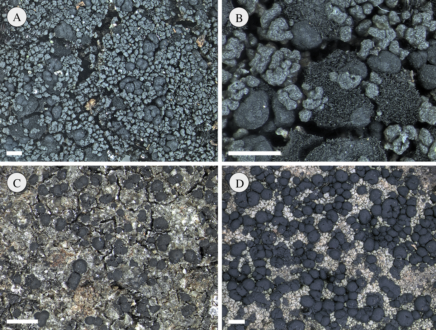
Fig. 5. New Tasmanian species of Micarea. A & B, M. oreina habit (A) and detail, showing black globose apothecia and cushions of Stygonema (B); C, M. sandyana; D, M. saxicola. Scales = 1 mm. Photographs: J. Jarman.

Fig. 6. Asci of Tasmanian species of Micarea, with amyloid parts stippled. A & B, Pilocarpaceae-type where the amyloid tholus is pierced by a narrow channel lacking a darker border: M. oreina (A), M. sandyana (B). C & D, prasina-type, where the penetrating channel has a darker-staining border: M. rubiginosa (C), M. saxicola (D). Scale = 10 µm.
Apothecia immarginate, scattered, strongly convex to subglobose, 0·2–0·8 mm diam., black and glossy. Hymenium 60–80 µm thick, dilute green but darker green in the uppermost 10–20 µm, K−, N+ crimson-red, usually rather poorly delimited from the hypothecium. Paraphyses numerous, branched and anastomosed, rather lax in K, 1–1·7(–2) μm thick, not or only sometimes swelling to 2·5 µm at the apices. Asci broadly clavate, 45–65(–70) × 12–15(–20) μm, with an amyloid outer coat and a well-developed, amyloid tholus penetrated by a narrow, pale channel lacking a darker-staining tube. Ascospores ellipsoid, 1-septate, 11–13·2–15(–16·5) × (4–)4·5–5·5–6(–7) μm (n = 75). Hypothecium 80–105 µm thick, hyaline or patchily dilute olivaceous, K−, N± crimson-red, composed of hyphae 0·8–2 µm thick; ascogenous hyphae to 5 µm thick. Exciple well developed, c. 40 µm thick at base of apothecium, outer part greenish, K−, N+ crimson-red, soon reflexed, composed of branched and radiating hyphae 1–2 µm thick.
Pycnidia sessile amongst the areoles and resembling developing apothecia, 80–100 µm wide; wall K+ green intensifying. Conidia bacilliform, 3·8–5 × 0·8–1 µm; conidiogenous cells ampuliform to subcylindrical, 5–8 × 2–2·5 µm.
Chemistry
Thallus K−, KC+ red, C+ red, P−, UV−; apothecia C−; containing gyrophoric acid in the thallus only.
Etymology
The specific epithet refers to the montane ecology of this species.
Ecology and distribution
Micarea oreina is widespread and common in alpine Tasmania, occurring on large boulders, often overgrowing cushions of the moss Andreaea. Owing to the hardness and size of the rocks, most specimens tend to be scraped from their substratum and, consequently, the species tends to be under-represented in herbaria. Most collections are from Jurassic dolerite, the rock-type which predominates in the central and eastern part of Tasmania. The species appears to be uncommon on the Precambrian metamorphic rocks that characterize Tasmania's south-west. Associated lichens include Cystocoleus ebeneus (Dillwyn) Thwaites, Pseudephebe pubescens (L.) M. Choisy, Rimularia circumgrisea Kantvilas, R. aspicilioides Kantvilas, depauperate tufts of Siphula decumbens Nyl., Siphulastrum mamillatum (Hook.f. & Taylor) D. J. Galloway, Stereocaulon caespitosum Redinger, Umbilicaria cylindrica (L.) Delise ex Duby and U. umbilicarioides (B. Stein) Krog & Swinsc.
Remarks
Micarea oreina is well characterized by a combination of the thallus composed of globose areoles containing gyrophoric acid, the black globular apothecia pigmented with Cinereorufa-green, and the 1-septate ascospores. For decades this species has been collected and identified in Tasmania as Catillaria contristans (Nyl.) Zahlbr., a superficially similar species that occupies a similar habitat in the Northern Hemisphere, but which differs by having a non-micareoid photobiont (cells 10–20 µm wide), by lacking lichen substances, having Biatora-type asci, more robust paraphyses and somewhat narrower ascospores, 2·5–4·5 µm wide (Fletcher & Coppins Reference Fletcher, Coppins, Smith, Aptroot, Coppins, Fletcher, Gilbert, James and Wolseley2009). All Tasmanian specimens previously named Catillaria contristans have now been re-identified as Micarea oreina, with the exception of one (Mt King William I, Kantvilas 91/84 (E, HO)) which has Porpidia-type asci and represents an unknown species possibly allied to Bryobilimbia hypnorum (Lib.) Fryday et al.
The first hints that there was confusion (in Tasmania) regarding C. contristans were published by Andersen & Ekman (Reference Andersen and Ekman2005), who found that DNA-sequence data placed a Tasmanian specimen of that species in a clade together with M. peliocarpa and its relatives. That observation was reconfirmed recently by M. Svensson (unpublished, in. litt.) with further Tasmanian collections and it is essentially as a result of this work that all Tasmanian collections of C. contristans were re-examined anatomically.
Amongst Tasmanian species of Micarea, M. oreina displays some similarities to M. magellanica and M. isabellina, two chiefly terricolous species from the wet peatlands of western Tasmania. Both differ from M. oreina by having larger (mostly 14–26 × 3·5–6·5 µm), 3-septate ascospores, as well as chemically, containing alectorialic acid (C+ pink, P+ yellow) and thiophanic acid (C+ orange) respectively. Also, somewhat similar is M. pseudocoppinsii, which likewise has subglobose, ±immarginate apothecia and a thallus of pale granules containing gyrophoric acid. This species differs by having scattered soralia, 3-septate ascospores, and the C+ reaction, indicating gyrophoric acid, occurs in the apothecia as well as in the thallus. Another superficially similar species is M. flagellispora, which may occur on high altitude rocks but has a C−, P− thallus, very intensely internally pigmented apothecia and filiform ascospores.
Selected specimens examined. Australia: Tasmania: Mt King William I, 42°14′S, 146°08′E, 1300 m, 1984, G. Kantvilas 86/84 (HO); Crater Peak, 41°39′S, 145°56′E, 1200 m, 1984, G. Kantvilas 417/84 & P. James (BM, HO); Mt Sprent, 42°48′S, 145°58′E, 950 m, 1987, G. Kantvilas 30/87 p.p. (E, HO); Mt Saddleback, 41°24′S, 147°45′E, 1200 m, 1993, G. Kantvilas 93/93 (HO); St Patricks Head, 41°34′S, 148°14′E, 550 m, 1997, G. Kantvilas 118/97 (HO); Lots Wife, 42°57′S, 146°28′E, 1089 m, 2000, G. Kantvilas 466/00 (BG, HO); Lake Sidon, 41°48′S, 146°19′E, 1190 m, 2002, K. Felton s. n. (HO); Skullbone Plains, 42°02′S, 146°19′E, 1000 m, 2012, G. Kantvilas 80/12 (HO); Mt Field West summit, 42°39′S, 146°31′E, 1430 m, 2014, G. Kantvilas 138/14 (HO).
Micarea pallida Coppins & Kantvilas sp. nov.
MycoBank No.: MB 831235
Micareae alabastriti simili sed thallo acidum gyrophoricum destituto, acidum porphyrilicum continenti, asci tubo atriore amyloideo instructi et ascosporis parvioribus, 9·5–15 µm longis, 2·5–4 µm latis, differens.
Typus: Australia, Tasmania, south-eastern slopes of McGregor Peak, 42°59′S, 147°57′E, on trunks of Atherosperma moschatum in Atherosperma-dominated rainforest, c. 400 m altitude, 19 January 1989, G. Kantvilas 368/89 (HO—holotypus).
(Figs 1D & 2E)
Thallus effuse, areolate, greenish to greyish white, slightly glossy; areoles plane to slightly convex, 0·05–0·2 mm wide, to 80 µm thick, ecorticate but with a hyaline epinecral layer 4–7 µm thick, discrete, subcontiguous or confluent and forming a ±continuous crust, weakly delimited by a blackish prothallus; prothallus hyphae purple-brown, K+ intensifying purplish, N+ orange-red, 1·5–2 µm thick; photobiont cells micareoid, 4–7 µm diam. Cephalodia absent.
Apothecia numerous, scattered but sometimes confluent in groups of up to 7, immarginate, convex, adnate or somewhat constricted at the base, 0·15–0·25 mm diam., occasionally tuberculate and to 0·4 mm diam., pale fawn brown to pinkish, matt, epruinose, in section lacking crystalline inclusions and hyaline throughout. Hymenium 45–60 µm thick, hyaline, lacking a distinct epithecium. Paraphyses rather few, branched and anastomosing, 0·7–1 µm thick, not or only slightly swollen at the apices. Asci narrowly clavate, 42–52 × 10–13 µm, of the prasina-type, with an amyloid outer coat and a distinct, amyloid tholus penetrated by a narrow, pale channel with a darker-staining, amyloid tube. Ascospores ovate-fusiform, typically with rather acute apices, (1–)3-septate, (9·5–)10–11·6–13·5(–15) × (2·5–)2·8–3·1–3·5(–4) μm (n = 70). Hypothecium 50–85 µm thick, hyaline, composed of interwoven hyphae 0·7–1·3 µm thick; ascogenous hyphae short-celled, to 4 µm thick. Exciple lacking or very poorly developed.
Pycnidia not observed.
Chemistry
Thallus and apothecia K−, KC−, C−, P−, UV−; containing porphyrilic acid.
Etymology
The specific epithet refers to the pale colour of the apothecia.
Distribution and ecology
Micarea pallida is known only from south-eastern Tasmania where it has been recorded from deeply shaded habitats, on smooth-barked understorey trees, in wet gully remnants of cool temperate rainforest dominated by Atherosperma moschatum. This vegetation type tends to support a rather depauperate lichen flora in comparison to the more extensive tracts of Nothofagus-dominated rainforest in Tasmania's west and highlands. However, unusual lichens are often present in such forests, such as foliicolous taxa with affinities to those found in warm temperate and subtropical rainforests of the Australian mainland.
Remarks
This species is characterized by its pallid, C− apothecia and relatively small, 3-septate, ovate-fusiform ascospores, typically with rather acute apices. It is thus distinguished from M. alabastrites and its allies (such as M. subcinerea Brand & van den Boom (van den Boom & Brand Reference van den Boom and Brand2004)) which are consistently C+ red and have larger, fusiform ascospores. Furthermore, members of the M. alabastrites group have asci that lack a darker-staining tube structure in the tholus. The most similar species to M. pallida is M. ceracea, which also has pallid, C− apothecia and prasina-type asci. These two species are easily distinguished from each other by their thallus morphology: smooth and waxy in M. ceracea but areolate in M. pallida. In addition, M. ceracea contains perlatolic acid, has small, crystalline inclusions in the apothecia and larger ascospores, 10–21 × 3·5–6 µm.
Additional specimen examined. Australia: Tasmania: Bun Hill, Forestier Peninsula, 42°58′S, 148°56′E, 320 m, 1989, G. Kantvilas 336/89 (HO).
Micarea peliocarpa (Anzi) Coppins & R. Sant.
In Coppins & P. James, Lichenologist 11: 155 (1979).
Micarea peliocarpa is characterized by a greenish white, effuse to rather bullate or areolate thallus, whitish to greyish to grey-black, sometimes piebald apothecia, 0·1–0·4 mm wide, that react C+ reddish in squash (due to gyrophoric acid), and almost exclusively 3-septate, fusiform to oblong ascospores, 13–23 × 3·5–5·5 µm (see Coppins Reference Coppins, Smith, Aptroot, Coppins, Fletcher, Gilbert, James and Wolseley2009). It is similar to M. alabastrites and M. cinerea, both of which have C+ red apothecia and transversely septate ascospores, and the differences between these lichens are discussed by Coppins (Reference Coppins1983). The latter species differs by having larger, mainly 5–7-septate ascospores (see above). Separation from M. alabastrites is more problematic, although M. peliocarpa will usually display at least some greyish to greenish, N+ reddish apothecial pigment in at least some of the apothecia. The intensity of pigmentation appears to be related to exposure and the almost entirely pallid morphotypes tend to be restricted to extremely shaded habitats. Also related to M. peliocarpa is M. pseudocoppinsii, which differs by being sorediate.
Micarea peliocarpa is a widespread species, recorded from most temperate regions of the globe (Coppins Reference Coppins, Smith, Aptroot, Coppins, Fletcher, Gilbert, James and Wolseley2009). In Tasmania, it is widely distributed from lowland to subalpine elevations, occurring on smooth bark or wood in rainforest, tall wet eucalypt forest, open sclerophyll woodlands and moorlands. It occurs within stable forest interiors as well as at forest margins, or in forests regenerating after logging where it is not infrequent on charred or scorched wood.
Selected specimens examined. Australia: Tasmania: Lindsay River at bridge on Link Road, 41°19′S, 144°59′E, 170 m, 1993, G. Kantvilas 283/93 & J. Elix (HO); S of Hellyer Gorge, 41°18′S, 145°35′E, 530 m, 2003, G. Kantvilas 579/03 (HO); Government Huts, Mt Field NP, 42°41′S, 146°36′E, 1000 m, 2003, G. Kantvilas 748/03 (HO); W of Tahune Bridge in the Warra SST, 43°06′S, 146°41′E, 130 m, 2006, G. Kantvilas 147/06 (HO); Buxton River, in gorge near old weir, 42°15′S, 147°59′E, 30 m, 2008, G. Kantvilas 265/008 (HO); Gowan Brae, hilltop above Nive River, 42°02′S, 146°26′E, 850 m, 2014, G. Kantvilas 162/14 (HO). Victoria: Baw Baw NP, Mushroom Rocks, 37°53′S, 146°21′E, 1200 m, 2008, G. Kantvilas 141/08 (HO, MEL); Morewell NP, Fosters Gully, 38°21′S, 146°23′E, 2008, G. Kantvilas 163/08 (HO, MEL); Vale of Belvoir, 41°33′S, 145°53′E, 780 m, 2019, G. Kantvilas 67/19 (HO). New South Wales: Brown Mountain, Rutherfords Creek, 36°35′22″S, 149°26′44″E, 815 m, 2008, G. Kantvilas 120/08, J. Elix & P. McCarthy (HO).
Micarea prasinastra Coppins & Kantvilas sp. nov.
MycoBank No.: MB 831236
Micareae prasinae Fr. aliquantum similis et item thallo goniocystis constituto, sed pigmentosa destituto, acidum gyrophoricum continenti et ascosporis angustioribus, (0–)1-septatis, 7–11·5 µm longis, 1·8–3·5 µm latis differt.
Typus: Australia, Tasmania, Savage River Pipeline Road near 26 mile peg, 41°12′S, 145°19′E, 410 m altitude, on dry trunk of old Nothofagus cunninghamii in rainforest, 26 November 2003, G. Kantvilas 709/03 (HO—holotypus; E, UPS—isotypi).
(Figs 1E & 2F)
Thallus effuse, light grey-green, commonly with an olivaceous greyish tinge, composed of very fine goniocysts 12–18 µm diam., forming a continuous granular-sorediose crust; photobiont cells micareoid, 4–7 µm diam. Prothallus absent. Cephalodia absent.
Apothecia scattered, immarginate, convex to subglobose, basally constricted, 0·12–0·45(–0·8) mm diam., sometimes becoming tuberculate and to 0·65 mm diam., pallid pinkish to mottled grey-brown, matt. Hymenium 30–48 µm thick, hyaline, lacking a distinct epithecium. Paraphyses rather numerous, branched, 0·7–1(–1·5) μm thick, expanding to 1·5–2 µm at the apices. Asci narrowly clavate, 33–41 × 7–12·5 µm, of the prasina-type, with an amyloid outer coat and an amyloid tholus, penetrated by a narrow, pale channel with a darker-staining border or tube. Ascospores narrowly ovoid to clavate-fusiform, (0–)1-septate, (7–)7·5–9·0–11(–11·5) × 1·8–2·1–2·7(–3·5) μm (n = 110). Hypothecium 65–95 µm thick, hyaline, composed of loosely interwoven hyphae, 0·5–1 µm thick; ascogenous hyphae to 4 µm thick. Exciple inapparent, but at the base of the apothecium the hypothecial hyphae become outwardly orientated and appear, at low-power magnification, as a loose weft of white hyphae 0·8–1·2 µm thick.
Pycnidia scattered, sometimes numerous, whitish to pale grey-brown, sessile and immersed amongst the thallus granules or shortly stipitate, 50–110 µm wide, 0·1–0·2 mm tall. Conidia (microconidia) bacilliform, straight, 2·8–4 × 0·7–1 µm.
Chemistry
Thallus, apothecia and pycnidial walls K−, KC+ red, C+ red, P− (best observed in microscope squashes); containing gyrophoric acid but frequently only in trace concentrations.
Etymology
The specific epithet alludes to the superficial similarity of this species to Micarea prasina.
Ecology and distribution
Micarea prasinastra is locally common in Tasmania, where it is a true indicator species for old trees and old forests (Kantvilas & Jarman Reference Kantvilas and Jarman2012). It colonizes wood, flaky or fibrous bark, and rarely charcoal, on the dry faces or in sheltered hollows of the oldest, most massive trees. Mature Nothofagus cunninghamii trees in cool temperate rainforest and old-growth eucalypts in wet sclerophyll forest are common hosts. This new species occurs in associations with Arthonia apteropteridis Kantvilas & Vězda, Lecanactis abietina (Ach.) Körb., Lepraria finkii (B. de Lesd.) R. C. Harris, Sagenidium molle Stirt. and species of Chaenotheca. However, it typically forms very extensive, mostly ‘pure’ colonies, interrupted only by small tufts of Lepraria. Micarea prasinastra is also known from New Zealand.
Remarks
The goniocyst-like thallus, immarginate apothecia, prasina-type asci and small, 0–1-septate ascospores align Micarea prasinastra with the M. prasina group. Within this complex of species, it is distinguished by its thallus, where the goniocysts form a fine, ±sorediose crust rather than coralloid clusters, the presence of gyrophoric acid, the absence of any pigments, notably K+ violet Sedifolia-grey, and its relatively narrow ascospores. Using spot tests with C cannot be relied on to detect gyrophoric acid and even TLC can be difficult when the thallus is thin and poorly developed. The lack of pigments readily distinguishes M. prasinastra from M. byssacea, the most common member of the M. prasina group in Tasmania, and any dark coloration in tissues appears to be due to infection by a hyphomycete. Young apothecia often have a faintly ‘pruinose’ appearance due to the tips of paraphyses protruding through the surface of the disc. Occasional thalli growing over charcoal sometimes develop minute, flat areoles 60–80 µm wide that break down into scurfy, granular soredia. In all other respects, however, such individuals are identical to typical M. prasinastra as described above and are seen as variants responding to an unusual habitat. Within the M. prasina group, two other species contain gyrophoric acid. Micarea viridileprosa has a brighter green, rarely fertile thallus; when fertile it can be further distinguished by its broader ascospores (2·5–4·5 µm vs. mostly 1·8–2·7 µm) (van den Boom & Coppins Reference van den Boom and Coppins2001). Micarea levicula (Nyl.) Coppins has a very different appearance, with its goniocysts arranged to form finely coralloid structures up to 150 µm tall (Brand et al. Reference Brand, van den Boom and Sérusiaux2014).
Selected specimens examined. Australia: Tasmania: Little Fisher River, 41°45′S, 146°20′E, 880 m, 1982, G. Kantvilas 180/82 (E, HO); Anthony Road, 41°50·5′S, 145°37·5′E, 550 m, 1992, G. Kantvilas 564/92 (HO); Warra Creek, 43°05′S, 146°43′E, 250 m, 1996, G. Kantvilas s. n. (HO); Hartz Road, 43°11′S, 146°47′E, c. 400 m, 1998, J. Jarman s. n. (HO); W of Tahune Bridge in the Warra SST, 43°06′S, 146°40′E, 90 m, 2003, G. Kantvilas 51/03 (HO); Wandle River at bridge on Murchison Hwy, 41°22′S, 145°35′E, 380 m, 2003, G. Kantvilas 728/03 (E, HO); Cuvier Valley Track, 42°01′S, 146°05′E, 790 m, 2011, G. Kantvilas 157/11 (HO); Rabalga Track, 41°05′S, 145°22′E, 280 m, 2019, G. Kantvilas 91/19 (H, HO).—New Zealand: North Island: Tongariro National Park, 39°23′S, 175°27′E, c. 750 m, 1981, L. Tibell 13544 (UPS). South Island: S of the summit of Hunters Hills, 46°28′S, 169°21′E, 450 m, 1981, L. Tibell 10331 (UPS); Mt Aspiring NP, close to Cameron Flat, 44°10′S, 169°18′E, 360 m, 1981, L. Tibell 10596 (UPS).
Micarea prasinella (Jatta) I. M. Lamb
Lilloa 26: 413 (1953).—Biatorina [as ‘Biarotina’] prasinella Jatta, Boll. Soc. Bot. Ital. 1911: 257 (1911); type: Tasmania occid., Dubbil Barril, Mt Lyell Railway, on trunk of tree, 30 Dec. 1899, W. A. Weymouth 977 (FI—holotype; BM!, FI—isotypes).
Thallus effuse, pale green to light olive green, in places with a suffused brownish tinge, composed of scattered, granular areoles 40–60 µm wide, often interconnected by colourless prothalline hyphae; soralia sometimes present, pale whitish green where abraded, elsewhere brownish, C+ red, flattened to convex, rounded, 0·15–0·35 mm wide, sometimes becoming confluent and forming larger sorediose patches, more rarely thallus ±entirely sorediate; photobiont cells micareoid, 4–7 µm. Cephalodia few to abundant, brown to black, dark blue-grey when wet, tuberculate, 0·1–0·4 mm wide; photobiont ?Nostoc, with cells 4·5–8·5 × 4·5–6 µm in short chains.
Apothecia dark blue-grey to black, turbinate to shortly stalked, sometimes somewhat resembling the ascomata of a Calicium species, 0·15–0·6 mm diam., 0·3–0·5 mm in overall height, with the ‘stalk’ 0·1–0·3 mm tall and 80–160 µm wide, sometimes ±greyish tomentose, and the ‘cap’ convex, usually immarginate, occasionally appearing flatter and ±marginate, also sometimes greyish tomentose on the underside. Hymenium 50–70 µm thick, reddish brown, K+ reddish or reddish pink, N+ orange-red, typically with a distinctly defined, heavily pigmented epithecial layer 5–10 µm thick. Paraphyses slender, flexuose, sparingly branched and anastomosing, 0·7–1·2 µm thick, widening in the upper part to 2 µm; apices coated in pigment to comprise the epithecium. Asci 65–70 µm, narrowly clavate, sometimes with purple-brown pigment within the ascoplasm, with an amyloid outer coat and an amyloid tholus, penetrated by a narrow, pale channel lacking a darker-staining border or tube. Ascospores ellipsoid to ovoid-ellipsoid, (0–)1(–3)-septate, 12–18 × (4–)4·7–7 µm. Hypothecium opaque dark purple-brown, with some areas dark blackish grey, K+ greenish black, N+ reddish, composed of hyphae 1·3–2 µm thick; ascogenous hyphae to 6·5 µm thick. Exciple hyaline, 50–70 µm thick, with a dark red-brown to black outer layer 5–10 µm thick; hyphae outwardly radiating, branched and anastomosing, 1·5–2 µm thick, with those from the outermost part of the exciple having walls sometimes coated with a dark pigment and to 3 µm thick.
Pycnidia sessile, dark reddish brown, 40–50 µm diam. Conidia narrowly bacilliform, 6·7–7·7 × 0·8 µm, slightly pointed at each end.
Chemistry
Thallus and soralia K−, KC+ red, C+ red, P−; gyrophoric acid by TLC.
Ecology and distribution
With a range spanning Tasmania, New Zealand, southern Chile, the Pacific Northwest of North America, Japan and Britain, M. prasinella is an excellent example of the floristic and ecological links that exist between the cool, oceanic forests of these regions. This species mostly occurs on bark or wood, or overgrowing epiphytic bryophytes, typically in very wet, deeply shaded habitats. In Tasmania it is clearly very uncommon. The provenance of the Tasmanian type collection is rainforest, yet despite the extensive lichenological exploration of wet forests in Tasmania over the last three decades, the species has been found at only two further localities. At one, it grew on a rotting log in wet eucalypt forest regenerating after logging. At the second locality, M. prasinella was very prolific on a gravel bank in a highly disturbed, very wet site, suggesting it might also be a successful pioneer species.
Remarks
This very distinctive species is characterized best by its unique, Calicium-like apothecia. In some respects, this structure is an extreme development of the hypothecial extension or ‘foot’ seen in some other species of Micarea, such as forms of M. incrassata and M. lignaria (Coppins Reference Coppins1983), and the Tasmanian M. saxicola. Additional and very detailed descriptive notes are also provided by Lamb (Reference Lamb1953) who, however, failed to note the stalked nature of the apothecia. Jatta's type is in rather poor condition with relatively few apothecia which are rather compacted and have, at best, a very inconspicuous stalk.
Pigmentation of the apothecia tends to be very intense and difficult to characterize, and it is possible that a spectrum of several pigments is involved. In Tasmanian specimens, the hymenium and epithecium have a distinct pinkish hue that intensifies pinkish in K and reddish in N, suggestive of Atra-red. The hypothecium is purple-brown, K+ green-black, N+ reddish, suggestive of Melaena-red. No greenish, N+ crimson pigments are present. A specimen studied originating from New Zealand (Bartlett, Ruby Lake) contains only brown pigments that intensify in K and react N+ reddish brown.
Selected specimens examined. Australia: Tasmania: Queenstown, 42°05′S, 145°33′E, on soil on gravel bank, 200 m, 1984, G. Kantvilas 188/84 & P. James (BM, E, HO); Warra Creek, 43°05′S, 146°43′E, 175 m, 1996, G. Kantvilas s. n. (HO).—New Zealand: North Island: Townsons Kauri Reserve, 1976, D. M. Henderson (E). South Island: Canterbury, Nina Valley, W of Lewis River, 42°28′S, 172°23′E, 600–630 m, 1981, P. W. James (BM); Nelson, Lockett Range, 1260 m, 1982, J. Bartlett (UPS); Nelson, Ruby Lake, below Ruby Saddle, 900 m, 1982, J. Bartlett (HO, UPS).—Chile: X Region: Parque Nacional Vicente Perez Rosales, Lago Todos los Santos, Cordillera Bonete, 41°12′S, 72°17′W, 500–800 m, 1986, B. J. Coppins et al. 5009 (BM).—USA: Oregon: Lane County, H. J. Andrews Experimental Forest, 44°13·2′N, 122°15·5′W, 610 m, B. McCune 25337 (BG). Alaska: c. 240 km ESE of Anchorage, 60°33′N, 145°41′W, 100–300 m, 1989, G. Thor 8577 (E, S).
Micarea pseudocoppinsii Brand et al.
Lichenologist 46: 434 (2014).
For a full description of this species, see Brand et al. (Reference Brand, van den Boom and Sérusiaux2014). It has the following salient features: thallus effuse, patchy, in part minutely areolate or granular, pale greenish yellow, with roundish soralia to c. 1 mm wide that spread across the thallus; apothecia to 0·5 mm wide, dark grey to blackish, immarginate, convex, colourless within apart from greenish, N+ crimson-red pigment mainly in the upper part of the hymenium; ascospores narrowly ellipsoid (0–)3-septate, 13–20 × 4–5 µm; chemistry of gyrophoric acid (soralia and apothecial sections C+ red).
As noted by Brand et al. (Reference Brand, van den Boom and Sérusiaux2014), soredia are rare in Micarea and, amongst the Tasmanian species studied, are found only in M. prasinella, which also grows on soil and contains gyrophoric acid but has distinctive, turbinate to shortly-stalked, black apothecia with pinkish and purple-brown pigments. Brand et al. (Reference Brand, van den Boom and Sérusiaux2014) also noted similarities to M. peliocarpa and we would have probably filed our Tasmanian specimens provisionally under ‘M. aff. peliocarpa’ had M. pseudocoppinsii not been described recently.
Hitherto known only from Réunion (Brand et al. Reference Brand, van den Boom and Sérusiaux2014), Micarea pseudocoppinsii is recorded here for Tasmania for the first time on the basis of two highly ecologically disjunct, well-developed collections. At one locality, it grew on peat in buttongrass (Gymnoschoenus-dominated) moorland, in dryish underhangs along a disused gravel road. This vegetation type has been well investigated for Micarea and commonly supports three terricolous species: M. isabellina, M. magellanica and M. melaena. The scurfy, effuse, discontinuous thallus of M. pseudocoppinsii, with its scattered C+ red soralia, is not unlike that of Trapeliopsis granulosa (Hoffm.) Lumbsch, which also grows in such habitats. At the second locality, M. pseudocoppinsii grew on soil in a gap in open, dry eucalypt forest with a grassy understorey.
Specimens examined. Australia: Tasmania: at foot of Hamilton Moraine, along road from Lake Margaret, 41°59′S, 145°34′E, 600 m, 1998, G. Kantvilas 197/98 (HO); Cherry Tree Hill, 41°59′S, 148°08′E, 180 m, 2012, G. Kantvilas 334/12 (HO).
Micarea rubiginosa Coppins & Kantvilas sp. nov.
MycoBank No.: MB 831237
Micareae byssaceae similis et item thallo goniocystis granularibus constituto, sed K+ violaceum pigmentosum et substantias chemicalias destituto, apotheciis pigmentum rubella-aurantiacum continentibus, et ascosporis 0–1-septatis, 9·5–17 µm longis, 3·5–5·5 µm latis differt.
Typus: Australia, Tasmania, W of Tahune Bridge in the Warra SST, Coupe WR 0081, 43°06′S, 146°40′E, 90 m altitude, on wet sides of very old eucalypts in Eucalyptus obliqua-dominated wet forest, 14 May 2003, G. Kantvilas 95/03 (HO—holotypus).
(Figs 1F, 2G & 6C)
Thallus vivid bright green when fresh and moist, dull light greenish to somewhat glaucous greenish when dry, composed of goniocysts. Goniocysts granular, 15–25 µm wide and to 200 µm tall, not noticeably swelling in K, often becoming coralloid and isidia-like; photobiont cells micareoid, 4–7 µm diam.; mycobiont hyphae 0·8–1·5 µm thick. Prothallus absent. Cephalodia absent.
Apothecia scattered, immarginate, convex to subglobose, at first adnate and nestled amongst the goniocysts, sometimes becoming tuberculate and constricted at the base, 0·2–0·4 mm diam., increasing to 0·5 mm when tuberculate, dull orange-red to red-brown, not glossy. Hymenium 45–50 µm thick, hyaline with vertical streaks of dull orange to red-brown, K+ dilute yellow-orange, C+ orange pigment extending to the hypothecium. Paraphyses numerous, much branched, irregular in width, 0·7–1·5 µm thick, irregularly swollen at the apices to 2(–2·5) μm. Asci clavate, 38–50 × 13–15·5 µm, of the prasina-type, with an amyloid outer coat and an amyloid tholus penetrated by a narrow, pale channel with an indistinct, darker-staining border or tube. Ascospores oblong-ellipsoid to ovoid-ellipsoid, 0–1-septate, (9·5–)10–12·2–15(–17) × 3·5–4·3–5(–5·5) μm (n = 100). Hypothecium 70–95 µm thick, dilute orange to red-brown, K+ dilute yellow-orange, C+ orange, composed of irregularly arranged hyphae 1–2 µm thick; ascogenous hyphae short-celled, to 5 µm thick. Exciple reflexed, not well developed, composed of outwardly radiating hyphae 0·8–1·2 µm thick.
Pycnidia not observed.
Chemistry
No substances detected by TLC.
Etymology
The specific epithet alludes to the reddish colour of the apothecia.
Ecology and distribution
Micarea rubiginosa is locally abundant in Tasmania in old-growth wet sclerophyll forest dominated by Eucalyptus obliqua. It is a truly ‘old forest-old tree’ indicator species (Kantvilas & Jarman Reference Kantvilas and Jarman2012). Unlike M. byssacea, which is common in standing forests but is also able to survive or re-establish after logging on remnant standing trees, stumps or large logs, M. rubiginosa is known only from old-growth standing forest where it grows amongst bryophytes in deeply shaded habitats on the bases of trunks of the oldest, largest trees, as well as on large logs. It has also been recorded from the decaying leaf bases of the large rosette sedge, Gahnia grandis, which is common in the understorey of such forests. This new species is also known from Chile.
Remarks
The granular, goniocyst-like thallus, the prasina-type asci and 0–1-septate ascospores ally this species with the M. prasina group, where it is easily distinguished by the distinctive, bright orange-red colour of the apothecia, particularly pronounced in moist, fresh specimens seen in the field. The pigment approximates ‘Rubella-orange’ of Ekman (Reference Ekman1996), commonly found in several species of the genus Bacidia. When treated with C, microscope squashes of the apothecia intensify orange, probably due to a bleaching of the tissues. In contrast, M. byssacea, with which it frequently occurs and which has a similar green thallus composed of granular to coralloid goniocysts, is distinguished by having pallid to greyish apothecia with C+ violet, K+ violet Sedifolia-grey pigment. Chemical differences also aid in distinguishing M. rubiginosa in that other species with a somewhat similar thallus, such as M. byssacea, M. nowakii and M. prasinastra, all contain lichen substances.
Selected specimens examined. Australia: Tasmania: Boyd Lookout, 550 m, 1981, P. W. James s. n. (BM); Ben Ridge Road, 830 m, 1981, G. Kantvilas 1113/81C (HO); west of Tahune Bridge in the Warra SST, 43°06′S, 146°41′E, 120 m, 1999, G. Kantvilas 111/99, 221/99 (HO); ibid., 100 m, 2008, J. Jarman s. n. (HO); Florentine Bridge, 42°30′S, 146°27′E, 350 m, 2003, G. Kantvilas 630/03 (HO); Gordon River Road, W of Frodshams Pass, 42°49′S, 146°22′E, 580 m, 2019, J. Jarman s. n. (HO).—Chile: X Region: SW of Puerto Montt, c. 15 km NE of Pargua, c. 400 m, 1986, B. J. Coppins, D. J. Galloway, G. Guzman & P. W. James 6061 (E).
Micarea sandyana Kantvilas sp. nov.
MycoBank No.: MB 831240
Micareae ternariae (Nyl.) Vĕzda similis sed partibus omnibus parvioribus, praecipue ascosporis, 7–13·5 µm longis, 3·5–6 µm latis differt. Etiam M. argopsinosae similis sed a qua argopsinum destituto differt.
Typus: Australia, Tasmania, South Sister, 41°32′S, 148°10′E, on exposed dolerite boulders, 780 m altitude, 22 November 2007, G. Kantvilas 361/07 (HO—holotypus).
(Figs 2H, 5C & 6B)
Thallus effuse, rimose-areolate, dull brownish grey, rather unevenly lumpy and gnarled, to 200 µm thick, sometimes ±absent and endolithic; photobiont cells micareoid, 5–10(–12) μm diam. Prothallus lacking. Cephalodia absent.
Apothecia 0·2–0·7 mm diam., numerous, scattered, broadly adnate; disc jet black, at first plane, soon becoming convex to undulate; margin concolorous with the disc, barely evident in the youngest apothecia, soon becoming inconspicuous or excluded. Hymenium 50–60(–70) μm thick, intensely aeruginose blue-green, K+ intensifying, N+ crimson-red in the upper part, increasingly dilutely pigmented in the lower part. Paraphyses numerous, richly branched with occasional anastomoses, 1–1·5 µm thick; apices not expanded or widening to c. 2·5 µm and caked with pigment. Asci clavate, 31–40 × 11–16 µm, with an amyloid outer coat and an amyloid tholus penetrated by a narrow, pale channel lacking a darker-staining amyloid border or tube. Ascospores oblong-ellipsoid with broadly rounded apices, straight or slightly curved, (1–)3-septate, 7–11·0–13(–13·5) × (3·5–)4–4·6–5·5(–6) μm (n = 80), sometimes a little constricted at the septa. Hypothecium 60–100 µm thick, hyaline. Exciple reflexed, 25–40 µm thick, composed of branched, radiating hyphae, aeruginose blue-green at the outer edge and similarly streaked within.
Pycnidia immersed; conidia fusiform to cylindrical, 5–7 × 1·5 µm.
Chemistry
Containing no substances detectable by TLC.
Etymology
This species is named in honour of the British lichenologist, Sandy Coppins, in appreciation of the friendship and hospitality shown to the author when some of the work on this paper was being undertaken.
Ecology and distribution
This species is known only from three Tasmanian collections, each from rather different habitats. The type is from an exposed dolerite boulder on a coastal pinnacle that supports wet, sclerophyllous scrub. It grew in the vicinity of galvanized fencing and sheds associated with a communications installation, and the extent of influence of zinc-contaminated run-off on this species is unclear. Zinc certainly has an obvious, very localized impact on the saxi-colous flora in general (Kantvilas et al. Reference Kantvilas, Elix and Jarman2008), with the immediate surrounds being devoid of lichens and the near vicinity dominated by Lecanora polytropa (Ehrh.) Rabenh., with Acarospora veronensis A. Massal., Candelariella vitellina (Hoffm.) Müll. Arg., Lecidella sublapicida (C. Knight) Hertel and Trapelia coarctata (Sm.) M. Choisy also present.
The second locality for M. sandyana is also a coastal pinnacle supporting heathland, where the new species grew on consolidated soil in the shelter of a large basalt boulder. At the third locality, the new species grew together with M. micromelaena on loose pebbles on bare, gravelly soil in disturbed heathland. Clearly further collections will be required before the ecology of this species can be interpreted.
Remarks
The morphology and anatomy of the thallus, apothecia and ascospores place this taxon in the M. lignaria-M. ternaria group of Coppins (Reference Coppins1983) which, in Tasmania, also includes M. argopsinosa, M. isabellina and M. magellanica. These taxa differ from the new species by a combination of thallus chemistry and ascospore size. Of these taxa, Micarea sandyana is closest to M. argopsinosa, recently described from mainland Australia, which has similar small spores but contains argopsin (McCarthy & Elix Reference McCarthy and Elix2016a). The absence of lichen substances in the new species allies it to the widespread, temperate Northern Hemisphere taxon, M. ternaria. That species is ±identical to M. sandyana in every respect, except that it is incrementally larger in all its parts, and most significantly in its larger ascospores, 13–22(–24) × 3·5–5 µm (see Coppins (Reference Coppins1983) for description).
In the Tasmanian flora, apart from M. argopsinosa, the species most likely to be confused with M. sandyana is M. saxicola, because both have an areolate, brownish thallus and black, ultimately convex apothecia. The latter is usually readily distinguished, however, by the presence of cephalodia, the absence of an apothecial margin from the outset, and by its simple ascospores.
The specimens of M. sandyana differ starkly in their thallus morphology and habitat ecology, but are identical anatomically, and there seems little doubt as to their conspecificity. This conclusion was also reached by McCarthy & Elix (Reference McCarthy and Elix2016a) who examined the same specimens on an earlier occasion.
Additional specimens examined. Australia: Tasmania: Linda, 42°04′S, 145°36′E, 280 m, 1984, G. Kantvilas 259/84A & P. James (HO); The Nut (at summit), 40°46′S, 145°18′E, 143 m, 1999, G. Kantvilas 284/99 (HO).
Micarea saxicola Coppins & Kantvilas sp. nov.
MycoBank No.: MB 831241
Micareae incrassatae Hedl. similis sed substrato saxicola, cephalodiis Stigonema continentibus, ascosporis brevioribus et relative latioribus, 0(–1)-septatis, 7–18 µm longis, 4·5–7 µm latis differt.
Typus: Australia, Tasmania, Hartz Mountains, Arthur Tarn, 43°15′S, 146°46′E, 1050 m altitude, on dolerite boulders in subalpine heathland, 9 December 2002, G. Kantvilas 595/02 (HO—holotypus; E—isotypus).
(Figs 2I, 5D & 6D)
Thallus composed of confluent, convex to globose areoles, also with scattered areole-like cephalodia, without a discernible prothallus. Areoles grey-brown to grey-green when shaded, matt, 0·1–0·25(–0·4) mm diam.; in section outer hyphae with light brown walls, and an algal-free medulla often differentiated. Photobiont micareoid, with cells 4–7 μm diam. Cephalodia usually frequent, resembling areoles but darker in colour and with a verruculose surface, c. 0·2–0·4 mm diam., containing Stigonema with cells 7–12 µm wide.
Apothecia numerous, black, immarginate, plane at first, usually soon convex, ±adnate and often partly enveloped by surrounding areoles, sometimes becoming ±globose and basally constricted, 0·35–0·8(–1·5) mm diam., discrete to 2–3-confluent, occasionally forming larger clusters to 1·8 mm diam. Hymenium 45–60 µm thick, hyaline; epithecium c. 5–10 µm thick, olive green to dark grey-brown, K−, N+ crimson-red, sometimes also with traces of a reddish brown, K−, N+ orange-brown pigment. Paraphyses numerous, simple or sparingly branched, occasionally anastomosed, 1·2–1·7 μm wide, not swollen at apices. Asci clavate, 45–57 × 12–15(–17) μm, 8-spored, approximating the prasina-type, with an amyloid outer coat and an amyloid tholus penetrated by a pale channel with a darker-staining amyloid border with ±diverging sides. Ascospores 0(–1)-septate, mostly ellipsoid to ovoid-ellipsoid, (7–)8·5–11·1–14·5(–18) × 4·5–5·5–6·5(–7) μm (n = 90). Hypothecium 200–400 µm thick, dark reddish brown, K− or K+ intensifying reddish, N+ bright orange-brown, sometimes extending as a ‘root’ to the base of the surrounding areoles; hyphae mostly vertically orientated, or laterally outwardly orientated, 1·7–2·5 µm wide; ascogenous hyphae 2·5–4·5 µm wide. Exciple usually inapparent, but sometimes visible in young ± plane apothecia as a narrow, non-amyloid zone adjacent to the hymenium; hyphae outwardly radiating, branched, c. 1·5–1·7 µm wide.
Pycnidia not found.
Chemistry
No substances detected by TLC.
Etymology
The specific epithet is derived from the habitat of this new species.
Ecology and distribution
Micarea saxicola is a widespread and common species in Tasmania, albeit one with a rather narrow ecological niche. It occurs almost exclusively on Jurassic dolerite, the dominant rock type in central and eastern parts of the island, especially at higher elevations. The typical habitat of the new species is on boulders and smaller stones, particularly along walking tracks, in wet, eucalypt-dominated woodland or in cool temperate rainforest where the forest canopy tends to be rather broken and the ground surface disturbed (Fig. 7). Here M. saxicola forms very extensive patches (sometimes covering rocks or boulders in their entirety), associated with other early succession species such as Placopsis gelida (L.) Linds., Stereocaulon ramulosum (Sw.) Räusch., Trapelia coarctata (Sm.) M. Choisy and T. glebulosa (Sm.) J. R. Laundon. As a result, this species is frequently seen and collected. Away from these relatively disturbed habitats, such as on the dolerite boulder-fields that are extensive at higher elevations in Tasmania, the species is rare or absent, even though lichens in general may be very abundant.

Fig. 7. Habitat of M. saxicola: a walking track in cool temperate rainforest in Tasmania's central highlands. The new species is the dominant lichen on the dolerite boulders on the forest floor.
Remarks
Micarea saxicola is characterized by its saxicolous habit, a thallus of brownish, contiguous areoles, the presence of cephalodia that appear like darker areoles, and convex, black apothecia. The combination of the presence of cephalodia, ±unbranched paraphyses, and the absence of substances detectable by TLC suggest affinities to M. assimilata (Nyl.) Coppins and M. incrassata. However, M. incrassata has longer and relatively narrower ascospores, which are more frequently septate, and its cephalodia contain Nostoc rather than Stigonema. Furthermore, in Tasmania, M. incrassata has a completely different ecology and occurs on consolidated soil in lowland grassland. More similar to M. saxicola is the northern European M. subconfusa (Nyl.) Alstrup, which is also saxicolous and has cephalodia with Stigonema but has smaller ascospores, (6–)7–9·5 × 2·5–3·5 µm (Coppins Reference Coppins, Smith, Aptroot, Coppins, Fletcher, Gilbert, James and Wolseley2009). The ascospore dimensions of M. saxicola closely approximate those of M. subalpina of Coppins & Spribille (Reference Coppins and Spribille2004) and these two species share a similar internal anatomy and pigmentation. However, the terricolous M. subalpina has smaller apothecia, a non-micareoid photobiont, non-septate ascospores and lacks cephalodia. The bryicolous-terricolous M. pannarica Fryday from Campbell Island, New Zealand, differs from M. saxicola by its larger, 3-septate ascospores and P+ orange thallus (indicating pannarin) (Fryday Reference Fryday2004).
In the Tasmanian flora, M. saxicola is superficially most similar to M. flagellispora in that both occur on boulders in wet forest, have a prominent areolate thallus and black, convex apothecia. The latter can be distinguished macroscopically by its green (rather than brownish) thallus and anatomically by its filiform ascospores and intensely internally-pigmented apothecia.
The darker-staining amyloid tube in the asci of the new species is usually very prominent. In mature asci, this tube has diverging flanks which, in some preparations, resembles a Porpidia-type ascus (Fig. 5D).
In ‘typical’ specimens of M. saxicola, the hypothecium of the apothecia often extends to the base of the surrounding areoles, as is characteristic of M. incrassata (Coppins Reference Coppins1983, fig. 4C). However, other specimens of M. saxicola which do not exhibit this feature show two extreme variations. For example, the apothecia in Kantvilas 596/02 remain plane or become only weakly convex, whereas in Kantvilas 15/03 many apothecia are almost globose, sometimes forming blackberry-like clusters. The disposition and concentration of the greenish, N+ crimson epithecial pigment appears to depend on the exposure of the habitat. With increasing exposure, this becomes ever more intense, whereas in more shaded situations it may be absent completely and the epithecium contains only the same reddish brown pigment seen in the hypothecium. Septate ascospores tend to be uncommon and, in sections where they were encountered, all tended to be within the one ascus.
Selected specimens examined. Australia: Tasmania: Mt Wellington, 42°54′S, 147°14′E, 1963, P. James s. n. (BM, HO); Hartz Mountains NP, near the hut, 43°11′S, 146°45′E, 750 m, 1988, A. Aptroot 23107 (E, hb. Aptroot); Hartz Mountains, near start of route to Arthur and Emily Tarns, 43°15′S, 146°46′E, 980 m, 2002, G. Kantvilas 596/02 (E, HO); track to Mt Mueller, 42°47′S, 146°29′E, 830 m, 2003, G. Kantvilas 15/03 (E, HO); track to Lake Webster, 42°40′S, 146°35′E, 840 m, 2003, G. Kantvilas 508/03 (HO); Bluestone Tier, 42°28′S, 147°48′E, 510 m, 2003, G. Kantvilas 271/03 (HO); South Sister, 41°32′S, 148°10′E, 750 m, 2006, G. Kantvilas 316/06 (HO); Brown Mountain, 42°36′S, 147°31′E, 730 m, 2007, G. Kantvilas 236/07 (HO); slopes of Mt Albert, 41°21′S, 147°52′E, 850 m, 2008, G. Kantvilas 254/08 (HO); Gordon River Road, W of Frodshams Pass, 42°49′S, 146°22′E, 580 m, 2019, G. Kantvilas 73/19 (HO).
Micarea tubaeformis Coppins & Kantvilas sp. nov.
MycoBank No.: MB 831242
A Micarea flagellispora Coppins et Kantvilas areolis apotheciisque parvioribus, cephalodiis pallidioribus, conidiomatibus grandioribus tubaeformibusque, materia chemicali dissimili differt.
Typus: Australia, Tasmania, Line 7, c. 0·5 km E of Olga River, 42°51′S, 145°50′E, on Phyllocladus aspleniifolius in large forested Eucalyptus-Phyllocladus copse in buttongrass moorland, 75 m altitude, 7 March 1991, G. Kantvilas 31/91 (HO—holotypus; CANB, E—isotypi).
(Figs 2J & 8A-D)
Thallus effuse, greenish white to pale green, vivid green when fresh and moist, matt, composed of confluent, granular to ±globose areoles, (40–)50–140(–150) μm diam., that often proliferate and coalesce to form a rather thick crust, 0·3–0·5 mm thick; prothallus black, thin, visible between cracks in the thallus, occasionally dominant when the overlying covering of thallus areoles is thin or patchy, with hyphae c. 2–3 µm thick, embedded in dense, amorphous dark green (rarely also purplish) pigment, K−, N+ crimson-red; mycobiont hyphae 1·5–2 µm thick, faintly I+ blue after pretreatment with K; photobiont cells micareoid, 4–7 µm diam. Cephalodia usually numerous but inconspicuous, resembling large areoles, 80–120(–300) μm diam., pale, whitish grey to yellow-brown when dry, pale milky blue-grey and ±translucent when wet, containing unbranched filaments of Scytonema with cells c. 7–13 µm wide; mycobiont hyphae weakly amyloid.

Fig. 8. Micarea tubaeformis. A, general habit on the trunk of a rainforest undershrub, showing green areolate thallus over a conspicuous, black prothallus; B, habit, with apothecia and black, stalked pycnidia; C, detail of stalked, trumpet-shaped pycnidia; D, detail of young, weakly marginate apothecia. Scales = 1 mm. Photographs: J. Jarman.
Apothecia scattered or occasionally confluent, sometimes clustered owing to several apothecia arising from an older apothecium, black, (0·15–)0·3–0·6 mm diam., usually sessile and adnate but sometimes basally constricted (especially when growing on loose bark fibres and bryophytes), at first plane and weakly marginate, soon becoming convex to subglobose and immarginate; overmature apothecia sometimes with a thin, ±flexuose margin owing to a collapse of the hymenium. Hymenium 48–70 µm thick, dilute greenish grey, overlain by a dark greenish, K+ green intensifying, N+ crimson-red epithecium. Paraphyses numerous, branched, (1–)1·5–2 µm thick, sometimes expanding to 2·7 µm towards the apices; apices usually embedded in dense pigment. Asci 50–70 × 7–9·5 µm, cylindrical, with an amyloid outer layer and an amyloid tholus penetrated by a narrow, pale channel, sometimes with a darker-staining border or tube evident in younger asci. Ascospores spirally arranged in the ascus, filiform, flexuose, indistinctly 3–7-septate, (45–)47–66·5–91(–100) × 1–1·4–2 µm (n = 30). Hypothecium (50–)100–340 µm thick, purplish brown, K+ intensifying purplish, N+ orange-red; hyphae 1–1·7 µm thick; ascogenous hyphae to 4·5 µm thick. Exciple concolorous with adjacent parts of the hypothecium within, becoming hyaline in the outer part, lacking any crystalline inclusions, composed of radiating, branched hyphae 1·5–2·5(–3·5) μm thick, with highly gelatinized walls and remaining coherent in K.
Pycnidia usually present, conspicuous, stalked, funnel-shaped, c. 0·1–2 mm tall and 0·2–0·3 mm wide at base of stalk, gradually widening above to 0·3–0·6 mm, then abruptly flaring at the apex to 0·5–1·2 mm wide, essentially black but with the ‘stalk’ often covered in a thin, adpressed, grey-white tomentum, and the inner surface of the ‘funnel’ often with a whitish deposit of conidia; ostiolar pore at base of funnel 80–180 µm diam.; rim of flared apex often becoming radially split and torn, and sometimes with one or two proliferating pycnidia; pycnidial cavity in section ±heart-shaped, c. 600 µm tall and 400 µm wide; surrounding tissue (including stalk) with a mixture of purplish brown and greyish green pigments as in the apothecia; hyphae c.1·5–2 µm thick, but often seen (especially in K) to be surrounded by a gelatinous sheath (to 7 µm wide in K) that is densely pigmented in the outer part; hyphae of stalk vertically arranged in the centre, becoming more irregularly orientated towards the outer surface. Conidia (macroconidia) 52–78 × 0·8–1 µm, filiform, flexuose, non-septate or indistinctly 3–7 septate, produced in the subapical swelling of the stalk, immediately below the flared apex.
Chemistry
Thallus K−, KC−, C−, P−, UV−; containing 2´-O-methylsuperlatolic acid (major) and 2´-O-methylhyperlatolic acid (minor). The former compound is seen on developed TLC plates as a pale yellow spot running faster than perlatolic acid in solvent A (see Elix & David Reference Elix and David1991; Brodo & Elix Reference Brodo and Elix1993).
Etymology
The specific epithet refers to the distinctive trumpet-shaped pycnidia of the new species.
Ecology and distribution
Micarea tubaeformis is one of the most common epiphytic lichens in Tasmanian rainforest, particularly in communities of the thamnic or implicate rainforest types (nomenclature after Jarman et al. (Reference Jarman, Kantvilas and Brown1994)) that predominate on the infertile rock types of western Tasmania. These forest types have a dense shrubby understorey and M. tubaeformis is usually very abundant in shaded habitats on horizontal or ascending stems and limbs, especially those with smooth bark such as Anodopetalum, Anopterus, Cenarrhenes and Eucryphia (Fig. 3B). In such situations, it is commonly associated with Coccotrema cucurbitula (Mont.) Müll. Arg., Fissurina insidiosa C. Knight & Mitt., Leiorreuma exaltatum (Mont. & Bosch) Staiger, Lepra novae-zelandiae (Szatala) I. Schmitt et al., Opegrapha stellata C. Knight and depauperate thalli of the macrolichens Bunodophoron australe (Laurer) A. Massal., Leifidium tenerum (Laurer) Wedin, Pseudocyphellaria glabra (Hook.f. & Taylor) C. W. Dodge and P. multifida (Nyl.) D. J. Galloway & P. James. Less commonly it may also colonize papery or rough bark, wood or, occasionally, well-drained mounds of peaty soil or terricolous bryophytes. It is also frequently observed in wet, scrubby vegetation adjacent to such rainforests, and ranges from lowland to alpine elevations.
Remarks
Micarea tubaeformis is readily identified by its remarkable, large pycnidia that resemble the fruiting body of a Calicium. It is most closely related to M. flagellispora, which has apothecia that are almost identical in anatomy and pigmentation, and differ only in being slightly larger and with a more intensely pigmented exciple. The two species also have seemingly identical conidia and conidiogenous cells, except that in M. flagellispora these are borne in immersed to ±sessile, ±globose pycnidia, 80–200 µm diam. The pycnidial wall in both species contains the same greenish and purplish brown pigments as found in the epithecium and hypothecium of the apothecia. Specimens without pycnidia can, however, be readily identified. Micarea flagellispora has a more coarsely granular thallus with larger areoles (mostly 100–400 µm diam.), dark brown cephalodia and is chemically distinct in containing perlatolic acid. In addition, the two species have somewhat different ecological ranges, with M. flagellispora occurring mainly in open heathland, woodland or moorland habitats on the ground, rocks and on shrub bases, whereas M. tubaeformis is primarily a rainforest epiphyte that only rarely occurs on litter or peaty soil.
The ascospores of M. tubaeformis are very tightly coiled within the ascus, and readily fragment on release (in squash preparation) but remain coiled, making their measurement a challenge. Hence the actual ascospore dimensions may be slightly longer than those given.
Somewhat similar, stalked pycnidia are found in three other species of Micarea-like lichens that have recently been transferred to the genus Szczawinskia A. Funk (Holien & Tønsberg Reference Holien and Tønsberg2002). In S. tsugae A. Funk (syn. Micarea clavopycnidiata Brodo & Tønsberg) and S. leucopoda Holien & Tønsberg, the apex of the pycnidium is spathulate and not flared into a funnel-shaped structure, and the internal pigmentation is yellowish brown or greenish and never purplish (even in K) (Brodo & Tønsberg Reference Brodo and Tønsberg1994; Holien & Tønsberg Reference Holien and Tønsberg2002). Furthermore, although filiform, the conidia of S. tsugae are broader (2–2·5 µm) and more distinctly septate. Szczawinskia foliicola Holien & Tønsberg differs from all species under consideration in having broader ascospores (3–3·5 µm). The three Szczawinskia species also differ from the two Tasmanian species (M. tubaeformis and M. flagellispora) in their chemistry: no substances in S. tsugae, norstictic acid in S. leucopoda and an unidentified C+ red substance in the thallus of S. foliicola. It is possible that M. tubaeformis is related to the species of Szczawinskia but currently we prefer to retain it in the genus Micarea. Clearly, M. tubaeformis and M. flagellispora are very closely related to each other, even if the latter lacks stalked pycnidia. Furthermore, in neither species have we been able to observe an exciple with cellular hyphae or crystalline inclusions such as is found in Szczawinskia. To include these Tasmanian taxa in Szczawinskia would necessitate a broadening of the definition of that genus, and a reappraisal of some of the other species currently included within Micarea.
Selected specimens examined. Australia: Tasmania: Mt Read, 42°26′S, 146°57′E, 1894, L. Rodway s. n. (HO); Lake Pedder, 42°57′S, 146°10′E, 1965, G. C. Bratt & J. A. Cashin 2770 (HO); Mt Black, 41°46′S, 145°48′E, 600 m, 1965, G. C. Bratt & J. A. Cashin 2394 (HO); Serpentine River, 42°47′S, 145°59′E, 460 m, 1980, G. Kantvilas 37/80 (BM, HO); Hartz Mtns Rd, on plateau, 43°13′S, 146°16′E, 800 m, 1981, G. Kantvilas 524/81 & P. James (BM, HO); Sumac Road, Spur 2, S of Arthur River, 41°08′S, 145°02′E, 170 m, 1981, G. Kantvilas 348/81 (E, HO); road to Corinna, near Waratah, 600 m, 1982, G. Kantvilas s. n. (E, HO); Franklin River Road to Mt McCall, 42°22′S, 145°40′E, 520 m, 1984, G. Kantvilas 269/84 & P. James (BM, HO); Weindorfers Forest, 41°38′S, 145°56′E, 960 m, 1984, G. Kantvilas 374/84 & P. James (BM, E, HO); Davey River, 43°03′S, 145°59′E, 30 m, 1990, G. Kantvilas 166/90 (E, HO); 4 km N of Precipitous Bluff, 43°25′S, 146°36′E, 730 m, 1990, G. Kantvilas 114/90 (E, HO, NY, UPS); unnamed lake c. 1 km S of Pinders Peak, 43°31′S, 146°41′E, 920 m, 1990, G. Kantvilas 91/90 (E, HO); c. 12 km SE of Zeehan along Howards Rd, 41°56′S, 145°27′E, 280 m, 1992, M. Wedin 4282 (HO, UPS); Mt Hesperus, 43°07′S, 146°14′E, 1090 m, 2006, G. Kantvilas 454/06 (HO); Crest Range, 43°17′31″S, 146°30′26″E, 960 m, 2016, G. Kantvilas 209/16 (HO); Gordon River Road, W of Frodshams Pass, 42°49′S, 146°22′E, 580 m, 2019, G. Kantvilas 74/19 (HO).
Micarea viridileprosa Coppins & van den Boom
Lichenologist 33: 87 (2001).
See van den Boom & Coppins (Reference van den Boom and Coppins2001) for a description and discussion. Micarea viridileprosa is a member of the M. prasina group and has a thallus composed of bright green goniocysts, 12–18 µm wide, frequently with areas of K+ violet pigment, pallid apothecia and 0–1(–2)-septate ascospores, 8–12(–14) × 2·4–4 µm. Within this group, it is characterized by the C+ red reactions of the apothecia and thallus owing to the presence of gyrophoric acid. Tasmanian and Victorian specimens seen are from the fibrous basal stocking of old-growth eucalypts in wet forest.
Specimens examined. Australia: Tasmania: Adamsons Road, 100 m, 1981, G. Kantvilas 1069/81 (BM, HO); W of Tahune Bridge in the Warra SST, 43°06′S, 146°41′E, 2002, G. Kantvilas 245/02 (HO). Victoria: Otway Range, between Chapple Vale and Lavers Hill-Cobden Rd, 38°39′39″S, 143°20′01″E, 2008, V. Stajsic 4833 (HO, MEL).
Other Micareoid species studied
Brianaria sylvicola (Flot. ex Körb.) S. Ekman & M. Svensson
Lichenologist 46: 292 (2014).
This species is characterized by a pale bluish grey, unevenly verrucose thallus containing a non-micareoid photobiont with globose cells 5–12 µm or ellipsoid cells up to 15 × 10 µm, minute, black to bluish grey, ±globose apothecia, 0·2–0·5(–0·8) mm diam., with a ±hyaline hymenium, bluish green to greenish, N+ crimson-red in the uppermost and lowermost parts, a blackish green, N+ crimson-red hypothecium, sometimes also containing purplish brown, K+ purplish intensifying pigment, Psora-type asci in which the amyloid tholus has a diverging, more intensely amyloid ring structure (Fig. 9A), and ellipsoid to ovoid, 0(–1)-septate, (6–)7–10 × (2·5–)3·5–4 µm ascospores (see Coppins Reference Coppins1983; Czarnota Reference Czarnota2007, as Micarea). It occurs on soil and stones in very dry, sheltered, shaded microhabitats, especially around the roots of large, overturned trees in rainforest and wet eucalypt forest. This species is widespread in similar habitats in the temperate Northern Hemisphere (Coppins Reference Coppins, Smith, Aptroot, Coppins, Fletcher, Gilbert, James and Wolseley2009). A combination of photobiont type, apothecial pigmentation, ascus type and ascospore size and shape distinguishes B. sylvicola from several terricolous species of Micarea in Tasmania (e.g. M. almbornii and M. melaenida) and from Psilolechia clavulifera, which occurs in very similar habitats.

Fig. 9. Asci of Tasmanian Micarea-like lichens, with amyloid parts stippled. A, Psora-type (Brianaria sylvicola); B, Porpidia-type (Psilolechia clavulifera); C, Psora-type (Leimonis erratica). Scale = 10 µm.
Selected specimens examined. Australia: Tasmania: W of Tahune Bridge in the Warra SST, 43°06′S, 146°42′E, 100 m, 1997, G. Kantvilas 256/97 (E, HO); track to Moonlight Ridge, 43°28′S, 146°50′E, 400 m, 1997, G. Kantvilas 81/97 (HO); track to Wylds Craig, 42°30′S, 146°26′E, 650 m, 2003, G. Kantvilas 622/03 (E, HO); northern side of Hellyer Gorge, 41°17′S, 145°36′E, 300 m, 2003, G. Kantvilas 587/03 (HO); Gordon Road, E of Humboldt Divide, 42°45′S, 146°30′E, 440 m, 2016, J. Jarman s. n. (HO).
Brianaria tuberculata (Sommerf.) S. Ekman & M. Svensson
Lichenologist 46: 292 (2014).
Essentially identical to Brianaria sylvicola with respect to the photobiont, apothecial pigmentation and ascus type, but differing by having somewhat smaller apothecia, not exceeding 0·3 mm in width, and narrower ascospores, 6–10 × 2–2·5(–3) μm (see Coppins (Reference Coppins1983) and Czarnota (Reference Czarnota2007) for full descriptions). These differences are not as clear-cut in the Tasmanian specimen compared with the descriptions given in the European literature. This is a first record of B. tuberculata for Australia. The single specimen grew in a habitat similar to that of B. sylvicola, on dolerite stones in a sheltered underhang in wet forest.
Specimen examined. Australia: Tasmania: track to Wylds Craig, 42°29′S, 146°25′E, 850 m, 2014, G. Kantvilas 44/14 (HO).
Leimonis erratica (Körb.) R. C. Harris & Lendemer
Opuscula Philolich. 6: 151 (2009).
This species is very widespread, having been recorded from the temperate latitudes of Europe and North America, from Indonesia and New Zealand (Czarnota Reference Czarnota2007), as well as from mainland Australia (McCarthy Reference McCarthy2018; Elix et al. Reference Elix, McCarthy, Kantvilas and Archer2019). It occurs on the ground on pebbles or, more rarely, on old timber. It has the following salient characters: thallus scurfy; photobiont non-micareoid, with cells 8–16(–20) μm diam.; apothecia black, with a plane to convex disc and a persistent excipulum, typically greenish at the outer edge and colourless within; hypothecium dark brown; epithecium greenish, N+ crimson-red; asci approximating the Psora-type; and ascospores simple, (5–)6–8(–9) × 3–3·5(–4) μm. Comprehensive descriptions are provided by Rambold (Reference Rambold1989), Czarnota (Reference Czarnota2007), Coppins (Reference Coppins, Smith, Aptroot, Coppins, Fletcher, Gilbert, James and Wolseley2009) and Harris (Reference Harris2009).
Whereas specimens that accord closely with this concept of the species occur in Tasmania (e.g. Kantvilas 85/00), others deviate somewhat, especially with respect to apothecial pigments, notably in having an internally pigmented excipulum and a mixture of greenish and purple-brown hypothecial pigments. Such specimens also tend to have somewhat larger ascospores, (6–)8–11 × 3·5–5 µm, suggesting that an additional taxon might be involved. However, pending further collections and study, all material is tentatively ascribed to L. erratica.
The ascus type of Leimonis was not mentioned by Harris (Reference Harris2009) but was classified (and illustrated) as Psora-type by Ekman et al. (Reference Ekman, Andersen and Wedin2008). In the present study, it was found to be difficult to interpret. However, the intensely amyloid tholus has a darker-staining, broad, diverging tube structure (Fig. 9C) ±consistent with the Psora-type.
In Tasmania, L. erratica has been recorded from disturbed roadsides in wet heathland and forests, usually growing on pebbles together with Porpidia crustulata (Ach.) Hertel & Knoph and species of Rhizocarpon and Trapelia. Most specimens are relatively poorly developed with a highly reduced thallus, and although apothecia are abundant, well-formed asci with ascospores are relatively few.
Specimens examined. Australia: Tasmania: Tim Shea Quarry, 42°43′S, 146°28′E, 600 m, 1963, G. C. Bratt 937 (HO); Noyes Road, 43°08′S, 147°43′E, 20 m, 2000, G. Kantvilas 85/00 (HO); W of Savage River Pipeline Road, 41°13′S, 145°19′E, 450 m, 2003, G. Kantvilas 683/03A (HO); W of Tahune Bridge, in the Warra SST, 43°06′S, 146°41′E, 120 m, 2006, G. Kantvilas 279/06 (HO); Gordon River Road, Hermit Valley, 42°50′S, 146°08′E, 340 m, 2019, G. Kantvilas 69/19 (HO).
Psilolechia clavulifera (Nyl.) Coppins
Bull. Br. Mus. Nat. Hist., Bot. 11: 201 (1983).
This distinctive species grows in shaded, sheltered microhabitats and is widespread in temperate regions of both hemispheres (Coppins & Purvis Reference Coppins and Purvis1987). The undersides of rocky overhangs and the dry soil on the elevated root plates of upturned trees in wet forest are a typical habitat, although it also occurs on bark or wood in dry, sheltered microhabitats. It is characterized by the following: thallus scurfy, leprose or granular, whitish or pale greyish green, with photobiont cells oblong, mostly 6–12 × 2·5–4 µm, usually in short chains (Stichococcus); apothecia ± subglobose, immarginate, sometimes tuberculate, 0·1–0·3 mm wide, black, or greyish when in extreme shade; hypothecium colourless to dilute greenish, N+ crimson-red; hymenium greenish, N+ crimson-red in the upper part; asci of the Porpidia-type, with the tholus weakly amyloid and a central, dark-staining tube or ring structure with parallel or slightly diverging sides (Fig. 9B); ascospores simple ovoid to tear-shaped, 4–7 × 1·2–2·5 µm. Full descriptions and discussion are provided by Coppins (Reference Coppins1983), Coppins & Purvis (Reference Coppins and Purvis1987) and Gilbert et al. (Reference Gilbert, Coppins, Purvis, Smith, Aptroot, Coppins, Fletcher, Gilbert, James and Wolseley2009). Anatomical features such as the coherent hymenium, even after the addition of KOH, the size and form of the asci, and the shape and size of the ascospores, which tend to be retained in the ascus in squash preparations, are features shared by all species of Psilolechia, which are distinguished primarily by apothecial pigmentation and thallus chemistry.
In Tasmania, P. clavulifera has been infrequently collected but is likely to have been overlooked. Most collections are from cool temperate rainforest or old-growth sclerophyll forest. It grows on soil, rock or wood and is often associated with P. lucida and Brianaria sylvicola.
Selected specimens examined. Australia: Tasmania: Mt Mueller area, 500 m, 1981, G. Kantvilas 1030/81 (BM, HO); Hartz Mountains Rd, 43°12′S, 146°47′E, 520 m, 2002, G. Kantvilas 611/02 (HO); Lake Dobson, 42°41′S, 146°35′E, 1040 m, 2013, G. Kantvilas 5/13 (HO).
Psilolechia lucida (Ach.) M. Choisy
Bull. Mens. Soc. Linn. Lyon 18: 142 (1949).
Typically recognized by the bright, lemon yellow to yellow-green, leprose thallus containing rhizocarpic acid, similarly lemon yellow or pale orange-yellow, immarginate apothecia to 0·4 mm wide, Porpidia-type asci and simple, ovoid to tear-shaped ascospores, 4–7 × 1·2–2·5 µm. Coppins & Purvis (Reference Coppins and Purvis1987) provide a detailed description and discussion of this species, noting some of its variability. Firstly, two types of photobiont have been observed: Stichococcus, with ‘brick-shaped’, oblong cells, and a chlorococcoid photobiont with globose cells, 5–12 µm diam. Both photobionts have been observed in Tasmanian collections and occasional specimens appear to have both photobionts intermixed. Secondly, the species exhibits some chemical variation with respect to the compounds that occur together with the diagnostic rhizocarpic acid. Specimens with additional zeorin and specimens with additional unknown substances were recorded from Tasmania by Coppins & Purvis (Reference Coppins and Purvis1987). Additional bourgeanic acid was recorded in two specimens in the present study.
Of greater interest is the occurrence in Tasmania of populations with a whitish or pale greenish grey, leprose thallus and whitish to pale pink apothecia. Although superficially distinct, traces of rhizocarpic acid can be detected by TLC in such material and, on that basis, they are ascribed to P. lucida. Either Stichococcus or a chlorococcoid photobiont occurs in such specimens.
Psilolechia lucida is widespread and common in Tasmania and it occurs, like the related P. clavulifera, in dry, sheltered microhabitats. It has been collected from bark, wood, charcoal, soil and rock, in cool temperate rainforests as well as in drier, sclerophyllous vegetation. The dry, elevated root mats of windthrown forest trees is a particularly favoured habitat; here it colonizes soil, stones and exposed dry roots.
Selected specimens examined. Australia: Tasmania: Dee Lagoon, 42°16′S, 146°36′E, 690 m, 1964, G. C. Bratt & J. A. Cashin 1804 (HO); Little Fisher River, 41°45′S, 146°20′E, 880 m, 1984, G. Kantvilas 446/84 & P. James (BM, HO); Styx Road, Carpenter Creek, 42°47′S, 146°35′E, 560 m, 1984, G. Kantvilas 651/84 (HO); Cape Huay Track, 230 m, 1986, G. Kantvilas 135/86 (BG, HO); Mt Black Saddle, 41°47′S, 145°35′E, 520 m, 1989, G. Kantvilas 314/89 (HO); c. 1 km NW of Liapootah Power Station, 42°22′S, 146°30′E, 360 m, 1990, G. Kantvilas 306/90 (HO); Skullbone Plains, ridge SW of Kenneth Lagoon, 42°03′S, 146°20′E, 980 m, 2012, G. Kantvilas 690/12 (HO); Bluff River Gorge, 42°33′S, 147°41′E, 200 m, 2017, G. Kantvilas 373/17 (HO); Rabalga Road at the Big Tree, 41°03′S, 145°22′E, 230 m, 2019, G. Kantvilas 62/19 (HO).
Psilolechia purpurascens Coppins & Purvis
Lichenologist 19: 41 (1987).
This uncommon Tasmanian endemic species is known from the type collection, from the stump of a Nothofagus cunninghamii in subalpine Eucalyptus-dominated woodland, and one further collection (below), also corticolous, from a nearby locality. It differs from P. clavulifera by the presence of a dilute, purple-brown, K+ intensifying pigment in the upper part of the hymenium and the hypothecium. See Coppins & Purvis (Reference Coppins and Purvis1987) for a full description. This diagnostic pigment is particularly dilute in the additional specimen.
Specimen examined. Australia: Tasmania: track from Lake Webster to Lake Fenton, 42°39′S, 146°36′E, 1000 m, 2003, G. Kantvilas 510/03 (HO).
This project was commenced under an Overseas Members' Travel Grant from the British Lichen Society, which enabled GK to travel to, and work in, the United Kingdom. Subsequent financial support was obtained from Forestry Tasmania as part of its research programmes at the Warra SST, and from the Australian Biological Resources Study (ABRS) through the Bush Blitz Programme (jointly funded by BHP Billiton) and a Tactical Taxonomy Grant to GK. We thank the herbarium curators and collectors cited under ‘Materials and Methods’ for access to their specimens. Prof. Jack Elix assisted with chemical analyses of selected critical specimens. Dr Jean Jarman provided most of the photographs (attributed in captions) and brought the line drawings up to publication standard. We thank Drs Alan Fryday and Måns Svensson for timely discussion and encouragement at critical times. GK would further like to express his thanks to Sandy Coppins for her friendship and hospitality during several visits to the UK to work on this project.


