Background
Oxygen is one of the most frequently-used therapeutic agents in medicine. It is the most commonly-administered drug by prehospital personnel. Unlike other drugs, often it is used reflexively and not titrated to a measured level of need. In many ways, oxygen has ceased to be viewed as a drug. Oxygen is essential for normal cellular function, and medical practice has appropriately focused on avoiding hypoxia. Historically, however, there has been less appreciation for the dangers of hyperoxia. This has resulted in prehospital protocols that emphasize the use of oxygen uniformly, and at high levels. More recently, there is increasing evidence to suggest that too much oxygen can be of equal detriment in certain clinical scenarios. That is, oxygen, like other drugs, has a therapeutic window and indications that should guide its use.
Oxygen is a highly reactive molecule. As it is metabolized in the body, free radicals and other reactive oxygen species are produced. These byproducts of oxygen metabolism are neutralized by antioxidants, but, if overabundant, they can lead to oxidative stress, which is detrimental to cell function, results in DNA damage, and promotes cell death. Cells that have been recently deprived of oxygen are particularly vulnerable to toxicity from a rapid return of oxygen, resulting in reactive oxygen species. This concept is known as reperfusion injury. In addition to cell damage by free radicals, too much oxygen has been shown to worsen ventilation-perfusion mismatch, promote absorption atelectasis, and cause vasoconstriction increasing systemic vascular resistance, thus reducing blood flow to tissues in need. It is known to worsen hypercapnic respiratory failure and delay recognition of clinical deterioration.
In 2008, the British Thoracic Society ((BTS) London, England UK) published guidelines on the emergency use of oxygen in adults based on the current evidence.Reference O'Driscoll, Howard and Davison 1 These guidelines emphasize the use of oxygen to treat hypoxemia, not breathlessness, with a focus on titrated oxygen therapy. The titration goal depends on the patient's underlying respiratory physiology: for most patients, this goal is 94% to 98%; for patients with chronic obstructive pulmonary disease (COPD) and other conditions that result in hypercapnia, the goal saturation is reduced to a range of 88% to 92%; for particular patient subsets in whom higher than normal oxygen saturations are required (including critical patients with actual or impending respiratory failure), 100% oxygen saturation remains the goal.
The concept of titrated oxygen therapy has not been adapted widely in the out-of-hospital setting. Five years after the publication of these guidelines, high-flow oxygen without titration is still common practice in most Emergency Medical Services (EMS) systems. In addition to risking harm in some patients, unnecessary use of oxygen results in an increased cost to the system.Reference Macnab, Susak, Gagnon, Alred and Sun 2 There is substantial evidence to drive a change in practice, but implementation can be challenging. This report is a description of the implementation of a titrated oxygen protocol in a large urban-suburban EMS system serving a population over 10 million, and is also a discussion of the practical application of this out-of-hospital protocol through clinical vignettes.
Report
The Los Angeles (LA) County (Los Angeles, California USA) EMS system involves 32 municipal fire departments, one law enforcement agency, and 25 private ambulance companies. These agencies employ over 3,700 paramedics and an estimated 7,000 emergency medical technicians (EMTs), serving 88 cities that span 4,084 square miles with a population of over 10 million persons. The LA County EMS Agency provides oversight of providers operating within the county, establishes protocols and procedures, and designates specialty care centers. In 2011, there were approximately 542,742 EMS responses and 242,635 patient transports in the county; this includes approximately 2,700 patients treated for ST-segment elevation myocardial infarction (STEMI), 5,400 patients with suspected stroke, and over 65,000 trauma patients. 3 The vast majority of these patients received oxygen treatment in the field, as directed by LA County EMS protocols. Yet, few were noted to have been hypoxic prior to initiation of oxygen therapy. For example, 82% of patients with suspected stroke received high-flow oxygen, but only five percent had any documented hypoxia. Even among the 40,000 patients with complaint of shortness of breath, 70% were treated with oxygen, but only 30% had documented hypoxia. In such a large system, protocol change has the potential to affect hundreds of thousands of patients each year, avoiding unnecessary treatment, reducing cost, and possibly reducing morbidity and mortality that is associated with hyperoxia. The BTS guidelines offer a template adaptable to the out-of-hospital setting.
During initial assessment of a patient, paramedics determine the patients’ acuity and the need for critical intervention. This assessment includes evaluation of the respiratory status. Critical patients, those with actual or impending respiratory or cardiopulmonary arrest, require initiation of 100% oxygen. For patients determined to be stable for further evaluation, it is feasible for paramedics to assess their blood oxygen levels by pulse oximetry and initiate oxygen therapy based on the individual patient's need. Paramedics have the capacity for ongoing monitoring during transport and can adjust their interventions according to the patient's clinical status.
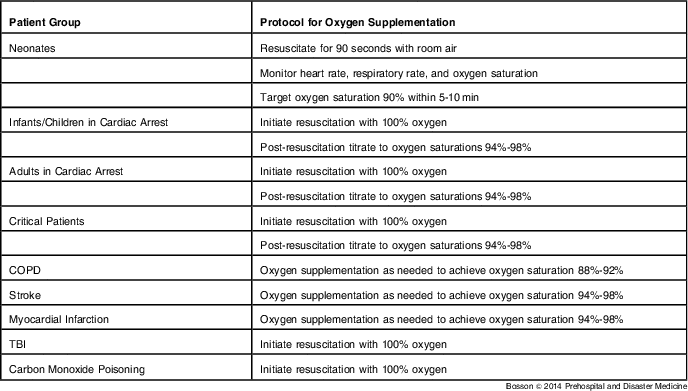
The LA County EMS Agency established titrated oxygen protocols that emphasize patient assessment to guide therapy. 4 Paramedics initiate immediate high-flow oxygen for critical patients and titrate oxygen therapy for all other patients. This protocol is simple to implement and to teach because it aligns with typical paramedic practice, and it is applicable to all patient encounters. Table 1 defines oxygen recommendations for particular clinical conditions emphasized in current guidelines.
Table 1 Recommendations for Use of Titrated Oxygen Supplementation in Select Patient Groups in the Out-of-Hospital Setting

Abbreviations: COPD, chronic obstructive pulmonary disease; TBI, traumatic brain injury.
Regardless of the patient's chief complaint, an initial assessment is made. If the patient has respiratory failure, or is at risk for respiratory failure, high-flow oxygen is initiated. In other cases, further assessment is required and oxygen therapy is begun based on the patient's level of need. Paramedics use pulse oximetry to guide therapy, taking into special consideration patients with chronic lung disease who are at risk for hypercapnic respiratory failure. Patients who are not at known risk for hypercapnia receive oxygen when needed, to the recommended goal of 94% to 98%, while patients with COPD are titrated to a lower oxygen saturation of 88% to 92%.
There are certain situations where the out-of-hospital setting limits application of oxygen titration. Paramedics in LA County currently do not have the capacity to administer albuterol with compressed air; their continuous positive airway pressure (CPAP) machines require high-flow oxygen to function, and they cannot administer blended oxygen during neonatal resuscitation and post cardiac arrest care. These limitations force adaption of the current guidelines to the reality of out-of-hospital care delivery. In LA County, neonatal resuscitation begins with room air for up to 90 seconds, and high-flow oxygen is added based on assessment of heart rate and response to ventilation. Traumatic brain injury (TBI) posed a particular dilemma during protocol development, because there is retrospective data that patients with the best neurologic outcomes had blood oxygen levels above normal, with partial pressures of 110 to 480 mm Hg.Reference Davis, Meade and Sise 5 Oxygen pressures above this were as harmful as hypoxia. Certainly, hypoxia is to be avoided in these patients. But titration to blood oxygen levels above 100 mm Hg requires blood gas analysis not available in most EMS systems. Therefore, the protocols continue to emphasize high-flow oxygen in patients with suspected TBI, given that hypoxia is highly detrimental and mild hyperoxia may be of benefit. Furthermore, use of oxygen in this population is unlikely to deviate from the core message of reserving high-flow oxygen for critical patients with actual or impending respiratory failure, because patients with significant TBI typically present altered and at risk for airway compromise. Finally, suspected carbon monoxide poisoning also requires special attention. Given the displacement of oxygen by the carbon monoxide on the hemoglobin molecules and the limits of pulse oximetry in assessing these patients, high-flow oxygen is recommended.Reference O'Driscoll, Howard and Davison 1
Implementation of titrated oxygen therapy is more difficult for Basic EMTs as they may not have pulse oximetry to guide treatment. In Los Angeles County, Basic Life Support (BLS) providers are not required to carry pulse oximeters. Guidelines were developed to assist providers in implementing titrated oxygen therapy based on the patient's clinical status, until pulse oximetry monitoring is available. This includes beginning high-flow oxygen in critical patients, providing supplemental oxygen by nasal cannula for stable patients who have signs or symptoms of dyspnea, increasing the oxygen flow as needed, and withholding oxygen therapy in stable patients without signs and symptoms of dyspnea.
The following clinical vignettes help to illustrate how titrated oxygen therapy protocol is implemented in paramedic practice.
Discussion of Clinical Application
Patient Scenario 1
Paramedics arrive to the home of a 50-year-old male with history of COPD complaining of shortness of breath and increased cough for one week. The patient had seen his primary care physician two days earlier and received nebulizer treatments and a course of steroids. Despite compliance with the treatment, the patient reports his symptoms are getting worse. Paramedics find the patient alert and oriented. He is tachypneic with accessory muscle use and talking in short sentences. Auscultation reveals diffuse wheezing with a respiratory rate of 28 breaths per minute and an oxygen saturation of 85% on room air.
Patients with COPD adapt to lower levels of oxygen within the body. While in healthy people, administration of oxygen leads to increase minute ventilation, in patients with COPD, hyperoxia has detrimental effects on the respiratory status. Multiple mechanisms are believed to contribute, including depression of ventilation due to abnormal chemoreceptors, worsening ventilation-perfusion mismatch from recruitment of poorly ventilated areas of the lungs, promotion of absorption atelectasis, and reduction of the buffering capacity for carbon dioxide. The result is hypercapnic respiratory failure. Studies evaluating inpatient management of patients with COPD have demonstrated increases in mortality, hospital length of stay, intensive care unit (ICU) admission, and ventilator requirements with the use of high-flow oxygen.Reference Joosten, Koh, Bu, Smallwood and Irving 6 , Reference Plant, Owen and Elliott 7 Similarly, studies in the out-of-hospital setting have shown that if the practice of beginning high-flow oxygen in the ambulance is continued in the emergency department, it is associated with increased length of stay, need for ventilator assistance, and risk of ICU admission.Reference Joosten, Koh, Bu, Smallwood and Irving 6 , Reference Wijesinghe, Perrin and Healy 8 A randomized controlled trial by Austin and colleagues of treatment of COPD in the out-of-hospital setting found an absolute mortality reduction of five percent for patients treated with titrated oxygen compared to those treated with high-flow oxygen (four percent and nine percent, respectively).Reference Austin, Wills, Blizzard, Walters and Wood-Baker 9 The relative risk for death in the titrated oxygen group was 0.42 (95% CI, 0.2-0.9), compared to the high-flow oxygen group. In addition to titrated oxygen therapy, continuous positive airway pressure (CPAP) is feasible in the out-of-hospital setting. Its use can decrease need for intubation and lower mortality.Reference Thompson, Petrie, Ackroyd-Stolarz and Bardua 10 The BTS guidelines support reduced oxygen saturation goals of 88% to 92% in patients at risk for hypercapnia, with consideration of CPAP if the condition persists despite treatment.
Paramedics note that this patient is hypoxic and recognize that his history of COPD places him at risk for hypercapnic respiratory failure. They begin albuterol treatment via nebulizer and administer supplemental oxygen via nasal cannula to a goal saturation of 88% to 92%. On reassessment, the patient demonstrates some improvement, but still has increased work of breathing and borderline oxygen saturation of 87% to 88% despite the nasal cannula at four liters per minute. The paramedics respond by initiating CPAP, using blended oxygen when available, and note a significant improvement in the patient's respiratory status en route.
Patient Scenario 2
Paramedics are called to an office building and arrive to find a 65-year-old male in moderate distress complaining of substernal chest pain that began 10 minutes prior. The patient is alert. He appears uncomfortable and diaphoretic. His vital signs are: blood pressure 152/90 mm Hg, heart rate 90 beats per minute, respiratory rate 22 breaths per minute, and oxygen saturation 96% on room air. An electrocardiogram (ECG) is performed and STEMI is noted in the inferior leads.
It was long believed that oxygen administration in the setting of acute myocardial infarction would result in increased oxygenation of the nutrient-deprived tissues, and thus, reduce myocardial damage. This was demonstrated initially in a swine model,Reference Cason, Wisneski and Neese 11 but its translation to humans remained unclear. Clinical practice developed from the logical belief that more inhaled oxygen, resulting in more oxygen in the blood, would lead to more oxygenation of ischemic tissues. While clinically plausible and anecdotally supported, the widespread use of oxygen in the setting of myocardial ischemia was not evidence-based. In a Cochrane review of three trials evaluating the effect of oxygen on mortality and pain in the setting of acute myocardial infarction, no trials demonstrated a benefit to supplemental oxygen.Reference Cabello, Burls, Emparanza, Bayliss and Quinn 12 Other reviews also have failed to find a benefit of oxygen treatment and suggest there may be harm.Reference Wijesinghe, Perrin, Ranchord, Simmonds, Weatherall and Beasley 13 , Reference Nicholson 14 There are multiple proposed mechanisms of harm, including raising systemic vascular resistance, blood pressure, heart rate, and cardiac oxygen consumption, as well as reducing blood flow to the ischemic myocardium through vasoconstriction.Reference Wijesinghe, Perrin, Ranchord, Simmonds, Weatherall and Beasley 13 , Reference Kones 15 , Reference Moradkhan and Sinoway 16 McNulty and colleagues demonstrated increased coronary resistance and decreased coronary blood flow during catheterization of 18 patients with coronary artery disease when 100% oxygen by face mask was administered.Reference McNulty, King and Scott 17 Adhering to the principle of first doing no harm, routine use of supplemental oxygen in patients with myocardial infarction is not supported by the literature. The BTS guidelines advise a titrated approach to oxygen therapy with a goal of 94% to 98%.
The paramedics place the patient on the monitor, administer aspirin and nitroglycerin with relief of the pain, and insert an intravenous fluid drip. Noting the patient's normal oxygen saturation ≥94%, they do not administer supplemental oxygen. After alerting the cardiac receiving center of the STEMI, they continue to monitor the patient for deterioration en route to the hospital.
Patient Scenario 3
Paramedics arrive at the home of a 72-year-old female. Family called 9-1-1 after she slumped in her chair and began slurring her speech. Paramedics find that she is having difficulty speaking and has moderate weakness of her right arm and right leg. Her vital signs and blood glucose are within normal limits.
Stroke results in areas of compromised blood flow in the brain. Oxygen, both normobaric and hyperbaric, has long been advocated as an important treatment in stroke. The rational is that increasing oxygen tensions in the ischemic penumbra (the area around the core region of infarction that is subject to reversible ischemia) can reduce the size of the final infarct territory. Hyperbaric oxygen for stroke has been demonstrated to be beneficial in animal studies, but not in humans.Reference Singhal 18 Some studies have found harm with oxygen therapy, postulated to be due to vasoconstriction or generation of free radicals leading to reperfusion injury.Reference Singhal 18 Ronning et al evaluated the effect of supplemental oxygen on 1-year survival and neurologic impairment after stroke. Using a quasi-randomized design, they compared patients with stroke treated with supplemental oxygen for 24 hours to a control group without oxygen therapy. The authors found that oxygen treatment did not benefit stroke victims and increased mortality among the patients with mild to moderate strokes, concluding that supplemental oxygen should not be routinely administered to these patients.Reference Ronning and Guldvog 19 This is supported by the BTS guidelines that advocate titrated oxygen therapy in stroke patients with a goal of 94% to 95% oxygen saturation.
Paramedics place the patient on the monitor with pulse oximetry. They note a normal oxygen saturation of 95% on room air and do not administer further supplemental oxygen. After alerting the hospital of their impending arrival, they continue to monitor the patient en route to the designated stroke center
Patient Scenario 4
Paramedics arrive on scene to find a 60-year-old female with history of congestive heart failure in severe respiratory distress. The patient is hypertensive, tachypneic, tachycardic, and using accessory muscles. Auscultation reveals crackles bilaterally. The monitor shows arterial fibrillation at a rate of 120. Recognizing the patient's critical status, paramedics immediately administer 100% oxygen via nonrebreather and sublingual nitroglycerin as they prepare for continuous positive pressure ventilation. However, the patient rapidly deteriorates to a pulseless electrical activity arrest. After 10 minutes of resuscitation, return of spontaneous circulation (ROSC) is achieved.
For critical patients (actual or impending arrest), 100% oxygen is supported in the guidelines. The role of oxygen during resuscitation remains uncertain. However, once ROSC is achieved, there is evidence that too much oxygen is harmful. Reperfusion injury is believed to occur because of an increase in oxidative stress with a buildup of reactive oxygen species. Neuronal tissue may be at increased risk during the vulnerable period after global hypoxia, causing normal endogenous antioxidants to be overwhelmed. Animal models have supported this, demonstrating improved neurologic outcome with rapid titration of oxygen after ROSC compared to 100% oxygen.Reference Neumar 20 - Reference Balan, Fiskum, Hazelton, Cotto-Cumba and Rosenthal 22 Studies in humans also have demonstrated harm with hyperoxia. A large retrospective study of patients treated in the ICU after ROSC found that both hyperoxemia and hypoxemia were associated with increased mortality compared with normal oxygen levels in the blood.Reference Kilgannon, Jones and Shapiro 23 Hyperoxemia also was associated with worse neurologic outcome at discharge and was an independent predictor of mortality. On multivariate analysis of the same cohort, excluding patients with hypoxia and obvious need for oxygen treatment, the authors found that each 100 mmHg increase in the highest arterial oxygen pressure measured after ROSC resulted in increased odds of in-hospital mortality (OR=1.24; 95% CI, 1.18-1.31).Reference Kilgannon, Jones and Parrillo 24 Current guidelines, including BTS and the American Heart Association ((AHA) Dallas, Texas USA) and European Resuscitation Council (Edegem, Belgium), recommend titrated oxygen supplementation in patients resuscitated after cardiac arrest.Reference Deakin, Nolan and Soar 25
The paramedics secure the patient's airway and assist ventilations, while continually monitoring the patient en route to the cardiac receiving facility. They are unable to titrate the oxygen level in the field and continue to administer 100% oxygen via bag-mask-ventilation. They know that the oxygen administration will be rapidly titrated, guided by pulse oximetry and blood gas analysis, once the patient arrives in the emergency department.
Patient Scenario 5
Paramedics respond to a construction site where a worker has fallen 20 feet onto cement. They find him altered with a Glasgow Coma Scale (GCS) of 11. His vital signs are: blood pressure of 90/60 mmHg, heart rate 118 beats per minute, respiratory rate 22 breaths per minute, and oxygen saturation 98% on room air. He has a hematoma to the right parietal region, multiple long-bone fractures, and an unstable pelvis.
As with other conditions resulting in tissue hypoxia, there is concern that hyperoxia in hemorrhagic shock may increase reactive oxygen species resulting in oxidative stress and worsening tissue damage.Reference Knight, Fry, Clancy and Pierce 26 There is currently no evidence, for the general trauma patient, that supplemental oxygen is of benefit in patients that are not hypoxic. Stockinger et al reviewed over 5,000 trauma patients and found that oxygen administration was associated with increased mortality, after adjustment for injury severity score, mechanism of injury, and age.Reference Stockinger and McSwain 27 However, TBI is more complicated. There is some evidence that patients with TBI may have improved neurologic recovery when partial pressures of oxygen in the blood are maintained at levels slightly above normal.Reference Davis, Meade and Sise 5 Most of the evidence supporting hyperoxia in TBI has not looked at patient-centered outcomes, and it remains unclear what affect oxygen supplementation has on neurologic outcome after TBI.Reference Knight, Fry, Clancy and Pierce 26 The BTS guidelines recommend supplemental oxygen use in all patients at risk for hypoxia, including those with major trauma and shock.
Paramedics assess the patient and are concerned for hemorrhagic shock given the altered mental status, hypotension, and unstable pelvis. Traumatic brain injury is also of concern. Despite the patient's normal oxygen saturation on room air, they administer 100% oxygen via nonrebreather. They perform c-spine immobilization and transport the patient rapidly to the nearest trauma center, establishing intravenous access en route.
Patient Scenario 6
Paramedics respond to the home of a 29-year-old gravida 4, para 3 female in active labor. On arrival, the paramedics note that the infant's head is crowning. The mother states that she has had prenatal care and that she is 32 weeks pregnant. They recognize that delivery is imminent and set up equipment for the delivery of a premature infant. The baby girl is born and is not spontaneously breathing. After quickly drying and stimulating the infant, paramedics note the heart rate remains below 100 beats per minute and the infant is gasping.
The newborn requiring resuscitation has invariably suffered an anoxic event. Some oxygen is needed to reverse hypoxic-induced apnea, but the amount necessary may only be 15% to 18% inspired oxygen.Reference Lefkowitz 28 , Reference Feet, Medbo, Rootwelt, Ganes and Saugstad 29 Oxidative stress forces the creation of free radicals, which have deleterious effects in neonates as they do in adults. The impact of such effects may be more serious in the newborn that is used to an in utero environment with lower oxygen tension.Reference Kattwinkel, Perlman and Aziz 30 There are now numerous studies in newborns and in newborn animal models showing increased survival and improved neurological outcome when resuscitation is initiated with room air versus 100% oxygen.Reference Ramji, Ahuja, Thirupuram, Rootwelt, Rooth and Saugstad 31 - Reference Wang, Anderson, Leone, Rich, Govindaswami and Finer 39 Blood oxygen levels of newborns may take over 10 minutes to achieve an oxygen saturation of >90%.Reference Rabi, Yee, Chen and Singhal 40 - Reference Dawson, Kamlin and Vento 42 In an international controlled trial of newborns resuscitated with room air or oxygen (RESAIR 2 study), there were no differences in oxygen saturations within 10 minutes whether the infants were resuscitated with 100% oxygen or not.Reference Saugstad, Rootwelt and Aalen 43 The initiation of 100% oxygen also delays the start of spontaneous respirations as well. Furthermore, visual assessment of cyanosis correlates poorly with actual oxygen saturation measurements.Reference O'Donnell, Kamlin, Davis, Carlin and Morley 44 Therefore, recent guidelines by the AHA and the Australian and New Zealand Resuscitation Councils suggest that pulse oximetry is recommended to monitor progress of resuscitation in newborns, and that regardless of gestational age, the “goal of oxygen administration should be to aim for those of healthy term babies” (Table 2).Reference Berg, Schexnayder and Chameides 45 - 48 For term infants, resuscitation should be initiated with room air, and oxygen supplementation should be added only if the oxygen saturations do not meet the timed targets. It is suggested to initiate room air resuscitation, or resuscitation with blended oxygen, if timed targets are not quickly achieved for preterm infants <32 weeks of gestation.Reference Kleinman, Chameides and Schexnayder 46 Both hypoxia and hyperoxia should be avoided.
Table 2 Recommended Targets for Oxygen Saturation of Newborns from Time of Birth[ Reference Rabi, Yee, Chen and Singhal 40 , Reference Dawson, Kamlin and Vento 42 , 48 ]

It is challenging to extrapolate the neonatal data to infants and children resuscitated post cardiac arrest. Animal studies do suggest that ventilation with 100% oxygen during and post arrest contribute to free-radical mediated reperfusion injury to the brain. In its 2010 guidelines, the AHA recommends that infants and children be resuscitated with 100% oxygen, but that post arrest, the oxygen should be titrated to maintain an oxygen saturation ≥94%.
The resuscitation of the newborn is both a low frequency and high impact event for the out-of-hospital provider. Development of simple protocols that embrace the evidence, allow for limitations in logistics, such as availability of blended oxygen, and allow for ease of use in a stressful situation is key. This is why the protocol developed stresses use of room air resuscitation for 90 seconds and then, if the patient's heart rate has not risen to appropriate levels (>100 beats per minute), resuscitation with 100% oxygen be initiated. The paramedic can monitor for oxygen saturations and later titrate to >90% saturation, as is appropriate.
Paramedics begin bag-mask ventilation with room air and place a pulse oximetry probe on the infant's right hand (preductal) to monitor oxygen saturation. Within five minutes, the infant's oxygen saturation reaches 90% and the infant begins spontaneous respirations. The heart rate increases to 140 beats per minute and the paramedics cease assisted ventilation and monitor respirations, oxygen saturation, and heart rate en route to the pediatric receiving facility.
Limitations
This report is not intended as a comprehensive review of the literature on oxygen therapy. This is one protocol adapted from the current guidelines. There may be other equally valid approaches to titrated oxygen therapy in the out-of-hospital setting. Although intended to be simple to implement and to adapt, this protocol developed for LA County EMS may not be generalizable to all EMS systems.
Conclusion
Based on current evidence, titration of oxygen supplementation in the out-of-hospital setting can improve outcomes for selected patients.