


Introduction
Tracheal aspiration is defined as the presence of saliva, secretions or food below the level of the vocal folds.Reference Simão, Alacid, Rodrigues, Albuquerque and Furkim1, Reference Brady, Hildner and Hutchins2 Although aspiration is one of the indications for tracheostomy, the presence of a tracheostomy tube in the larynx may cause aspiration in these patients.Reference Belafsky, Blumenfeld, Lepage and Nahrstedt3
Up to 70 per cent of tracheostomised patients will have tracheal aspiration; this is an important factor in decannulation failure in these patients. Thus, it is crucial to establish criteria to determine the appropriate time for tracheostomy tube removal, and thereby minimise the risk of aspiration.Reference Ajemian, Nirmul, Anderson, Zirlen and Kwasnik4, Reference Warnecke, Suntrup, Teismann, Hamacher, Oelenberg and Dziewas5
The causes of aspiration in tracheostomy patients are multifactorial and include: disuse atrophy of the laryngeal muscles, blunting of the reflexive cough, diminished proprioception, limitation of laryngeal elevation, decreased infraglottic pressure, pressure exerted by the cuff that may compress the proximal oesophagus and residual effects from the prolonged use of narcotics. Given this complexity of causal factors, an objective method for evaluating aspiration in patients with tracheostomy has not yet been well defined, making it difficult to assess its true incidence.Reference Belafsky, Blumenfeld, Lepage and Nahrstedt3–Reference Donzelli, Brady, Wesling and Craney8
There are several tests available for the objective evaluation of swallowing; these tests have variable sensitivity, specificity and predictive values.Reference Meyers9 The fibre-optic endoscopic evaluation of swallowing was introduced by Langmore et al.Reference Langmore, Schatz and Olsen10 It involves passing a flexible endoscope through the nasal passages, with the purpose of viewing swallowing dynamics with stained food.Reference Hiss and Postma11, Reference Aviv, Murry, Zschommler, Cohen and Gartner12 It has the advantages that it can be performed at the bedside, and repeated several times to provide information on the anatomy and physiology of the larynx.Reference Hiss and Postma11, Reference Langmore, Schatz and Olson13
The original Evans blue dye test to assess swallowing in tracheostomised patients was introduced by Cameron et al.Reference Cameron, Reynolds and Zuidema14 in 1973. The test is performed by placing 4 drops of 1 per cent solution to Evans blue dye on the back of the patient's tongue every 4 hours. The patient is then placed on a suctioning programme via the tracheostomy tube for 48 hours, and the secretions are monitored for the presence of the blue dye. The modified Evans blue dye test introduces a slight variation on the original examination. In addition to the simple administration of drops containing blue dye, the modified Evans blue dye test involves the administration of food materials with the dye. The advantages of the modified Evans blue dye test are its relative ease of administration, and the lack of need for special expertise in endoscopy and for expensive endoscopic equipment.Reference Belafsky, Blumenfeld, Lepage and Nahrstedt3
Because of the large impact of aspiration on the morbidity and mortality of tracheostomised patients, and given the differences of cost-effectiveness that the tests for the early detection of aspiration represent, it is important to define which test is closest to the ideal for better follow up of these patients. This study aimed to evaluate the sensitivity and specificity of the modified Evans blue dye test compared to the fibre-optic endoscopic evaluation of swallowing to detect aspiration in tracheostomised patients.
Materials and methods
This observational accuracy study, included 17 patients hospitalised for respiratory complications, subjected to prolonged intubation, and for this reason, tracheostomised. The patients had been admitted to the Special Care Unit at the Northern Regional Hospital (Sobral, Northeast of Brazil), from January 2015 to February 2016.
The instruments used in data collection were the fibre-optic endoscopic evaluation of swallowing and the modified Evans blue dye test, methods already used in hospitalised patients at this hospital.
The fibre-optic endoscopic evaluation of swallowing was performed and interpreted by the same professionals: an ENT doctor and a speech and language pathologist. The patient remained in bed in an upright sitting position (90°). A flexible fibre-optic endoscope was introduced into one nostril, with no topical anaesthetic and no nasal vasoconstrictor. The scope was advanced into the oropharynx to enable visualisation of the laryngeal structures.
We initiated the evaluation of swallowing with the administration of 20 ml of a substance with a honey-like consistency, dyed with blue food colouring. The honey-like consistency was obtained using 2.4 g of Resource® Thicken Up™ thickening agent for 100 ml of water. The patients were assessed at the moment the food was offered (0 minutes), and then 2 hours and 4 hours later.
The fibre-optic endoscopic evaluation of swallowing result was considered positive when food below the true vocal folds was observed at any of these three assessment intervals. The modified Evans blue dye test result was considered positive when the presence of blue-tinged food during tracheal suctioning was observed, at any of the three assessment intervals.
During swallowing, the tracheostomy was digitally locked to maintain the infraglottic pressure closest to a normal physiological state. The cuff was deflated throughout the study, thus avoiding interference caused by this compression.
Means and standard deviations of the following independent variables were calculated: age (years), length of speech and language therapy (days), intubation time (days), and duration of tracheostomy plus mechanical ventilation (days). In order to validate the modified Evans blue dye test as a tool for diagnosing aspiration, and compare it to the fibre-optic endoscopic evaluation of swallowing (the ‘gold standard’), the sensitivity, specificity, and positive and negative predictive values were calculated. Logistic regression analysis was used to identify a possible relationship between the independent variables (continuous) and the dependent variable of fibre-optic endoscopic evaluation of swallowing.
The Epi Info program (version 6.04) and SPSS software (version 17.0 for Windows; SPSS, Chicago, Illinois, USA) were used for the tabulation and statistical analysis of data. The fibre-optic endoscopic evaluation of swallowing was considered the gold standard, and we compared the prevalence rates of aspiration with those revealed by the modified Evans blue dye test. Sensitivity and specificity rates were assessed. We also analysed whether there was a relationship between aspiration and the length of speech and language therapy, intubation time, and duration of tracheostomy plus mechanical ventilation.
Results
The mean age of patients was 60.2 ± 21.0 years. Fifty-two per cent of the subjects were male. The mean length of speech and language therapy was 8.6 ± 4.8 days. The mean intubation time was 11.1 ± 2.9 days. The mean duration of tracheostomy plus mechanical ventilation was 14.35 ± 12.10 days (Table 1).
Table 1. Sample characterisation

SD = standard deviation
Aspiration was detected in 10 out of 17 patients when assessed by the fibre-optic endoscopic evaluation of swallowing; of these, 1 had aspiration when evaluated by the modified Evans blue dye test. Thus, the modified Evans blue dye test had a sensitivity of 10.0 per cent and specificity of 100.0 per cent for the detection of aspiration (Table 2). There were no statistically significant relationships between patients with aspiration (as determined by a positive fibre-optic endoscopic evaluation of swallowing result) and: length of speech and language therapy, intubation time, or duration of tracheostomy plus mechanical ventilation (Table 3).
Table 2. Validity of modified Evans blue dye test for diagnosing aspiration

Table 3. Comparative analysis of study variables with positive fibre-optic endoscopic evaluation of swallowing result
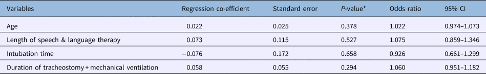
*Based on logistic regression. CI = confidence interval
Discussion
The high prevalence of aspiration in tracheostomised patients greatly increases the possibility of complications such as aspiration pneumonia. Methods to effectively and objectively assess aspiration in these patients are of fundamental importance in order to determine the best time to remove the tracheostomy tube, minimising its effects on swallowing.
Belafsky et al.Reference Belafsky, Blumenfeld, Lepage and Nahrstedt3 evaluated 30 tracheostomised patients using the modified Evans blue dye test and the fibre-optic endoscopic evaluation of swallowing, and found a sensitivity of 82 per cent and a specificity of 38 per cent for the modified Evans blue dye test (Table 4). Winklmaier et al.Reference Winklmaier, Wust, Plinkert and Wallner6 studied 30 patients with head and neck cancer post-treatment using the modified Evans blue dye test (with saliva and water), and observed a sensitivity of 95.24 per cent and specificity of 100 per cent. In our study, we found a sensitivity of only 10 per cent for the modified Evans blue dye test.
Table 4. Summary of studies assessing modified Evans blue dye test and fibre-optic endoscopic evaluation of swallowing or videofluoroscopic swallowing study
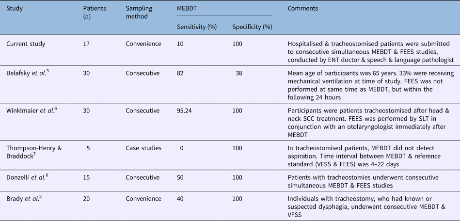
MEBDT = modified Evans blue dye test; FEES = fibre-optic endoscopic evaluation of swallowing; SCC = squamous cell carcinoma; SLT = speech and language therapist; VFSS = videofluoroscopic swallowing study
In contrast to our study, the fibre-optic endoscopic evaluation of swallowing in the study by Belafsky et al.Reference Belafsky, Blumenfeld, Lepage and Nahrstedt3 was not performed at the same time as the modified Evans blue dye test, but within the following 24 hours, which may decrease the probability of a positive (gold standard) result for the fibre-optic endoscopic evaluation of swallowing. This long interval between tests only identified aspirations that occurred after swallowing, and failed to assess those that occurred before and during swallowing. Yet the difference in sensitivity in relation to the findings by Winklmaier et al.Reference Winklmaier, Wust, Plinkert and Wallner6 may also be related to the fact we did not perform fibre-optic endoscopic evaluation of swallowing concomitantly with the modified Evans blue dye test, as the former was carried out immediately after swallowing. Discrepancies between our results and those of Winklmaier et al.Reference Winklmaier, Wust, Plinkert and Wallner6 may also have been the result of differences in the consistency of the substance offered and the structural alterations present in the patients with head and neck cancer.
The specificity value of our study is in agreement with the findings of Winklmaier et al.,Reference Winklmaier, Wust, Plinkert and Wallner6 and Thompson-Henry and Braddock.Reference Thompson-Henry and Braddock7 The latter investigated five patients with the fibre-optic endoscopic evaluation of swallowing or videofluoroscopy (both gold standard) and the modified Evans blue dye test, and found a specificity of 100 per cent (Table 4).
Donzelli et al.Reference Donzelli, Brady, Wesling and Craney8 assessed 15 patients with various neurological diseases, using food with an uncontrolled consistency or volume. The authors found aspiration with the fibre-optic endoscopic evaluation of swallowing in eight patients. Four of these patients had positive modified Evans blue dye test results, showing a false-negative rate of 50 per cent. These results are in agreement with the results of Brady et al.,Reference Brady, Hildner and Hutchins2 who evaluated 20 patients with various neurological diseases using 10 ml of food in nectar-like and pureed consistencies with the modified Evans blue dye test and videofluoroscopy. They found a 50 per cent false-negative rate for the modified Evans blue dye test. We found a 90 per cent false-negative rate in patients evaluated with the modified Evans blue dye test.
In our study, patients were evaluated at three different time points (0 minutes, 2 hours and 4 hours) after receiving 20 ml of a substance with a honey-like consistency. This long period of assessment, associated with a greater amount of food offered, simplifies the observation of small amounts of food aspirated after swallowing because of food stasis in the pharyngolaryngeal region. Post-swallowing aspirations are difficult to observe with the modified Evans blue dye test, because frequently only a small amount of food is aspirated, and this may blend in with tracheal secretions, hampering detection. These observations may explain the differences between our findings and those reported by Donzelli et al.,Reference Donzelli, Brady, Wesling and Craney8 who did not specify the type or amount of food, or how many times the patients were assessed. This could also explain the differences with the findings by Brady et al.,Reference Brady, Hildner and Hutchins2 who offered up to 10 ml of food, and there was no sequential evaluation after the offer of food. The homogeneity of our sample, in which all patients had pulmonary disease, with preserved sensitivity in the laryngeal region, differs from samples in the studies by Donzelli et al.Reference Donzelli, Brady, Wesling and Craney8 and Brady et al.,Reference Brady, Hildner and Hutchins2 and may explain the considerable discrepancy in findings (Table 4).
In the patients with positive aspiration findings, shown by fibre-optic endoscopic evaluation of swallowing, there were no statistically significant associations with length of speech and language therapy, orotracheal intubation time, or duration of tracheostomy with mechanical ventilation. The absence of a significant relationship with the length of speech and language therapy may be related to the short period of time in which these patients were accompanied by a speech and language pathologist. Regarding intubation time, we observed that the intubation-to-tracheostomy standard transition protocol, which is 10 days, was followed. Mechanical ventilation associated with tracheostomy may not have presented a significant relationship, as some authors report that the tracheostomy tube does not influence the dynamics of laryngeal movements.Reference Terk, Leder and Burrell15
Compared to previously published studies on this topic, our study is distinct in terms of the homogeneity of patients’ pathology. No patients presented a pathology that altered laryngeal sensitivity. The assessment involved a reasonable amount of food and the evaluation comprised a long follow-up period, thereby facilitating the detection of aspiration after swallowing. Furthermore, the chosen substance consistency was honey-like, and therefore different from the studies that used liquid. This increased the probability of detection for both tests, as this consistency easily adheres to the tracheal region.
• The high prevalence of aspiration in tracheostomised patients increases the possibility of complications such as aspiration pneumonia
• The modified Evans blue dye test had a sensitivity of 10.0 per cent and specificity of 100.0 per cent for detecting aspiration
• This dye test is simple and inexpensive, and does not require prior knowledge in endoscopy
• The dye test may be used for initial screening in all tracheostomised patients, to evaluate aspiration
Conclusion
The modified Evans blue dye test is simple and inexpensive to carry out, and it does not require prior knowledge in endoscopy. It may very well be used as an initial screening test in all tracheostomised patients for the evaluation of aspiration. Our results have shown that the modified Evans blue dye test can be useful for determining when to request the fibre-optic endoscopic evaluation of swallowing. Studies with a greater number of patients are needed to confirm the ideal objective test in this class of patients.
Acknowledgements
The authors would like to thank patients and staff at the Northern Regional Hospital, Sobral-Ceará for their participation and co-operation during this study. This project was funded by the Federal University of Ceará, Sobral Unit – Research Initiative Grant.
Competing interests
None declared


