Introduction
Otitis externa is characterised by inflammation of the external auditory canal, and constitutes a large proportion of presentations to both primary care and otolaryngology clinics worldwide.Reference Rowlands, Devalia, Smith, Hubbard and Dean1 The annual prevalence in the USA is 0.4 per cent.Reference Guthrie2 The most common pathogens known to cause otitis externa are Pseudomonas aeruginosa and Staphylococcus aureus.Reference Ninkovic, Dullo and Saunders3, Reference Walshe, Rowley and Timon4
For mild otitis externa, topical acidifying drops may be sufficient. However, for the majority of otitis externa cases, topical combined antimicrobial and steroid drops are recommended.Reference Hannley, Denneny and Holzer5 Aminoglycoside (neomycin), polymyxin B and steroid drops are used most frequently because of low costs and availability compared with quinolone drops. Oral antibiotics are reserved for severe cases and base of skull osteomyelitis. Increasing antimicrobial resistance is a global problem; resistant organisms are becoming more frequently reported in otitis externa patients.Reference Walshe, Rowley and Timon4, Reference Davies and Davies6
Literature investigating the microbiology and antimicrobial resistance in otitis externa in the UK is limited to a single paper published in 2008.Reference Ninkovic, Dullo and Saunders3 This study aimed to expand current microbiological understanding and identify changes in antimicrobial resistance in the UK.
Materials and methods
Ear swabs taken from patients with otitis externa presenting to the secondary care rapid access clinic at The University Hospital of South Manchester NHS Foundation Trust, from 1 January 2015 to 30 March 2016, were analysed. The rapid access clinic receives referrals from primary care and the emergency department. Microbiological samples are taken from all new otitis externa presentations and repeated if clinically indicated.
Overall, 302 swabs were collected from 217 patients (100 male, 117 female) who were diagnosed with otitis externa by senior house officers. The patients’ average age was 49.7 years (range, 3.9–95.5 years). Forty bilateral and 45 repeat swabs were taken. Patients who had otitis media, base of skull osteomyelitis or alternative ear pathologies were excluded. Patients aged under three months of age were also excluded. The sensitivities and resistances of 10 swabs that cultured P aeruginosa were unavailable and so were not included in the susceptibility results.
All swabs were collected by senior house officers and processed in the Public Health England microbiology laboratory at our hospital. Columbia blood agar, chocolate agar, cystine lactose electrolyte deficient agar, Sabouraud agar, staph/strep agar and fastidious anaerobe agar plates were inoculated with the swabs. The organisms were identified using standard microbiological techniques. Antimicrobial susceptibilities were determined by the Vitek® automated system, using European Committee on Antimicrobial Susceptibility Testing (‘EUCAST’) criteria to categorise the antimicrobials as susceptible, resistant or intermediate.
Results
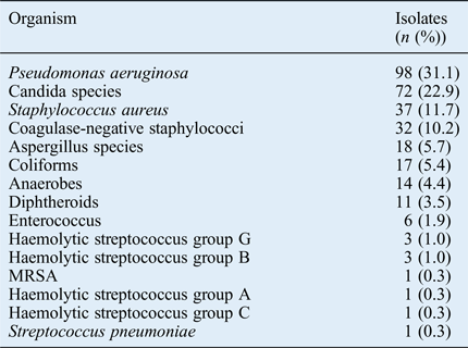
In total, 302 swabs were analysed. Of these, 232 swabs grew organisms, with the most common growth being bacterial (71.4 per cent) followed by fungal (28.6 per cent) (Figure 1). In all, 315 organisms were isolated, with the most frequent being P aeruginosa (Table I). Of the bilateral swabs (n = 40), 34 (85.0 per cent) grew the same organism. A total of 45 repeat swabs were taken; 24 (53.3 per cent) of these cultured the same organisms.

Fig. 1 Swab result categorised by organism type.
Swabs identifying P aeruginosa, S aureus, anaerobes, haemolytic streptococci, Streptococcus pneumoniae and methicillin-resistant S aureus (MRSA) were tested for antimicrobial sensitivity. P aeruginosa was sensitive to ciprofloxacin in 97.7 per cent of cases and gentamicin in 78.4 per cent (Table II). Every S aureus species isolated was sensitive to gentamicin (Table III).
Table II Pseudomonas aeruginosa susceptibility*

Data represent numbers (and percentages) of swabs.
* n = 88
Table III Staphylococcus aureus susceptibility*

Data represent numbers (and percentages) of swabs.
* n = 37
Anaerobes were 100 per cent sensitive to metronidazole on the nine occasions tested. Haemolytic streptococcus groups A, C and G were all 100 per cent sensitive to penicillin and clarithromycin. The single isolate of S pneumoniae was sensitive to penicillin. One swab containing MRSA was cultured and was sensitive to doxycycline.
Discussion
Studies investigating the microbiology of otitis externa have been published worldwide (Table IV).Reference Ninkovic, Dullo and Saunders3, Reference Walshe, Rowley and Timon4, Reference Jayakar, Sanders and Jones7–Reference Kiakojuri, Omran, Jalili, Hajiahmadi, Bagheri and Shahandashti12 The vast majority of the studies identified either P aeruginosa or S aureus to be the most common bacterial pathogens causing otitis externa.Reference Ninkovic, Dullo and Saunders3, Reference Walshe, Rowley and Timon4, Reference Jayakar, Sanders and Jones7–Reference Dibb11 However, recently, Kiakojuri et al. found that bacillus species were the most frequent pathogen causing otitis externa in Iran.Reference Kiakojuri, Omran, Jalili, Hajiahmadi, Bagheri and Shahandashti12 Studies from the UK, Ireland and the USA have a higher proportion of P aeruginosa isolates compared to developing countries.
• Otitis externa is most frequently caused by Pseudomonas aeruginosa, Staphylococcus aureus and candida species
• Rates of gentamicin-resistant P aeruginosa are increasing
• Quinolone antibiotics remain effective for treating P aeruginosa
Table IV Worldwide comparison of microbiology of otitis externa

By far the largest study investigating the microbiology of otitis externa patients was a multicentre study conducted in the USA. Roland and Stroman found the most frequent pathogen to be P aeruginosa (38 per cent), followed by Staphylococcus epidermidis (9.1 per cent) and S aureus (7.8 per cent).Reference Roland and Stroman9 Swabs containing S epidermidis at our centre were either reported as coagulase-negative staphylococci or skin flora. The current study bears similarities to the findings of Roland and Stroman regarding the distribution of bacterial organisms found.
This study supplements the current literature surrounding the microbiology of otitis externa in the UK. When compared with the previous UK study, published in 2008,Reference Ninkovic, Dullo and Saunders3 the current study identified a similar distribution of bacterial isolates in otitis externa cases. However, the proportion of fungal isolates was significantly higher in the current study (Table IV). Fungal isolates were most commonly identified in Argentina and most infrequently found in the USA (Table IV). It is known that otomycosis occurs more frequently in mild and wet climates, which may account for these findings.Reference Amigot, Gomez, Luque and Ebner10 A prospective trial identifying the predisposing factors of otomycosis is required at our centre. Appropriate treatment of otomycosis is essential, as the use of topical antibiotics is known to propagate further fungal growth.Reference Hannley, Denneny and Holzer5
No previous studies have documented the outcomes of taking bilateral or repeated ear swabs in otitis externa patients. We have demonstrated that there is a high likelihood that patients presenting with bilateral otitis externa will have the same pathogenic organism in both ears; hence, culturing a single swab may be sufficient. However, patients who re-present with otitis externa may require additional microbiological cultures because of the high likelihood of a different pathological organism.
Rising antimicrobial resistance worldwide could be one of the greatest challenges currently facing the medical profession. Identification of resistant organisms is therefore essential to guide appropriate antimicrobial use.Reference Davies and Davies6 Alarmingly, MRSA is becoming more commonly cultured in otitis externa.Reference Walshe, Rowley and Timon4
Studies from Roland and Stroman,Reference Roland and Stroman9 and Ninkovic et al.,Reference Ninkovic, Dullo and Saunders3 demonstrated that P aeruginosa is more frequently sensitive to ciprofloxacin than gentamicin: 99 per cent versus 96 per cent, and 100 per cent versus 98.5 per cent, respectively. The current study reveals a much higher proportion (78.4 per cent) of P aeruginosa species resistant to gentamicin. In addition, 97.7 per cent of P aeruginosa organisms were sensitive to ciprofloxacin, which is consistent with previous studies. The sensitivities of S aureus reported in the current study echo those documented previously showing high levels of sensitivity to gentamicin and flucloxacillin.Reference Ninkovic, Dullo and Saunders3, Reference Roland and Stroman9
Systematic review has demonstrated the efficacy of topical antibiotics for otitis externa. Without therapy, resolution occurs in 15 per cent of patients, compared with 65–80 per cent of patients receiving topical antimicrobials with or without corticosteroids.Reference Rosenfeld, Singer, Wasserman and Stinnett13 Topical drops containing aminoglycoside and steroid have historically been the mainstay of treatment for otitis externa. Recent evidence in the form of meta-analysis has shown quinolone monotherapy to be superior in treating otitis externa.Reference Mösges, Nematian-Samani, Hellmich and Shah-Hosseini14 Quinolones have mainly Gram-negative and some Gram-positive cover, making them the ideal agent to treat P aeruginosa. Although quinolones are more expensive, they reduce the risk of ototoxicity and local hypersensitivity compared with aminoglycosides.Reference Hannley, Denneny and Holzer5 In many otitis externa patients, ototoxic drops should be avoided when canal debris and swelling prohibits assessment of tympanic membrane perforation.
Topical antibiotic drops are only effective if the ear canal is clear of debris or a Pope ear wick is inserted. It has been suggested that many cases of resistant otitis externa in primary care are caused by a lack of canal clearing before the application of topical antibiotics.Reference Hannley, Denneny and Holzer5
Current UK opinion advocates using aminoglycosides for short periods as first-line therapy in patients with obvious infection, regardless of tympanic patency.Reference Phillips, Yung, Burton and Swan15 Conversely, American guidance recommends the use of quinolone antibiotics rather than aminoglycosides, because of similar efficacy and reduced ototoxicity.Reference Rosenfeld, Schwartz, Cannon, Roland, Simon and Kumar16 Future UK antibiotic guidance should reflect the current literature, which demonstrates the efficacy and safety of quinolones and the increasing antibiotic resistance to aminoglycosides in the UK.Reference Rosenfeld, Singer, Wasserman and Stinnett13
The patterns of prevalence of different organisms causing otitis externa and antimicrobial resistance differ worldwide. This study has demonstrated that in our population the most frequent causative pathogen in otitis externa is P aeruginosa. Increasing resistance of P aeruginosa to gentamicin may necessitate changes in prescribing guidance, favouring more effective antimicrobials such as quinolones. Studies are required to identify whether similar resistance patterns are apparent worldwide.


