


Introduction
Just over 110 years after its first description, Chagas disease, caused by the protozoan Trypanosoma cruzi, is still a serious public health problem, affecting approximately 8 million people worldwide, mainly in 21 Latin American countries (Echeverria and Morillo, Reference Echeverria and Morillo2019; Santana et al., Reference Santana, Oliveira, Barros de Castro and Pereira2020). Trypanosoma cruzi has intraspecific genetic heterogeneity, which is grouped into six Discrete Typing Units (DTUs) from TcI to TcVI, and Tcbat based on phylogenetic, molecular, biochemical and biological markers (Zingales et al., Reference Zingales, Andrade, Briones, Campbell, Chiari, Fernandes, Guhl, Lages-Silva, Macedo, Machado, Miles, Romanha, Sturm, Tibayrenc, Schijman and Second Satellite2009; Zingales, Reference Zingales2018) and five TcI subgenotypes have recently been described (TcIa–TcIe) (Cura et al., Reference Cura, Mejia-Jaramillo, Duffy, Burgos, Rodriguero, Cardinal, Kjos, Gurgel-Goncalves, Blanchet, De Pablos, Tomasini, da Silva, Russomando, Cuba, Aznar, Abate, Levin, Osuna, Gurtler, Diosque, Solari, Triana-Chavez and Schijman2010; Gómez-Hernández et al., Reference Gómez-Hernández, Rezende-Oliveira, Nascentes, Batista, Kappel, Martinez-Ibarra, Contreras, Lages-Silva and Ramírez2011).
The TcI genotype is widely distributed in northern South America, Central America and Mexico, and is responsible for the majority of the clinical manifestations of Chagas disease in Mexican patients (Monteon et al., Reference Monteon, Triana-Chavez, Mejia-Jaramillo, Pennignton, Ramos-Ligonio, Acosta and Lopez2016). Mexican strains of T. cruzi I (TcIa) have distinct biological characteristics such as metacyclogenesis, growth, infectivity and they also differ in their ability to invade cells and cause infection (Gómez-Hernández et al., Reference Gómez-Hernández, Rezende-Oliveira, Nascentes, Batista, Kappel, Martinez-Ibarra, Contreras, Lages-Silva and Ramírez2011, Reference Gómez-Hernández, Perez, Rezende-Oliveira, Barbosa, Lages-Silva, Ramirez and Ramirez2019; Barbosa et al., Reference Barbosa, Carvalho Costa, Desiderio, Ferreira, Silva, Hernandez, Santos, Trevisan, Bovi, Rodrigues, Machado, Ramirez, de Oliveira and da Silva2019a, Reference Barbosa, Gomez-Hernandez, Rezende-Oliveira, Da Silva, Rodrigues, Tiburcio, Ferreira, Rodrigues, Yoshida and Ramirez2019b).
With the success obtained after the implementation of control measures for the triatomine bug vector (also called ‘kissing bugs’), other forms of transmission, such as congenital, have posed a new challenge in the implementation of research and public health policies. This form of transmission is a problem that persists in endemic countries and is common in non-endemic countries (Buekens et al., Reference Buekens, Cafferata, Alger, Althabe, Belizan, Carlier, Ciganda, Dumonteil, Gamboa-Leon, Howard, Matute, Sosa-Estani, Truyens, Wesson and Zuniga2013; Bustos et al., Reference Bustos, Milduberger, Volta, Perrone, Laucella and Bua2019). Congenital transmission is of great importance due to the global spread of Chagas disease, and despite the low rate of transmission through this route, it represents a growing and neglected public health problem, in non-endemic areas especially (Liempi et al., Reference Liempi, Castillo, Carrillo, Munoz, Droguett, Galanti, Maya and Kemmerling2016; Droguett et al., Reference Droguett, Carrillo, Castillo, Gomez, Negrete, Liempi, Munoz, Galanti, Maya and Kemmerling2017; Volta et al., Reference Volta, Perrone, Rivero, Scollo, Bustos and Bua2018; Carlier et al., Reference Carlier, Altcheh, Angheben, Freilij, Luquetti, Schijman, Segovia, Wagner and Albajar Vinas2019; Kemmerling et al., Reference Kemmerling, Osuna, Schijman and Truyens2019). The rate of maternal-fetal transmission varies from 1 to 8% across the American continent (Santana et al., Reference Santana, Oliveira, Barros de Castro and Pereira2020).
During congenital transmission of T. cruzi, the placenta plays an important role and, in some cases, to prevent infection of the fetus by the parasite. However, some factors are closely involved in the vertical transmission of T. cruzi, such as the strain of the parasite, the immune responses of the mother and fetus. Some researchers consider that changes in the maternal-fetal interface, such as microdetachments, may favour transmission of the parasite (Carlier et al., Reference Carlier, Sosa-Estani, Luquetti and Buekens2015; Liempi et al., Reference Liempi, Castillo, Carrillo, Munoz, Droguett, Galanti, Maya and Kemmerling2016; Bustos et al., Reference Bustos, Milduberger, Volta, Perrone, Laucella and Bua2019; Kemmerling et al., Reference Kemmerling, Osuna, Schijman and Truyens2019).
The T. cruzi strains, such as the Colombian strain (TcI), exhibit almost 100% incidence of placental parasitism, while the Y strain (TcII) infects only 17% of placentas. In addition, both strains differ in the location of amastigotes in the placental parenchyma, while the Colombian strain amastigotes show increased tropism in placental vascular regions. The VD strain (TcVI), in turn, exhibits greater infectivity in human placental explants and placenta-derived epithelial cell lines, in comparison to the Y strain. Thus, it is clear that the parasite genotype plays an important role in placental tropism and, consequently, in the success (or otherwise) of congenital transmission (Medina et al., Reference Medina, Castillo, Liempi, Herbach, Cabrera, Valenzuela, Galanti, de Los Angeles Curto, Schijman and Kemmerling2018; Kemmerling et al., Reference Kemmerling, Osuna, Schijman and Truyens2019).
The placenta is an organ that develops during embryogenesis and is responsible for supplying nutrients to the fetus and providing hormones, growth factors and immunological protection against pathogens needed during pregnancy (Mor et al., Reference Mor, Aldo and Alvero2017; Rios et al., Reference Rios, Campos, Menon, Zago and Garg2020). This organ has immunological functions capable of initiating an innate immune response by releasing immune mediators such as pro-inflammatory cytokines, chemokines, reactive oxygen and nitrogen intermediates, and antimicrobial peptides (Carlier and Truyens, Reference Carlier and Truyens2015). Trophoblast cell turnover is an important antiparasitic mechanism involved in the innate immune system, eliminating pathogens before their placental spread (Liempi et al., Reference Liempi, Castillo, Carrillo, Munoz, Droguett, Galanti, Maya and Kemmerling2016; Kemmerling et al., Reference Kemmerling, Castillo, Liempi, Medina, Carrillo, Droguett, Maya and Galanti2017).
The rate of congenital transmission of T. cruzi is low, and studies indicate that the placenta plays an important role in preventing transmission of this parasite (Rendell et al., Reference Rendell, Gilman, Valencia, Galdos-Cardenas, Verastegui, Sanchez, Acosta, Sanchez, Ferrufino, LaFuente, Abastoflor Mdel, Colanzi and Bern2015; Kemmerling et al., Reference Kemmerling, Osuna, Schijman and Truyens2019). It has been demonstrated that multiple Mexican strains show distinct biological behaviour in vivo, ex vivo and in vitro, even though they belong to the same TcIa genotype (Gómez-Hernández et al., Reference Gómez-Hernández, Rezende-Oliveira, Nascentes, Batista, Kappel, Martinez-Ibarra, Contreras, Lages-Silva and Ramírez2011, Reference Gómez-Hernández, Perez, Rezende-Oliveira, Barbosa, Lages-Silva, Ramirez and Ramirez2019; Barbosa et al., Reference Barbosa, Carvalho Costa, Desiderio, Ferreira, Silva, Hernandez, Santos, Trevisan, Bovi, Rodrigues, Machado, Ramirez, de Oliveira and da Silva2019a, Reference Barbosa, Gomez-Hernandez, Rezende-Oliveira, Da Silva, Rodrigues, Tiburcio, Ferreira, Rodrigues, Yoshida and Ramirez2019b). The present study aimed to investigate the infectivity and modulation of immune responses in human placental explants infected with Mexican strains of T. cruzi Ia.
Materials and methods
Parasites
The T. cruzi INC-5 and Ninoa strains from Mexico belonging to the TcIa genotype were used (Gómez-Hernández et al., Reference Gómez-Hernández, Rezende-Oliveira, Nascentes, Batista, Kappel, Martinez-Ibarra, Contreras, Lages-Silva and Ramírez2011, Reference Gómez-Hernández, Perez, Rezende-Oliveira, Barbosa, Lages-Silva, Ramirez and Ramirez2019). These strains are maintained by the cryopreservation bank of the parasitology discipline of the Federal University of Triângulo Mineiro (UFTM). The parasites were grown at 28°C in liver infusion tryptose medium (Sigma-Aldrich, St. Louis, MO, USA) supplemented with 10% inactivated fetal bovine serum (FBS) (LCG Biotecnologia, Cotia, SP, Brazil). Metacyclic forms of growing cultures were purified on a DEAE cellulose column, as described previously (Yoshida et al., Reference Yoshida, Teixeira and Sbravate1986).
Trypomastigotes
VERO CCL-81 cells were infected with 105 metacyclic forms of the Mexican strains of T. cruzi. After approximately 72 h, trypomastigotes were released after lysis of the host cells. They were separated from the cell debris by low-speed centrifugation (500g). The parasites were isolated from the supernatant by centrifugation at 3500g in Roswell Park Memorial Institute (RPMI) 1640 medium (without FBS; Sigma-Aldrich) containing 50 mm HEPES buffer (Gibco, Grand Island, NY, USA), 2 mm L-glutamine (Gibco), 50 mm β-mercaptoethanol (Gibco) and 40 μg mL−1 gentamicin (Neoquímica, Anápolis, GO, Brazil), and quantified using a Neubauer chamber (Kasvi, São José dos Pinhais, PR, Brazil) (Castillo et al., Reference Castillo, Villarroel, Duaso, Galanti, Cabrera, Maya and Kemmerling2013; Liempi et al., Reference Liempi, Castillo, Duaso, Droguett, Sandoval, Barahona, Hernandez, Galanti, Maya and Kemmerling2014; Medina et al., Reference Medina, Castillo, Liempi, Herbach, Cabrera, Valenzuela, Galanti, de Los Angeles Curto, Schijman and Kemmerling2018).
Culture of placental explants and infection with T. cruzi trypomastigotes
Human term placentas were obtained from women who had uncomplicated pregnancies in the surgical centre of the Hospital of Clinics of the UFTM, with the following exclusion criteria: severe fetal and placental changes, intrauterine infection, positive serologies (e.g. HIV, hepatitis B and C), presence of any obstetric changes or maternal comorbidities. All selected pregnant women freely signed an informed consent form.
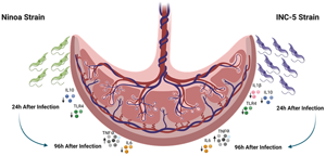
The placentas were processed in sterile phosphate-buffered saline (PBS) (80.0 g NaCl, 11.6 g Na2HPO4, 2.0 g KH2PO4, 2.0 g KCL, q.s.p. to 10 L pH to 7.0) for approximately 30 min, and aseptically dissected with the aid of a stereoscopic microscope to remove endometrial remains and fetal membranes. The tissues were collected in a buffered solution with cold PBS and processed for no more than 30 min after delivery. The placentas were processed for villous dissection: the volume of villus explants was approximately 3 mm3 and the explants were then co-cultured with T. cruzi trypomastigotes (1 × 10⁵ mL−1) in RPMI 1640 medium supplemented with 10% inactivated FBS serum (Sigma-Aldrich) and 40 μg mL gentamicin (Neoquímica) for 24 and 96 h, at 37°C in a humidified atmosphere with 5% CO2 (Duaso et al., Reference Duaso, Rojo, Cabrera, Galanti, Bosco, Maya, Morello and Kemmerling2010; Medina et al., Reference Medina, Castillo, Liempi, Herbach, Cabrera, Valenzuela, Galanti, de Los Angeles Curto, Schijman and Kemmerling2018). After this period, they were washed 3× with PBS to remove non-adherent parasites. Finally, the villi were collected for morphological, molecular and immunological analyses, and the supernatants were stored at −80°C for subsequent measurement of cytokines.
Histopathology
The placental villus explants were fixed in formaldehyde with 10% 0.1 M phosphate buffer (pH 7.3) for 24 h and then dehydrated with absolute alcohol, diaphanized in xylol, embedded in paraffin and sectioned to 5 μm. Paraffin sections were stained with haematoxylin–eosin for histological analysis (Rojo et al., Reference Rojo, Castillo, Duaso, Liempi, Droguett, Galanti, Maya, Lopez-Munoz and Kemmerling2014). Changes caused by the parasite to the structure of the placental villi were evaluated, such as disorganization and trophoblast detachment (Duaso et al., Reference Duaso, Rojo, Cabrera, Galanti, Bosco, Maya, Morello and Kemmerling2010; Medina et al., Reference Medina, Castillo, Liempi, Herbach, Cabrera, Valenzuela, Galanti, de Los Angeles Curto, Schijman and Kemmerling2018).
RNA extraction and complementary DNA (cDNA) synthesis
RNA extraction from placental explants was performed using a Promega RNA extraction kit (SV Total RNA Isolation System; Promega Corporation, Madison, WI, USA) according to the manufacturer's instructions. cDNA was synthesized from 5 μg of total RNA using oligonucleotides (primers) complementary to the poly-A tail characteristic of messenger RNA (mRNA), thus producing a purer cDNA, exclusively from mRNA. Synthesis was performed using a Reverse Transcriptase Kit (GoScript™ Reverse Transcriptase; Promega).
At the end of the synthesis, 40 μL of cDNA were obtained, which was stored at −20°C until amplification by the polymerase chain reaction technique in real time. Reverse transcription of the samples was performed in a Mastercycler®Nexus Gradient thermocycler (Eppendorf AG, Hamburg, Germany). The concentration of cDNA (ssRNA) was assessed by spectrophotometry using a NanoDrop® 2000 (Thermo Fisher Scientific, Inc., Waltham, MA, USA) using 2 μL of the solution obtained from the extraction. The purity of the RNA was considered satisfactory when the absorbance ratio at 260 and 280 nm was close to 2.0.
T. cruzi RT-qPCR
PCR primers used to amplify the selected genes were designed using PrimerExpress (Applied Biosystems, Foster City, CA, USA) (Table 1). Reactions were set up in a total volume of 20 μL using 3 μL of cDNA, 10 μL SYBRGreen I master mix (Qiagen, Hilden, Germany) and 0.8 μL of each gene-specific primer (Table 1). Reactions were performed using the Step One Plus real-time PCR system (Applied Biosystems). The cycling conditions were as follows: one cycle of 95°C for 10 min; 40 cycles of 95°C for 15 s and 60°C for 1 min with a single fluorescence measurement.
Table 1. Sequences of primers used to amplify T. cruzi genes by RT-qPCR

a Mejía-Jaramillo et al. (Reference Mejía-Jaramillo, Fernández, Palacio and Triana-Chávez2011).
b Tao et al. (Reference Tao, Yang, Wang, Wang, Luo, Tang, Dong and Ma2007).
The sample was considered valid when the internal control (human β-actin) was efficiently amplified and was considered positive for T. cruzi when the cycle threshold (Ct), which is the first cycle of the PCR reaction where fluorescence is detected, was <40. Sample normalization was performed using T. cruzi Ct and an internal control from the same sample, using the following formula: R = 2−ΔΔCt [R = 2−(Ct T. cruzi–Ct β-actin)]. The results generated a T. cruzi relative mRNA value, which was comparable between all samples in this study.
Cytokine gene expression
Expression of the IL-1β, TNF-α, IL-10 and TLR-4 genes was evaluated by real-time PCR using the StepOnePlus equipment (Applied Biosystems). The reference gene β-actin was used as an endogenous control. The final volume of the reactions was 10 μL and contained 5 μL of TaqMan Universal Mastermix (2×), 4.5 μL of cDNA (100 ng) and 0.5 μL of TaqMan® Gene Expression Assays (20 × ) (Applied Biosystems). The experiment was carried out under the following PCR conditions: 50°C for 5 min, 95°C for 10 min, and 40 amplification cycles of 95°C for 15 s and 60°C for 1 min. For the relative quantification of genes, the 2−ΔΔCt method was used (Livak and Schmittgen, Reference Livak and Schmittgen2001), where Ct is the threshold cycle at which the level of fluorescence reaches a sufficient quantity for sample detection.
Enzyme-linked immunosorbent assay (ELISA)
The concentrations of IL-1β, TNF-α and IL-6 were measured in supernatants from cultures of placental villus explants and BeWo cells by capture ELISA (BD Biosciences, San Jose, CA, USA), according to the manufacturer's instructions. Absorbance was determined by subtracting the absorbance at 570 nm from the absorbance at 450 nm using an Inspire microplate reader (Perkin-Elmer, Waltham, MA, USA). Cytokine concentrations were calculated by 5PL regression analysis of absorbance values obtained for the recombinant cytokines, and the results were expressed in picograms per millilitre. The sensitivity of the tests ranged from 2 to 20 pg mL−1. The coefficients of variation within and between trials were <10% (Barbosa et al., Reference Barbosa, Carvalho Costa, Desiderio, Ferreira, Silva, Hernandez, Santos, Trevisan, Bovi, Rodrigues, Machado, Ramirez, de Oliveira and da Silva2019a).
Statistical analysis
Statistical analysis was performed using StatView software (Abacus Corporation, Baltimore, MD, USA) and GraphPad Prism v.5.0 (GraphPad Software, La Jolla, CA, USA). Normality was checked using the Kolmogorov–Smirnov test, and Kruskal–Wallis one-way ANOVA (F) test was used to compare three or more independent samples, and the Mann–Whitney U test was used to compare two samples. The results were considered statistically significant at P < 0.05.
Results
Trypomastigotes of Mexican strains of T. cruzi infect human placental explants
The strains Inc-5 and Ninoa of T. cruzi (TcIa) were able to infect human placental explants. Initially, parasitism caused by the strains was evaluated 24 h after infection. There was a slight increase in parasitism caused by the Inc-5 strain when compared to the Ninoa strain (Fig. 1A). At 96 h, the strain Inc-5 showed a slight increase in parasitism when compared to the 24 h infection; whereas the Ninoa strain showed no differences between the times of infection (Fig. 1B and C).

Fig. 1. Quantification of T. cruzi parasitism by RT-qPCR in placental explants. Mexican strains were incubated in a culture with placental explants, the parasitism was measured by RT-qPCR through the relative quantification of samples. (A) Parasitism between Inc-5 and Ninoa strains within 24 h. (B) Comparison of the parasitism of the Inc-5 strain at 24 and 96 h. (C) Comparison of parasitism of the Inc-5 strain at 24 and 96 h. Statistical analysis was performed with the Mann–Whitney test, where *P < 0.05.
Strains Inc-5 and Ninoa of T. cruzi induce histological changes in infected chorionic villi
The presence of histological changes in placental explants was investigated after confirming the infective capacity of the strains studied. Tissue lesions such as destruction and detachment of the trophoblast and displacement of the basal lamina (arrows, Fig. 2B, C, E and F) were found in all infected placental explants 24 and 96 h after infection, in a similar way by which both strains were evaluated. Structures such as trophoblasts and villous stroma of uninfected explants showed preserved morphology (Fig. 2A and D).

Fig. 2. Mexican strains of T. cruzi induce tissue damage from placental explants. (A) Uninfected placental explant with 24 h of incubation. (B) Placental explant infected with the Inc-5 strain after 24 h incubation. (C) Placental explant infected with the Ninoa strain after incubation for 24 h of incubation. (D) Uninfected placental explant with 96 h of incubation. (E) Placental explant infected with the Inc-5 strain after 96 h of incubation. (F) Placental explant infected with the Ninoa strain after 96 h of incubation. The arrows show syncytiotrophoblast detachment caused by T. cruzi. Haematoxylin–eosin stain. 10 μm bar scale.
Modulation of cytokine gene expression in placental explants infected with Mexican T. cruzi strains
Evaluation of gene expression showed that the Mexican strains of TcIa differentially modulated signalling pathways involved in the production of cytokines (Fig. 3).

Fig. 3. Relative expression of cytokine mRNA in placental explants. Cytokines and TLR-4 receptor expressed after 24 h of incubation in HPE infected with Mexican strains of T. cruzi. (A) Expression of the IL-1β gene within 24 h of incubation. (B) Expression of the IL-1-gene within 24 h of incubation. (C) Expression of the TLR-4 gene within 24 h of incubation. (D) TNFα gene expression within 24 h of incubation. Statistical analysis was performed using the Kruskal–Wallis test, where *P < 0.05.
The strains Inc-5 and Ninoa of T. cruzi modulated the synthesis of IL-1β and IL-10 in the initial period after infection. Human placental explants infected with the Inc-5 strain showed a significant reduction in the expression of IL-1β (P = 0.0079), when compared to the control group (Fig. 3A) 24 h after infection. IL-1β expression in explants infected with the Ninoa strain was also lower, similar to that observed with the Inc-5 strain, although not statistically significant (P = 0.2222).
Explants infected with the Ninoa strain showed null expression of TLR-4 and IL10, and explants infected with the Inc-5 strain showed considerable downregulation of IL10 (P = 0.0476) and TLR-4 (P = 0.00159) (Fig. 3B and C). TNF-α, on the other hand, exhibited increased expression, and no significant difference was observed in explants infected by both Inc-5 (P = 0.1032) and Ninoa (P = 0.3333) strains (Fig. 3D).
IL-1β expression in the Inc-5 (P = 0.1429) and Ninoa (P = 0.6905) strains; IL-10 Inc-5 (P = 0.9999) and Ninoa (P = 0.9999); TNF-α Inc-5 (P = 0.9999) and Ninoa (P = 0.4444); TLR-4 Inc-5 (P = 0.9999) and Ninoa (P = 0.7222) showed no significant difference 96 h after infection; the expression of IL-10, TNF-α and TLR-4 was null (0.00).
Quantification of TNF-α, IL-6 and IL-1β in supernatants from human placental explants infected with T. cruzi Inc-5 and Ninoa strains
In view of the results found in terms of gene expression of the pro-inflammatory cytokines IL-1β, IL-10 and TNF-α in placental explants infected with the strains Inc-5 and Ninoa, the concentrations of TNF-α, IL-6 and IL-1β were quantified in culture supernatants 24 h after infection.
The concentration of TNF-α was significantly higher in the supernatants of the explants of placentas infected with the Inc-5 (P = 0.079) and Ninoa (P = 0.0079) strains than in the control group (Fig. 4A). The concentration of IL-6 was significantly lower in explants infected with Inc-5 (P = 0.0079) and Ninoa (P = 0.0317) strains than in the control group; this reduction was more evident in explants infected with the Inc-5 strain (Fig. 4B). However, no significant difference was observed in terms of the concentration of IL-1β in the Inc-5 (P = 0.8413) and Ninoa (P = 0.3095) strains at 24 h after infection (Fig. 4C).

Fig. 4. Dosage of IL-1β, TNF-α and IL-6 cytokines in the culture supernatant by ELISA. Supernatants were collected after 24 h of infection in placental explants with T. cruzi strains. (A) TNFα. (B) IL-6. (C) IL-1β. Statistical analysis was performed using the Kruskal–Wallis test, where *P < 0.05.
Discussion
The placenta is an organ that plays important roles in the health and development of the fetus; when it is infected by pathogenic microorganisms, intense inflammatory responses can put the life of the mother and child at risk, often resulting in the death of one or the other (Guttmacher et al., Reference Guttmacher, Maddox and Spong2014; Soares et al., Reference Soares, Varberg and Iqbal2018). Congenital transmission is an important route of contamination by T. cruzi and occurs in approximately 5% of children born to infected mothers, with regional variation in the rate of transmission, and is important in countries that are not part of Latin America due to the internationalization of Chagas disease (Carlier et al., Reference Carlier, Sosa-Estani, Luquetti and Buekens2015; Soriano-Arandes et al., Reference Soriano-Arandes, Angheben, Serre-Delcor, Trevino-Maruri, Gomez and Jackson2016; Messenger and Bern, Reference Messenger and Bern2018). Studies on parasites from different regions of Mexico (T. cruzi Mexican strains) show that these parasites may be able to cause placental and/or transplacental disease, which is important for understanding interactions involving the parasite and human placenta.
In congenital transmission, interactions between the parasite and chorionic villi are essential for the microorganism to invade and cause infection. It is evident that the placenta can play an extremely important role in the course of infection in order to prevent or allow the parasite to settle and for the inflammatory process to develop. Injuries caused by the presence of T. cruzi in placental tissue can favour congenital transmission, and if these are very intense, it can contribute to the occurrence of abortion (Castillo et al., Reference Castillo, Munoz, Carrillo, Liempi, Gallardo, Galanti, Maya and Kemmerling2017; Kemmerling et al., Reference Kemmerling, Castillo, Liempi, Medina, Carrillo, Droguett, Maya and Galanti2017).
There is evidence that strains of T. cruzi are more adapted for, and may have a greater capacity to generate transplacental infections. Certain genotypes of the parasite show significant tropism for the human placenta, capable of infecting and resulting in higher parasitaemia (Messenger and Miles, Reference Messenger and Miles2015; Messenger and Bern, Reference Messenger and Bern2018). In our study, Mexican strains of T. cruzi TcIa were employed, and among the T. cruzi genotypes studied in a murine model, TcI showed greater parasitism in placental tissue (Carlier et al., Reference Carlier, Sosa-Estani, Luquetti and Buekens2015; Medina et al., Reference Medina, Castillo, Liempi, Herbach, Cabrera, Valenzuela, Galanti, de Los Angeles Curto, Schijman and Kemmerling2018). However, the genotypes often related to congenital human transmission are TcIV and TcV (del Puerto et al., Reference del Puerto, Nishizawa, Kikuchi, Iihoshi, Roca, Avilas, Gianella, Lora, Velarde, Renjel, Miura, Higo, Komiya, Maemura and Hirayama2010; Gonzalez et al., Reference Gonzalez, Ortiz and Solari2010). Tissue parasitism in the placenta can often be considered virtual, as the nests of amastigotes are small and difficult to observe under conventional microscopy, whereas in striated muscle tissue, these structures are able to accommodate hundreds of parasites.
The ability of placental cells to defend against parasite infection is very effective, generating several changes both in the epithelium and in immune responses, and it is not clear whether the parasites use these response mechanisms in their favour. A number of in vitro studies have shown that placental villi can modulate T. cruzi infection (Luján et al., Reference Luján, Triquell, Sembaj, Guerrero and Fretes2004). This was observed when no significant difference in parasitism was observed in our study 24 and 96 h after infection, suggesting that replication may somehow compensate for the absence of those parasites killed by structural changes involving infected villi, such as flaking of the syncytiotrophoblast.
Basal renewal of the placental villus epithelium can be altered (accelerated or decreased) in response to various stimuli. Infection with T. cruzi generally stimulates epithelial turnover, which consists of a mechanism that aims to ensure the integrity of the trophoblast, which is considered part of the innate immune system. The parasite binds to the trophoblast, and as these cells are continuously eliminated, adherent parasites are also eliminated (Liempi et al., Reference Liempi, Castillo, Duaso, Droguett, Sandoval, Barahona, Hernandez, Galanti, Maya and Kemmerling2014, Reference Liempi, Castillo, Carrillo, Munoz, Droguett, Galanti, Maya and Kemmerling2016; Kemmerling et al., Reference Kemmerling, Castillo, Liempi, Medina, Carrillo, Droguett, Maya and Galanti2017, Reference Kemmerling, Osuna, Schijman and Truyens2019). In the present study, it was not possible to observe parasites or T. cruzi amastigote nests in placental tissue, but they were detected by RT-qPCR because of the technique's high sensitivity and specificity. However, changes such as syncytiotrophoblast detachment and destruction of the basal lamina in placental tissues were observed 24 h after incubation, and after 96 h of incubation, the tissue damage was further aggravated. As previously mentioned, tissue parasitism in the placenta can often be considered virtual, as the nests of amastigotes are small and difficult to observe using conventional microscopy but can be detected by molecular techniques such as RT-qPCR. In addition, detachment is one of the mechanisms of placental defence in an attempt to prevent congenital infections. It is widely described that syncytiotrophoblast detachment resulting from T. cruzi infection can cause abortion (Duaso et al., Reference Duaso, Rojo, Cabrera, Galanti, Bosco, Maya, Morello and Kemmerling2010; Medina et al., Reference Medina, Castillo, Liempi, Herbach, Cabrera, Valenzuela, Galanti, de Los Angeles Curto, Schijman and Kemmerling2018; Liempi et al., Reference Liempi, Castillo, Medina, Rojas-Pirela, Araneda, Maya, Parraguez and Kemmerling2021).
Pregnancy is characterized by an anti-inflammatory and pro-inflammatory profile and is, therefore, of an immunomodulatory nature. The first trimester of pregnancy, especially in the last few weeks, is characterized by a pro-inflammatory phase that is associated with implantation and placentation. Inflammation at the implant site is mainly characterized by the presence of IL-6, IL-8, IL-15 and TNF-α, which are derived from both endometrial stromal cells and infiltrated immune cells. The second trimester is characterized by an anti-inflammatory stage associated with fetal growth and development of the Th2 type. The third trimester constitutes a pro-inflammatory phase of the Th1 type, which is responsible for the initiation of delivery. Studies have shown that the signalling pathway for the pro-inflammatory nuclear factor-κB initiates labour and is crucial for the continued progress of labour and delivery. Childbirth is characterized by an influx of immune cells into the myometrium to promote the resurgence of an inflammatory process. This pro-inflammatory environment promotes contraction of the uterus, expulsion of the baby and rejection of the placenta (Abrahams et al., Reference Abrahams, Bole-Aldo, Kim, Straszewski-Chavez, Chaiworapongsa, Romero and Mor2004; Duaso et al., Reference Duaso, Rojo, Cabrera, Galanti, Bosco, Maya, Morello and Kemmerling2010; Mor et al., Reference Mor, Aldo and Alvero2017; Ander et al., Reference Ander, Diamond and Coyne2019).
IL-6 is a cytokine that, depending on the stimulatory situation, can exert pro- or anti-inflammatory effects and sometimes has an effect similar to that of hormones in order to maintain homoeostasis. Decrease in, or even deficiency of IL-6, can affect the innate and adaptive immune responses during the infectious process caused by different microorganisms (viruses, bacteria and parasites) (Hunter and Jones, Reference Hunter and Jones2015). The modulation of this cytokine by parasites may be one of the mechanisms of immune system evasion, and the T. cruzi Mexican strain (TcI) can significantly modulate levels IL-6 when compared to uninfected controls in an attempt to decrease inflammatory cytokines that generally predominate in the term placenta, which represents part of the delivery process. IL-6 can also activate macrophages in an alternative way, and thereby inhibit the ability of these cells to eliminate pathogens (Hunter and Jones, Reference Hunter and Jones2015).
TNF-α has an inflammatory action and is considered to be a pivotal factor in the activation of the Th1 cytokine cascade (pro-inflammatory profile). Each quarter of the gestational period has a variation in the pattern of immune responses, with variations in the expression of TNF-α, suggesting that this cytokine plays specific roles during each period of pregnancy. It is important to note that changes in TNF-α production can increase the risk of obstetric complications (recurrent abortion, gestational diabetes mellitus, hypertensive syndromes and fetal growth deficiency) (Haider and Knofler, Reference Haider and Knofler2009; Carpentier et al., Reference Carpentier, Dingman and Palmer2011; Brogin Moreli et al., Reference Brogin Moreli, Cirino Ruocco, Vernini, Rudge and Calderon2012; Basu et al., Reference Basu, Agamasu, Bendek, Salafia, Mishra, Benfield, Prasad and Mikhail2016).
Infection of mouse placenta by T. cruzi induces an increase in TNF-α synthesis, compromising the maintenance of pregnancy and directly reflecting on increased fetal mortality (Mjihdi et al., Reference Mjihdi, Truyens, Detournay and Carlier2004). In humans, increased production of TNF-α induced by the presence of T. cruzi in the placenta can result in the birth of children with low weight, congenital transmission or abortion, depending on the intensity of the placental lesion (dependent strain) (Torrico et al., Reference Torrico, Solano, Guzman, Parrado, Suarez, Alonzo-Vega, Truyens, Carlier and Torrico2005). Trypanosoma cruzi Mexican strains induce an increase in both expression and production of TNF-α, which may reflect the low prevalence of congenital transmission of Chagas disease observed in Mexico, especially in terms of the birth of children with low weight, without congenital transmission or abortion, which often justifies the lack of statistical data regarding this route of transmission.
IL-1β is a pro-inflammatory cytokine produced during the inflammatory process, especially in autoimmune diseases. This cytokine is processed from the pro-IL-1β molecule, which is converted to IL-1β by caspase 1 via the inflamosome (Ren and Torres, Reference Ren and Torres2009; Lawlor et al., Reference Lawlor, Feltham, Yabal, Conos, Chen, Ziehe, Graß, Zhan, Nguyen, Hall, Vince, Chatfield, D'Silva, Pang, Schroder, Silke, Vaux, Jost and Vince2017). Inflamosomes are a complex of intracellular proteins that act in the regulation of inflammation and play important roles in innate immunity and the activation of IL-1β.
Infection of placental villi by trypomastigotes induces increased levels of TNF-α, IL-1β, IL-8 and IL-6, which helps to eliminate the parasite, but can cause tissue damage if inflammation is exacerbated (Castillo et al., Reference Castillo, Munoz, Carrillo, Liempi, Gallardo, Galanti, Maya and Kemmerling2017); IL-1β is responsible for increased myosin heavy chain gene transcription and, consequently, hypertrophy of cardiomyocytes in Chagasic cardiomyopathy (Petersen and Burleigh, Reference Petersen and Burleigh2003). However, it has been observed that the Mexican strains of T. cruzi appear to suppress immune responses during placental infection, since the expression of IL-1β was significantly lower in placental explants infected by the Inc-5 strain. Genetic diversity and specific biological behaviour of different T. cruzi strains (Gómez-Hernández et al., Reference Gómez-Hernández, Rezende-Oliveira, Nascentes, Batista, Kappel, Martinez-Ibarra, Contreras, Lages-Silva and Ramírez2011) can trigger a specific immune response in Chagas disease.
Trypanosoma cruzi is able to activate evasion mechanisms that are important for its survival in host cells, and modulation of the synthesis of pro-inflammatory cytokines is one of the means of avoidance. In the present study, downregulation of IL-1β and IL-6 was observed, suggesting an avoidance mechanism (strain-dependent) that could prevent activation of the innate immune system, which results in activation of cytokine synthesis for the adaptive response. However, other mechanisms are activated, such as apoptosis pathways (caspase-3 and caspase-8) and, when significantly activated, they can induce abortion (Lemmers et al., Reference Lemmers, Salmena, Bidere, Su, Matysiak-Zablocki, Murakami, Ohashi, Jurisicova, Lenardo, Hakem and Hakem2007; Castillo et al., Reference Castillo, Munoz, Carrillo, Liempi, Gallardo, Galanti, Maya and Kemmerling2017; Lawlor et al., Reference Lawlor, Feltham, Yabal, Conos, Chen, Ziehe, Graß, Zhan, Nguyen, Hall, Vince, Chatfield, D'Silva, Pang, Schroder, Silke, Vaux, Jost and Vince2017).
TLRs are the first line of defence in the innate immune response against pathogens. Trypanosoma cruzi is recognized by TLR-2, TLR-4, TLR-7 and TLR-9. TLR-2 and TLR-4 recognize mucin-like glycoproteins anchored to the surface of T. cruzi and are considered the main mediators of immune responses. TLR-2 and TLR-4 induce the production of IL12, TNF-α and nitric oxide. The literature indicates that infection by the parasite is related to elevated expression and activation of TLR-2 and TLR-4, which leads to the activation of signalling pathways and the positive regulation of genes involved in innate immune responses (Hayden and Ghosh, Reference Hayden and Ghosh2004; Mukherjee et al., Reference Mukherjee, Karmakar and Babu2016; Olmos-Ortiz et al., Reference Olmos-Ortiz, Flores-Espinosa, Mancilla-Herrera, Vega-Sánchez, Díaz and Zaga-Clavellina2019).
However, intense tissue damage triggered by the parasite may not only interfere with the expression of the TLR-4 receptor, but also with the expression of pro- and anti-inflammatory cytokines in the placenta. Studies have not observed differences in the expression of TNF-α, IL-1β, IL-6, IL-10 and IFN-γ in cultured human placental chorionic villi infected with Toxoplasma gondii, and lower expression of IL-10 may be partially related to less-effective antiparasitic responses (Duaso et al., Reference Duaso, Rojo, Cabrera, Galanti, Bosco, Maya, Morello and Kemmerling2010; Castillo et al., Reference Castillo, Munoz, Carrillo, Liempi, Gallardo, Galanti, Maya and Kemmerling2017). It is important to emphasize that IL-10-deficient mice present impairment in the expansion and function of CD8+ T cells and in the synthesis of IL-12, demonstrating the important role of IL-10 in the balance of pro-inflammatory activity in T. cruzi infection (Pino-Martinez et al., Reference Pino-Martínez, Miranda, Batalla, González-Cappa and Soto2019). These data appear to justify the findings observed in this study, since Inc-5 and Ninoa strains triggered important lesions in placental tissue, with lower expression of immune response mediators 24 and 96 h after infection.
A previous study by our group investigated the biological behaviour (in vivo and in vitro) of Mexican strains of TcIa under different experimental conditions (Gómez-Hernández et al., Reference Gómez-Hernández, Rezende-Oliveira, Nascentes, Batista, Kappel, Martinez-Ibarra, Contreras, Lages-Silva and Ramírez2011, Barbosa et al., Reference Barbosa, Carvalho Costa, Desiderio, Ferreira, Silva, Hernandez, Santos, Trevisan, Bovi, Rodrigues, Machado, Ramirez, de Oliveira and da Silva2019a, Reference Barbosa, Gomez-Hernandez, Rezende-Oliveira, Da Silva, Rodrigues, Tiburcio, Ferreira, Rodrigues, Yoshida and Ramirez2019b). In this study, ex vivo infection of human placental villi was performed to better understand parasitic interactions involving Mexican T. cruzi strains and the human placenta. It was concluded that the strains of TcIa establish parasitism in placental tissue, modulation of the intact placental immune system, and cause intense detachment of the syncytiotrophoblast, a fact that may be more associated with abortion and premature birth events than the congenital transmission itself, justifying the low rate of this transmission mechanism involving this genotype.
Acknowledgements
Priscila Silva Franco, for providing training to obtain the villus explants.
Author contributions
C.G.B., C.G.H., M.V.S., K.R.O., E.A.V.F., V.R.J. and L.E.R. conceived and designed the study. C.G.B., C.G.H., M.V.S., K.R.O., P.T.M.F., A.C.M.O., C.S.D., T.M.C.C., I.K.P.S., L.K.A.S., C.J.F.O., J.R.M., V.R.J. and L.E.R. conducted and performed the experimental process. C.G.B., C.G.H., M.V.S., K.R.O., F.R.H., T.M.C.C., V.R.J. and L.E.R. wrote and reviewed the article.
Financial support
National Incentive Program for Research in Parasitology Basic/2010, Coordenação de Aperfeiçoamento de Pessoal de Nível Superior (CAPES).
Conflict of interest
None.
Ethical standards
This study was approved by the Ethics and Research Committee of the UFTM, Uberaba, Minas Gerais, Brazil (protocol no. 56529416.1.0000.5154), and all participants provided written informed consent.


