


Introduction
Parasites are diverse, ubiquitous and functionally important components of ecosystems (Hudson et al., Reference Hudson, Dobson and Lafferty2006; Kuris et al., Reference Kumar, Stecher and Tamura2008; Carlson et al., Reference Carlson, Hopkins, Bell, Doña, Godfrey, Kwak, Lafferty, Moir, Speer, Strona, Torchin and Wood2020). It is thus increasingly realized that parasites can pose major challenges for conservation biology, disease management and zoonoses in our changing world (Stephens et al., Reference Skovgaard and Buchmann2016; Schmeller et al., Reference Santos, Ferreira and Zuanon2020). However, many parasites themselves may be more prone to extinction than their free-living hosts (Carlson et al., Reference Carlson, Hopkins, Bell, Doña, Godfrey, Kwak, Lafferty, Moir, Speer, Strona, Torchin and Wood2020). Conservation of non-charismatic taxa such as parasites is poorly appreciated but nevertheless important in view of their ecological and evolutionary roles (Dougherty et al., Reference Dougherty, Carlson, Bueno, Burgio, Cizauskas, Clements, Seidel and Harris2016; Okamura et al., Reference Okamura, Hartikainen, Schmidt-Posthaus and Wahli2018). In addition, poor sampling compromises knowledge of patterns of host use (e.g. host specificity) and of parasite diversity (Poulin, Reference Pérez and Fabré2014; Okamura et al., Reference Okamura, Hartikainen, Schmidt-Posthaus and Wahli2018), thus greatly diminishing our understanding of organismal interactions and the tree of life. Parasite diversity has been shown to correlate with the diversity of potential hosts (Kamiya et al., Reference Jorge and Poulin2014) and would therefore be expected to peak in biodiversity hot spots. However, a recent investigation of spatial congruence between vertebrate host and helminth parasite diversity using global datasets highlights that poor knowledge of parasite diversity in regions of highest host species richness can obscure this relationship (Jorge and Poulin, 2018). Detecting and mapping global parasite diversity is therefore fundamental for identifying geographical hot spots of diversity, potential disease emergence, predicting and mitigating the impacts of climate change on the distributions of parasites and hosts, and improved understanding of biodiversity (Carlson et al., Reference Carlson, Hopkins, Bell, Doña, Godfrey, Kwak, Lafferty, Moir, Speer, Strona, Torchin and Wood2020).
The Amazon River system has served as a cradle of diversification with lineages originating and accumulating over geological time in a biogeographic core of Neotropical diversity, as particularly documented for fishes (Reis et al., Reference Rambaut2016). The high diversity of this freshwater fauna reflects both ancient association with continental freshwaters since the formation of South America and enrichment by a number of marine-derived lineages via marine incursions from the Eocene to the Miocene (Bloom and Lovejoy, Reference Bloom and Lovejoy2017). The presence of varied invertebrate and vertebrate hosts in this Neotropical cradle of diversification suggests that affiliated parasitic taxa may have diversified in concert with their hosts over time. The aim of this study was to begin to address this issue by examining a range of freshwater fish from the Amazon Basin for infections of an early diverging and poorly sampled clade of myxozoan parasites – the Malacosporea.
Myxozoans are microscopic obligate cnidarian endoparasites with complex life cycles, exploiting invertebrate (annelids and bryozoans) and vertebrate (primarily fish) hosts in freshwater and marine environments, comprising some 2600 species worldwide (Okamura et al., Reference Okamura, Hartikainen, Schmidt-Posthaus and Wahli2018). Together with the monotypic Polypodium hydriforme, myxozoans form the Subphylum Endocnidozoa in the Phylum Cnidaria (Giribet and Edgecombe, Reference Giribet and Edgecombe2020). There are two major myxozoan clades: the relatively speciose Myxosporea and the species-poor Malacosporea (Fiala et al., Reference Fiala, Bartošová-Sojková, Okamura, Hartikainen, Okamura, Gruhl and Bartholomew2015). Malacosporeans have only been observed in freshwater environments and evidence to date indicates they exclusively use freshwater bryozoans (Class Phylactolaemata) as definitive hosts and fish as intermediate hosts.
The five described malacosporean species are variously assigned to the genera Buddenbrockia and Tetracapsuloides (Patra et al., Reference Okamura, Hartigan and Naldoni2018). Tetracapsuloides bryosalmonae is the causative agent of proliferative kidney disease, an emerging disease exacerbated by warming waters and changing climates (Okamura et al., Reference Okamura and Gruhl2011; Borgwardt et al., Reference Borgwardt, Unfer, Auer, Waldner, El-Matbouli and Bechter2020). Tetracapsuloides bryosalmonae infects a range of wild and farmed salmon and trout (Family Salmonidae) in Europe and North America (Hedrick et al., Reference Hedrick, MacConnell and de Kinkelin1993; Skovgaard and Buchmann, Reference Schmeller, Courchamp and Killeen2012). Molecular data have identified 12 undescribed malacosporeans (Bartošová-Sojková et al., Reference Bartošová-Sojková, Hrabcová, Pecková, Patra, Kodádková, Jurajda, Tyml and Sibylle2014; Hartikainen et al., Reference Hartikainen, Gruhl and Okamura2014; Naldoni et al., Reference Murphy, Gerth, Pauk, Konstantinidis and Arismendi2019) most of which have been detected in fish kidney. These include kidney infections in 17 fish species in the family Cyprinidae (Bartošova-Sojková et al., Reference Bartošová-Sojková, Hrabcová, Pecková, Patra, Kodádková, Jurajda, Tyml and Sibylle2014; Hartikainen et al., Reference Hartikainen, Gruhl and Okamura2014; Naldoni et al., Reference Murphy, Gerth, Pauk, Konstantinidis and Arismendi2019) and single species in the families Percidae (Bartošova-Sojková et al., Reference Bartošová-Sojková, Hrabcová, Pecková, Patra, Kodádková, Jurajda, Tyml and Sibylle2014) and Nemacheilidae (Naldoni et al., Reference Murphy, Gerth, Pauk, Konstantinidis and Arismendi2019). Nearly all malacosporeans have been detected in Europe and North America, exceptions being the detection of a Buddenbrockia species in bryozoan hosts in Borneo (Hartikainen et al., Reference Hartikainen, Gruhl and Okamura2014) and an early observation of Buddenbrockia in a Brazilian bryozoan host (Marcus, Reference Lisnerová, Fiala, Cantatore, Irigoitia, Timi, Pecková, Bartošová-Sojková, Sandoval, Luer, Morris and Holzer1941). Our knowledge of host species and of malacosporean diversities and distributions is thus highly biased and a hidden malacosporean diversity may await discovery. In view of the poor sampling of malacosporeans in tropical environments, we used molecular tools to survey for malacosporean infections in the kidney of a diversity of fishes in the Amazon Basin. Here we report the results of our survey, explore their implications and provide the first insights on malacosporean infections in fishes in the Southern Hemisphere.
Materials and methods
Sample collection
The kidney of 146 fish specimens belonging to 21 Amazonian fish species of 12 families was screened for the presence of malacosporean DNA (Tables 1 and 2). The fish were caught near the Brazilian municipalities of Santarém from the Tapajós River and from Manaus on the Amazon River (Fig. 1) in May (high water season) of 2018. The areas sampled near Manaus are some ~600 km upstream from the areas sampled near Santarém. The fish were caught by local fishermen using fishing nets and fishhooks and transported live to a nearby field laboratory, where they were euthanized by severance of the spinal cord. The methodology of the present study was approved by the Ethics Research Committee of the Federal University of São Paulo (CEUA N 92090802140), in accordance with Brazilian law (Federal Law No. 11794, dated 8 October 2008). Approximately 27 mm3 of tissue was immediately dissected from the posterior portion of the kidney and fixed in 99% ethanol for the molecular analysis. The fish sampling and access to the genetic data were authorized by the Brazilian Ministry of the Environment (SISBIO n° 44268-9 and SISGEN No. A656D8E, respectively).

Fig. 1. Sampling localities in the Amazon Basin.
Table 1. Fish sampled from the Amazon Basin infected with Buddenbrockia sp. E

bp, base pairs.
Details are provided on the region of collection of fish (near Santarém or Manaus), fish according to common name and species (and family and order), prevalence of infection (and number of infected individuals/number of fish examined), small subunit ribosomal DNA (ssrDNA) sequence lengths and GenBank accession numbers, and closest malacosporean species in GenBank (and % similarity according to BLASTn search). Notations (F) and (BS) refer to species identified by Fiala et al. (Reference Fiala, Bartošová-Sojková, Okamura, Hartikainen, Okamura, Gruhl and Bartholomew2015) and Bartošová-Sojková et al. (Reference Bartošová-Sojková, Hrabcová, Pecková, Patra, Kodádková, Jurajda, Tyml and Sibylle2014), respectively.
Table 2. Fish sampled from the Amazon Basin in which infections were not detected by PCR

Details are provided on the region of collection of fish (near Santarém or Manaus), common and species (and family) names of fish, and number sampled.
DNA extraction, polymerase chain reaction amplification, sequencing and species identification
The DNA was extracted using a DNeasy® Blood & Tissue kit (QIAGEN Inc., California, USA), following the manufacturer's instructions. Malacosporean-specific mala-f and mala-r primers (Grabner and El-Matbouli, Reference Grabner and El-Matbouli2010) were used in polymerase chain reactions (PCRs) of all samples, amplifying approximately 680 pair of bases (bp) of the small subunit ribosomal DNA (ssrDNA) to confirm infection. The positive samples were then selected to conduct further PCRs (Fig. 2). Malacosporean-specific budd-f and budd-r primers (Grabner and El-Matbouli, Reference Grabner and El-Matbouli2010) were used in the first-round PCR giving a product of approximately 1800 bp. The product was then used as a template in the second round of PCRs. In two hemi-nested reactions, the primers budd-f and mala-r amplified a fragment of approximately 1500 bp, and mala-f and budd-r amplified approximately 1050 bp. Sequencing of these two partially overlapping fragments provided a contig of approximately 1800 bp. When the hemi-nested PCR failed, the PCR product from the first-round PCR (budd-f /r) was reamplified in a nested PCR using the primers mala-f and Malac18S_R3 (Hartikainen et al., Reference Hartikainen, Gruhl and Okamura2014), amplifying a fragment of approximately 900 bp (Fig. 2). The primers used here are conserved and amplify both of the major lineages of malacosporeans – Buddenbrockia and Tetracapsuloides (Supplementary Fig. S1).

Fig. 2. Schematic representation of part of the partial small subunit ribosomal DNA (ssrDNA) of Buddenbrockia. sp. E, which shows locations of PCR primers and the approximated (~) bases pair (bp) amplified using the various combinations of primers.
The PCR was carried out with a final volume of 25 μL, composed of 10–50 ng of extracted DNA, 0.2 pmol of each primer, 12.5 μL of DreamTaqGreen PCR Mater Mix (all reagents from Invitrogen, Thermo Fisher Scientific, USA) in an Eppendorf AG 22331 Hamburg Thermocycler (Eppendorf, Hamburg, Germany).
The original cycling conditions were used for mala-f and mala-r primers as described by Grabner and El-Matbouli (Reference Grabner and El-Matbouli2010). For runs using all other primer combinations, an initial denaturation stage at 95°C for 5 min was followed by 40 cycles of denaturation at 95°C for 45 s, annealing at 61°C for 45 s, extension at 72°C for 145 s, finishing with an extended elongation stage at 72°C for 8 min.
PCR products were electrophoresed in 2.0% agarose gel in TBE buffer (0.045 m Tris-borate, 0.001 m EDTA pH 8.0), stained with SYBRsafe® (Invitrogen) and analysed by a MiniBis Pro Transilluminator. The size of the amplicons was estimated by comparison with the 1 kb Plus DNA Ladder (Invitrogen). After purification of the PCR products using the QIAquick PCR Purification Kit (QIAGEN Inc., California, USA), sequencing was carried out using the same primer pairs used in the amplification stage, and with the BigDye® Terminator v3.1Cycle Sequencing Kit (Applied Biosystems™) in a 3500 DNA sequencing analyser (Applied Biosystems, USA) at the Helixxa Company (Paulinia City, São Paulo State, Brazil). Basic Local Alignment Search Tool (BLAST) searches were performed to verify the similarity of the sequences obtained in this study with other sequences available in the GenBank database (Altschul et al., Reference Altschul, Madden, Schaffer, Zhang, Zhang, Miller and Lipman1997). Phylogenetic analysis was performed using the malacosporean sequences from Fiala et al. (Reference Fiala, Bartošová-Sojková, Okamura, Hartikainen, Okamura, Gruhl and Bartholomew2015) and Patra et al. (Reference Okamura, Hartigan and Naldoni2018), all available in GenBank. Aurelia aurita (AY039208) and Hydra magnipapillata (HQ392522) sequences were used as the outgroup. Nucleotide sequences were aligned using ClustalW within BioEdit version 7.0.9.0 (Hall, Reference Hall1999) and alignments improved manually. Phylogenetic analysis was conducted using maximum likelihood in the PhyML 3.0 implemented via the web server (http://www.atgc-montpellier.fr/phyml/) (Guindon et al., Reference Guindon, Dufayard, Lefort, Anisimova, Hordijk and Gascuel2010), with topology assessed by bootstrapping with 1000 replicates, using the GTR + I + G model of evolution. The resulting tree was visualized with FigTree v1.3.1 (Rambaut, Reference Poulin2008). Only bootstrap values above 50 were considered well supported. The data for families of host fish were obtained from Fiala et al. (Reference Fiala, Bartošová-Sojková, Okamura, Hartikainen, Okamura, Gruhl and Bartholomew2015), Patra et al. (Reference Okamura, Hartigan and Naldoni2018), Naldoni et al. (Reference Murphy, Gerth, Pauk, Konstantinidis and Arismendi2019) and Fishbase (Froese and Pauly, Reference Froese and Pauly2013). A second alignment, including only the malacosporean sequences generated in this study, obtained from different fish species, was used to produce a pairwise dissimilarity matrix using MEGA 7.0 (Kumar et al., Reference Kamiya, O'Dwyer, Nakagawa and Poulin2016). All new sequences were deposited in GenBank (accession numbers are provided in Table 1). We employed the accepted range of variation for identifying malacosporean species as <1% sequence dissimilarity of ssrDNA (Bartošová-Sojková et al., Reference Bartošová-Sojková, Hrabcová, Pecková, Patra, Kodádková, Jurajda, Tyml and Sibylle2014; Hartikainen et al., Reference Hartikainen, Gruhl and Okamura2014; Atkinson et al., Reference Atkinson, Bartošová-Sojková, Whipps, Bartholomew, Okamura, Gruhl and Bartholomew2015).
Results
Of the total of 146 fish from 12 different families examined from the Amazon Basin in Brazil (Tables 1 and 2), 13 specimens belonging to six families were positive by PCR for malacosporean infection and were thus subject to ssrDNA sequencing (Table 1 and Fig. 3). The sequences generated ranged from 755 to 1732 bases pair (bp) and the genetic divergence among these sequences ranged from 0 to 0.3% (Table 3). Blastn searches of the sequences generated here against the NCBI GenBank database returned Buddenbrockia species with similarities ranging from 97.45 to 98.16% (Table 1). This surprising result implies that we detected only one malacosporean species infecting fish from various families. We therefore refer to this Amazonian Buddenbrockia as Buddenbrockia sp. E, following on with the sequential alphabetical lettering used to designate malacosporean species yet to be described (Fiala et al., Reference Fiala, Bartošová-Sojková, Okamura, Hartikainen, Okamura, Gruhl and Bartholomew2015). Buddenbrockia sp. E clustered in the Buddenbrockia clade as a sister species of Buddenbrockia sp. A in our molecular phylogenetic analysis (Fig. 4).
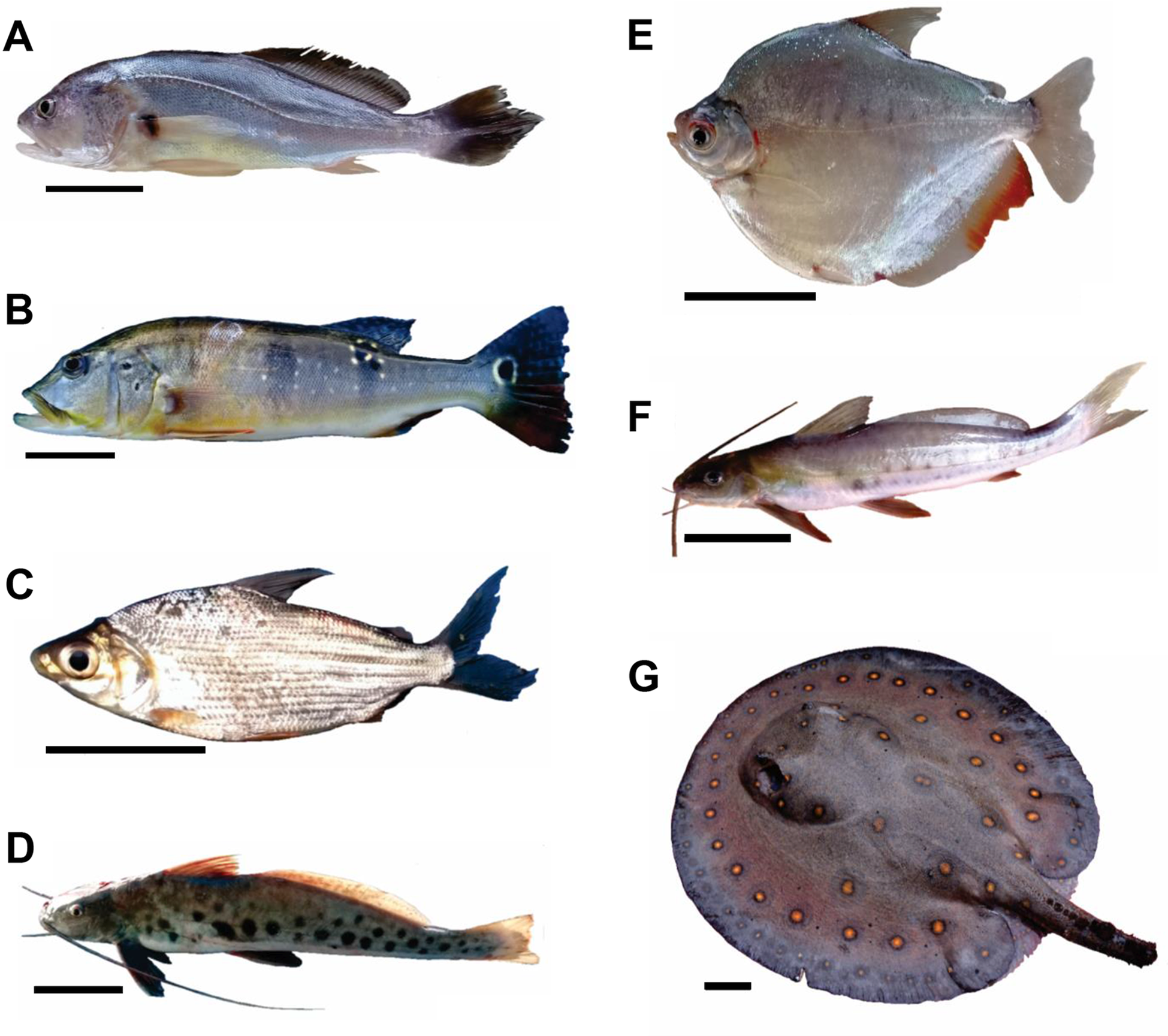
Fig. 3. Fish species infected by Buddenbrockia sp. E. (A) Plagioscion squamosissimus. (B) Cichla pinima. (C) Curimata inornata. (D) Calophysus macropterus. (E) Mylossoma aureum. (F) Pimelodina flavipinnis (G) Potamotrygon motoro. (A–D) Fish collected near Santarém. (E–G) Fish collected near Manaus. Scale bar: 5 cm.

Fig. 4. Maximum likelihood tree showing the relationship between Buddenbrockia sp. E and all malacosporean species reported in Fiala et al. (Reference Fiala, Bartošová-Sojková, Okamura, Hartikainen, Okamura, Gruhl and Bartholomew2015) plus Tetracapsuloides vermiformis and Buddenbrockia bryozoides described in Patra et al. (Reference Okamura, Hartigan and Naldoni2018) using sequences deposited in GenBank based on partial small subunit ribosomal DNA (ssrDNA). Fish host families are listed on the right for each malacosporean species when known. Information for Buddenbrockia sp. E in bold. Numbers above nodes indicate bootstrap confidence levels from maximum likelihood. Values for weakly supported nodes (<50) are not shown.
Table 3. Dissimilarity matrix for small subunit ribosomal DNA (ssrDNA) of Buddenbrockia sp. E sequenced from fish species 1–7

Fish species identified in the first column. The upper triangle shows nucleotide differences in relation to the number of bases compared. The lower triangle shows % pairwise distances.
Infection prevalences ranged from some 6 to 100% for fish species with infected individuals (Table 1) but most sample sizes were small. Among fish species sampled in both regions, two (Potamotrygon motoro and Plagioscion squamosissimus) showed infections, but not in both regions (Santarém and Manaus). For the interesting case of the stingray, P. motoro, infection was detected in one of two individuals sampled in the Manaus region, but not in 22 individuals sampled downstream in the Santarém region.
Discussion
Incidence of infection of Amazonian fishes
Here we report the first case of a malacosporean infecting fish in the Southern Hemisphere. This is not unexpected given the observation of a Buddenbrockia species in freshwater bryozoans in São Paulo State (Marcus, Reference Lisnerová, Fiala, Cantatore, Irigoitia, Timi, Pecková, Bartošová-Sojková, Sandoval, Luer, Morris and Holzer1941). Unexpectedly, however, our molecular phylogenetic analysis demonstrated that only a single species of Buddenbrockia, referred to here as Buddenbrockia sp. E, infected a wide range of Amazonian fish hosts. Infections were often detected even when sampling <5 fish per species (in four of seven fish species, Table 1). Infections were not detected in nine fish species from Santarém and in seven fish species from Manaus, although sample sizes were generally very low (Table 2). The seven fish species identified to harbour infections of Buddenbrockia sp. E belong to six families and five orders (Table 1). All six families (Sciaenidae, Cichlidae, Curimatidae, Pimelodidae, Serrasalmidae and Potamotrygonidae) and four of the five orders (Cichliformes, Siluriformes, Characiformes and Myliobatiformes) are new records for malacosporean infections. We note that fish hosts include species with varying ecologies – e.g. P. squamosissimus is a benthic carnivore in deep waters feeding on fish and shrimp; Calophysus macropterus inhabits channels and floodplain lakes and consumes fish and invertebrates; the toothless characin Curimata inornata feeds on detritus and surface films (Santos et al., Reference Ruppert, Fox and Barnes2006; Pérez and Fabré, Reference Patra, Hartigan, Morris, Kodádková and Holzer2009). Our results therefore are inconsistent with the infection of fish with common ecologies.
Knowledge of malacosporean host specificity is still incipient, in keeping with the generally poor sampling and biased reporting of parasite occurrence in general (Dallas et al., Reference Dallas, Huang, Nunn, Park and Drake2017). Nevertheless, the current picture suggests that Buddenbrockia sp. E is characterized by a remarkable ability to infect a broad range of fish. Buddenbrockia sp. E infections have been detected in five families of teleosts and one family of chondrichthyans (Potamotrygonidae). In particular, Buddenbrockia sp. E was detected in the kidney of four of 13 fish species from the Santarém region and from three of 10 fish species from the Manaus region (total number of fish sampled = 146). Only two fish species examined were common to both regions: the freshwater stingray (P. motoro) and pescada (P. squamosissimus).
Our study only found one malacosporean species despite the use of general malacosporean primers for detection. Several previous studies using the same primers in temperate regions show higher malacosporean diversity, although direct comparisons are difficult due to different evolutionary histories of fish host fauna and relative sampling effort. Five malacosporean species were detected in six of nine fish species sampled (total number of fish sampled = 117) from the River Stour (UK) (Naldoni et al., Reference Murphy, Gerth, Pauk, Konstantinidis and Arismendi2019) and five malacosporeans were detected in nine of 10 species of fish sampled (total examined = 48) from the River Dyje in the Czech Republic (Bartošová-Sojková et al., Reference Bartošová-Sojková, Hrabcová, Pecková, Patra, Kodádková, Jurajda, Tyml and Sibylle2014). In the current study, seven of 21 species of fish sampled from sites some 600 km apart in the Amazon Basin yielded only the one malacosporean species with comparative detection methods.
The detection of a single species is somewhat surprising, as the collective data do not support the general expectation that parasite diversity will mirror host diversity in biodiversity hot spots. Such a result may arise from an incomplete sampling of Neotropical parasites, although a study on helminth parasite diversity in tropical freshwater fish also suggested a weak trend that helminth species richness may be higher in certain areas in the north temperate region, and that overall helminth species richness in tropical freshwater fish appears to be relatively poor (Choudhury and Dick, Reference Choudhury and Dick2000). However, general inferences of such patterns must be tempered by appreciation that further sampling in the Amazon could reveal a different picture.
Evolutionary scenarios
The detection of Buddenbrockia sp. E in the freshwater stingray is the first report of a malacosporean infection in a cartilaginous fish. If this is a true host, facilitating parasite spore production, the position of Buddenbrockia sp. E in our molecular phylogeny may reflect a relatively recent acquisition of cartilaginous fish hosts. Thus, stingrays may have been incorporated as hosts at some point after their inferred Eocene invasion of the Amazon Basin (Bloom and Lovejoy, Reference Bloom and Lovejoy2017). Similarly, croakers would have been infected following their invasion. This pattern of host use suggests that the Buddenbrockia sp. E lineage could have been present in South America long before marine incursions introduced host lineages now utilized.
There are, however, alternative explanations to the scenario proposed above. These include a relatively recent introduction and spread of a generalist parasite. It could also be the case that stingrays are accidental hosts, which would then demonstrate a capacity to infect fish characterized by substantially differing physiologies. These issues could variously be resolved with broader sampling, the use of more general primers and more in-depth profiling of genetic variation of Buddenbrockia sp. E.
We stress that evolutionary scenarios based on the patterns of host use in the present day must always be tempered with appreciation that parasites may undergo unseen shifts in host use over time that can muddy the waters (De Baets et al., Reference De Baets, Dentzien-Dias, Upeniece, Verneaujj and Donoghue2015; Okamura and Gruhl, Reference Naldoni, Adriano, Hartigan, Sayer and Okamura2019). A better understanding of myxozoan diversities and patterns of host use can help to evaluate, add perspective to and refine proposed evolutionary scenarios regarding early host use (e.g. Kodádková et al., Reference Kodádková, Bartošová-Sojková, Holzer and Fiala2015; Holzer et al., Reference Holzer, Bartošová-Sojková, Born-Torrijos, Lövy, Hartigan and Fiala2018; Lisnerová et al., Reference Kuris, Hechinger, Shaw, Whitney, Aguirre-Macedo, Boch, Dobson, Dunham, Fredensborg, Huspeni, Lorda, Mababa, Mancini, Mora, Pickering, Talhouk, Torchin and Lafferty2008).
Is malacosporean diversity really low in the Amazon Basin?
It was surprising to detect sequences characterising only one malacosporean species infecting fish species sampled from a diversity of fish families in a biodiverse region. One explanation is that many Neotropical malacosporeans may have diverged sufficiently to preclude amplification using the primers employed in this study, despite the primers being suitable for characterising broad malacosporean diversity in temperature regions (Supplementary Fig. S1). This could be investigated by histology and ultrastructure to confirm the presence of malacosporean stages in kidney material not amplified by the primers employed here. Characterising kidney metagenomes might then prove helpful. The alternate explanation, that malacosporeans are indeed species poor in the Neotropics, is supported by the apparent absence of closely related species that might have amplified in concert with Buddenbrockia sp. E. The broad host range of Buddenbrockia sp. E provides some support for the hypothesis that host heterogeneity may limit the evolution of specialist parasite lineages (Gibson et al., Reference Gibson, Baffoe-Bonnie, Penley, Lin, Owens, Khalid and Morran2020) which is based on theoretical expectations regarding the evolution of niche width. Nevertheless, many parasite radiations are apparently linked with relative host diversities (e.g. Kamiya et al., Reference Jorge and Poulin2014). Indeed, this pattern has been noted for myxozoans with the species-rich myxosporeans using diverse polychaete (some 8000 extant species; Ruppert et al., Reference Reis, Albert, Di Dario, Mincarone, Petry and Rocha2004) and oligochaete (some 3500 and extant species; Ruppert et al., Reference Reis, Albert, Di Dario, Mincarone, Petry and Rocha2004) hosts and the species-poor malacosporeans exploiting depauperate freshwater bryozoan hosts (<100 species; Massard and Geimer, Reference Marcus2008; Okamura et al., Reference Okamura, Hartikainen, Schmidt-Posthaus and Wahli2018). The invertebrate host use of Buddenbrockia sp. E is entirely unknown. Wood and Okamura (Reference Stinson, Atkinson and Bartholomew2017) identify nine freshwater bryozoan species that could serve as hosts in the Amazon Basin with further sampling revealing some four new species and one previously unrecorded from the region (Wood and Okamura, unpub. data). Finally, it is entirely possible that other malacosporeans were simply not sampled in view of low sample sizes, biased sampling (e.g. poor sampling of younger fish by fishermen) and the limited period of sampling.
Further insights and caveats
For such a limited study, our data highlight various issues and questions in addition to those raised above. Notably, evidence that malacosporeans may take advantage of fish invading freshwater habitats (stingrays and croakers) from a marine environment suggests a considerable facility for incorporating new vertebrate hosts – a scenario further supported by myxosporeans that utilize non-fish hosts (e.g. shrews, mole and ducks) and the repeated adoption of diverse amphibians (Hallett et al., Reference Hallett, Atkinson, Bartholomew, Székely, Okamura, Gruhl and Bartholomew2015; Hartigan et al., Reference Hartigan, Wilkinson, Gower, Streicher, Holzer and Okamura2016). Possibly this is promoted if species already have a wide host range or a rather unspecific host invasion strategy.
It is important to note that ascribing fish hosts based on molecular detection of Buddenbrockia sp. E infections is potentially confounded. Such detection identifies putative hosts but confirmation of hosts requires demonstration of spore development or undertaking transmission trials. Naldoni et al. (Reference Murphy, Gerth, Pauk, Konstantinidis and Arismendi2019) gained evidence for spore development (by ultrastructure) in fish belonging to three of the seven species from the River Stour in which malacosporean infections were detected by molecular methods. Bartošová-Sojková et al. (Reference Bartošová-Sojková, Hrabcová, Pecková, Patra, Kodádková, Jurajda, Tyml and Sibylle2014) report detecting malacosporean developmental stages and/or spores in 36% of all fish samples and that malacosporean DNA was detected in half of these microscopically positive results. Microscopic detection of spores can be variously compromised, e.g. if infections are immature or spores are sparse. These results suggest that a reasonable proportion of fish (if not all) identified in this study to harbour Buddenbrockia sp. E infections act as true hosts. In theory, infecting a broad range of dead-end hosts should be selected against. Notably, spore development has been observed even in non-native fish when infected by a generalist strain of the myxosporean Ceratonova shasta (Stinson et al., Reference Stephens, Altizer, Smith, Alonso Aguirre, Brown, Budischak, Byers, Dallas, Jonathan Davies, Drake, Ezenwa, Farrell, Gittleman, Han, Huang, Hutchinson, Johnson, Nunn, Onstad, Park, Vazquez-Prokopec, Schmidt and Poulin2018).
Conclusions and future work
This study highlights our limited knowledge of malacosporean diversity and patterns of host use – in keeping with our generally poor knowledge of parasite diversity (Dobson et al., Reference Dobson, Lafferty, Kuris, Hechinger and Jetz2008; Poulin, Reference Pérez and Fabré2014; Okamura et al., Reference Okamura, Hartikainen, Schmidt-Posthaus and Wahli2018) and parasite host range (Dallas et al., Reference Dallas, Huang, Nunn, Park and Drake2017). Improving such knowledge for malacosporeans is likely to become ever more difficult due to the many challenges faced by residents of freshwater environments, including overexploitation, pollution, flow modification, habitat destruction or degradation, and invasion by exotic species (Dudgeon et al., Reference Dudgeon, Arthington, Gessner, Kawabata, Knowler, Lévêque, Naiman, Prieur-Richard, Soto, Stiassny and Sullivan2006). As a result of such challenges, some 28% of freshwater fish are at risk of extinction and on the IUCN Red List (IUCN, Reference Hudson, Dobson and Lafferty2019), and affiliate taxa (parasites and symbionts) are therefore threatened with co-extinction along with their endangered freshwater hosts (Dunn et al., Reference Dunn, Harris, Colwell, Koh and Sodhi2009; Carlson et al., Reference Carlson, Burgio, Dougherty, Phillips, Bueno, Clements, Castaldo, Dallas, Cizauskas, Cumming, Doña, Harris, Jovani, Mironov, Muellerklein, Proctor and Getz2017). Amplicon sequencing applied to environmental samples and archived material (e.g. museum specimens) across global scales may be one means of retrieving insights on the hidden diversity of malacosporeans and other parasites before many vanish (e.g. Hartikainen et al., Reference Hartikainen, Bass, Briscoe, Knipe, Green and Okamura2016; Harmon et al., Reference Harmon, Littlewood and Wood2019; Murphy et al., Reference Massard and Geimer2020).
Supplementary material
The supplementary material for this article can be found at https://doi.org/10.1017/S0031182020002322.
Acknowledgements
The authors thank two reviewers whose comments helped to improve our manuscript, Antônio Sousa Figueira for support of fieldwork in Santarém; local fishermen (Fernando Dias de Souza and Francisco dos Santos Pinto in Santarém and Edson Silva Araujo in Manaus) for their knowledge of fish availability and provision of material for study; Dr Maria Isabel Müller for kidney samples of Potamotrygon motoro; Professor Dr Frank Raynner Vasconcelos Ribeiro for help with fish species identification; and PhD student Marcos Sidney Brito de Oliveira for the photographs of fish presented in this study.
Financial support
This study was supported by the São Paulo Research Foundation (FAPESP) (grants # 2016/22047-7) to E.A.A., and also was partially financed by the Coordination for the Improvement of Higher Education Personnel (CAPES), Brazil, Finance Code 001. E.A.A. received a research productivity grant from the Brazilian Fostering Agency CNPq (grant # 301886/2016-4). J.N. was supported by a postdoctoral scholarship from FAPESP (grant # 2014/22700-7). L.L.C. was supported by CAPES/FAPESPA (grant # 88881.160660/2017-01). B.O. received support from the Visiting Researcher Programme – FAPESP (grants # 2015/19463-6 and # 2018/01425-9).
Conflict of interest
None.
Ethical standards
The methodology of the present study was approved by the Ethics Research Committee of the Federal University of São Paulo (CEUA N 92090802140), in accordance with Brazilian law (Federal Law No. 11794, dated 8 October 2008).


