Introduction
A few cases of bisphosphonate-associated osteonecrosis of the temporal bone have been reported.Reference Polizzotto, Cousins and Schwarer1, Reference Froelich, Radeloff, Köhler, Mlynski, Müller and Hagen2 Bisphosphonate-associated osteonecrosis of the jaws (both the maxilla and the mandible) is more common, with hundreds of cases described in the literature.Reference Marx3–Reference Mavrokokki, Cheng, Stein and Goss5 Bisphosphonate-associated osteonecrosis of the jaws is defined as an area of exposed bone in the jaws, of more than eight weeks' duration, in a patient taking oral or intravenous bisphosphonate. Important exclusions are previous radiotherapy to the head and neck and malignant disease in the area.
Risk factors are an age over 55 years, intravenous bisphosphonates, immunocompromise from steroid use or diabetes, and bone-invasive jaw procedures. Approximately 75 per cent of all cases of jaw osteonecrosis follow tooth extraction or similar procedures.Reference Mavrokokki, Cheng, Stein and Goss5 It has been estimated that the risk of jaw osteonecrosis following dental extraction in patients receiving intravenous bisphosphonate for malignancy ranges from 1 in 9 to 1 in 15.Reference Mavrokokki, Cheng, Stein and Goss5 The risk for patients taking oral bisphosphonate for osteoporosis is much less: of the order of 1 in 300 to 1 in 1000. Ninety-five per cent of all patients receiving bisphosphonate take the drug in oral form for osteoporosis.Reference Mavrokokki, Cheng, Stein and Goss5, Reference Vudiniabola, Pirone, Williamson and Goss6 Given that most oral bisphosphonates are prescribed for benign conditions, the risks and benefits need to be carefully explained to the patient.
In this paper, we present two cases of bisphosphonate-associated osteonecrosis of the temporal bone component of the external auditory canal.
Case reports
Case one
A 64-year-old man with an 11-year history of immunoglobulin G kappa multiple myeloma presented in December 2009 with a 7-month history of persistent exposed bone in his right external auditory canal (Figure 1). This had first been noted after an ear infection in April 2008 and had initially been diagnosed as keratosis obturans, but had failed to respond to simple non-surgical dressings with Kenacomb Otic (Sigma Pharmaceuticals, Croydon, Victoria, Australia).

Fig. 1 Fibre-optic scope views of the right external auditory canal for patient one, showing persistent ulceration.
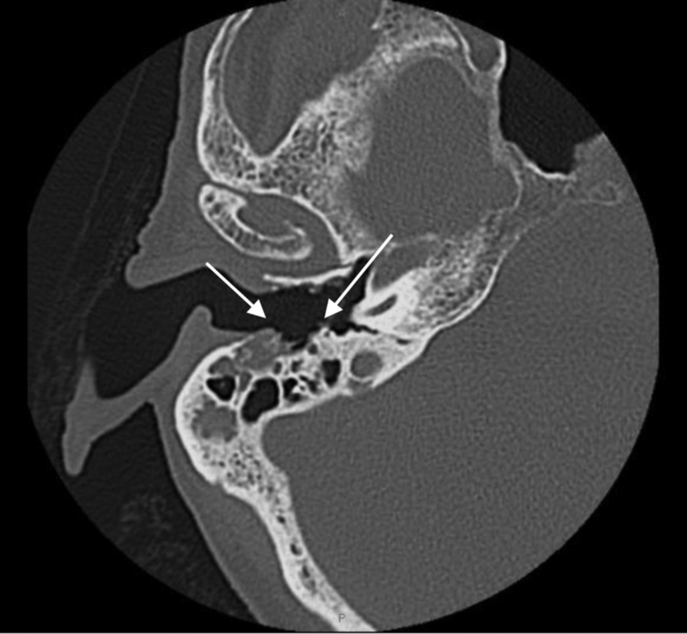
Although an initial magnetic resonance imaging scan had shown no pathology, subsequent computed tomography (CT) scans in July and September 2010 demonstrated a soft tissue density in the right ear canal with irregularity of the underlying bone (Figure 2). The CT scan also showed an unrelated, 18 × 7 mm, lytic bone defect in the right lateral mass of the atlas, consistent with a myelomatous deposit.

Fig. 2 Axial computed tomography scan of the right mastoid for patient one, showing increased sclerosis (arrow) in the bony wall of the external auditory canal.
The patient's multiple myeloma had been first diagnosed in 1997, and had been treated with intermittent melphalan and prednisolone between 1998 and 2001. Following disease progression in 2003, sodium clodronate 1600 mg daily had been commenced and the patient had elected to continue with further intermittent monthly melphalan (12 mg daily for four days) and prednisolone (25 mg daily for 4 days), declining an offer of alternative, more intensive therapy. As he had been in an apparent plateau phase for some months, and because of concerns regarding the persistence of his auditory canal lesion, the patient's oral chemotherapy and sodium clodronate had been discontinued five months previously, in July 2009.
The presumed myelomatous lesion in the right lateral mass of the atlas was treated with 20 Gy radiotherapy, with shielding of the right external auditory canal. At the most recent follow-up examination, three months after radiotherapy completion, the patient was noted to have no change in the degree of right external ear canal bone exposure, and had a healthy mouth with no signs of exposed bone in the jaws.
Case two
A 72-year-old woman was commenced on alendronate 70 mg weekly for osteoporosis (Z score, −2.6; T score, −0.3), in November 2006. She had no history of fractures, diabetes or prednisolone use, nor of trauma or surgery to the ear canal. The only past medical history was a partial gastrectomy in 1991 for gastric cancer.
In April 2010, she presented with right ear irritation, and was diagnosed with otitis externa and subsequently treated with Otodex (Sanofi-Aventis Australia, Macquarie Park, New South Wales, Australia), ear toilet and Kenacomb Otic. At review the following week, the tympanic membrane appeared healthy and the extent of debris in the ear canal had improved; however, a new area of exposed bony outgrowth on the floor of the right external auditory canal was discovered, prompting referral to an otorhinolaryngologist.
A biopsy of the right ear canal, performed in June 2010, was reported as showing osteomyelitis with necrotic bone, together with keratinised epithelium with no evidence of malignancy. Tests for blood inflammatory markers (i.e. erythrocyte sedimentation rate, C-reactive protein and white cell count) had normal results. A CT scan showed destruction of the right tympanic bone. The patient was managed with microsuction and Kenacomb Otic ear drops.
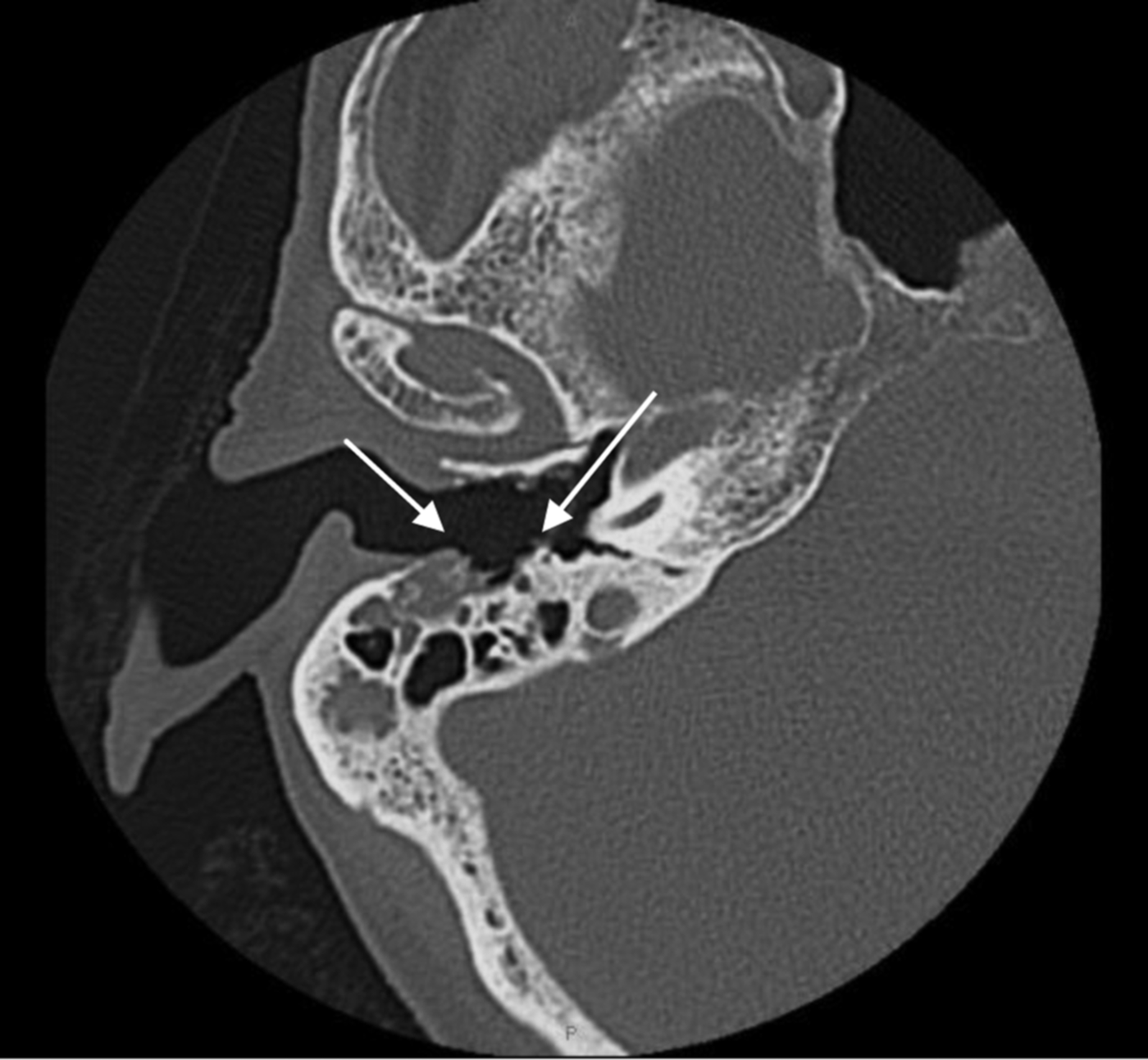
A subsequent CT scan of the right temporal bone performed in February 2011 revealed mild progression of destruction posteriorly into the mastoid air cells and facial nerve canal (Figure 3).

Fig. 3 Axial computed tomography scan of the right petrous temporal bone for patient two, showing osteonecrosis of the anterior, posterior and inferior wall of the external auditory canal with extension into the mastoid (arrows).
In June 2011, examination of the external auditory canal revealed persistent debris and exposed bony defect, with an intact facial nerve. Bisphosphonate-related osteonecrosis of the temporal bone was diagnosed and the patient's alendronate was ceased.
In July 2011, she underwent a right modified radical mastoidectomy with canal wall reconstruction to remove the bony sequestrum.
Discussion
The two cases presented above are consistent with bisphosphonate-associated osteonecrosis of the temporal bone. Both cases presented as an ear infection failing to resolve with topical therapies. In case one, the underlying lesion in the deep external auditory canal had been present for eight months. There was no evidence of myeloma in the auditory canal, although a new lytic lesion was discovered in the patient's right lateral atlas. Although he had previously received radiotherapy to the head and neck, this had been commenced five months after the exposed bone had first been noticed. The radiotherapy dose had been low, at 20 Gy, compared with the usual 55 Gy or more associated with mandibular osteoradionecrosis, and the ear canal had been shielded successfully by the radiotherapist.
Bisphosphonate-associated osteonecrosis of the jaws has some similarities to osteoradionecrosis of the jaws.Reference Vudiniabola, Pirone, Williamson and Goss6 Both present as exposed bone with varying degrees of pain. However, osteoradionecrosis is usually confined to the mandible, with maxillary cases being exceedingly rare. Osteoradionecrosis may occur in the temporal bone, which is similar to the mandible in its dense cortical bone arrangement and poor blood supply.Reference Vudiniabola, Pirone, Williamson and Goss6 The development time of osteoradionecrosis ranges from 7.5 to 20 years.Reference Vudiniabola, Pirone, Williamson and Goss6 Typically, osteoradionecrosis of the temporal bone presents later than that of the mandible, which may relate to the fact that invasive ear canal procedures are uncommon whereas tooth extraction from the mandible is common.Reference Vudiniabola, Pirone, Williamson and Goss6
• Bisphosphonate-associated osteonecrosis should be part of the differential diagnosis of temporal bone lesions
• Taking a relevant drug history is important, especially regarding bisphosphonate usage, type and duration
• The risks and benefits of bisphosphonates should be discussed with patients
• Surgery may be indicated for removal of erosive bony deposits
The development of osteonecrosis of the jaws is associated with drug potency. Most commonly, bisphosphonate-associated jaw osteonecrosis occurs with the potent, nitrogen-containing bisphosphonates zoledronic acid, pamidronate and alendronate. Our first case involved sodium clodronate, a weaker, non-nitrogen-containing oral bisphosphonate. Although this drug is much weaker than the nitrogen-containing bisphosphonates, cases of clodronate-associated osteonecrosis of the jaws have now been reported.Reference Crépin, Laroche, Sarry and Merle7
Osteonecrosis of the jaws is also associated with the duration of bisphosphonate treatment.Reference Woo, Hellstein and Kalmar8 The minimum duration has been reported as six months.Reference Marx3 The risk posed by oral bisphosphonates for non-cancer patients increases after five years.Reference Barasch, Cunha-Cruz, Curro, Hujoel, Sung and Vena9 Our second case was found to have osteonecrosis of the external auditory canal only 3.5 years after commencing alendronate. Bisphosphonates become incorporated into bone without being degraded; the half-life of alendronate is approximately 12 years.Reference Lin, Russell and Gertz10 A growing number of patients are now receiving bisphosphonates, and this has implications for the future. Our first case had been taking clodronate, a relatively weak bisphosphonate; however, he had taken it for six years (with a total dose of over 3300 g) and thus had received a significant overall dose over a substantial period of time.
We draw these case studies to the attention of otorhinolaryngologists so that when they discover an area of exposed bone in the external ear, they will consider bisphosphonate-associated osteonecrosis in the differential diagnosis. Specific enquiry should be made regarding the use of bisphosphonates by the patient, as this will have implications for how such lesions should be managed.
Acknowledgements
We acknowledge the contribution of the following clinicians to the presented two cases: Dr N Barker, general medical practitioner; Dr T Cook, consultant physician; Dr J Harrison, otorhinolaryngologist; Dr J Tomich, otorhinolaryngologist; and Dr M Borg, radiation oncologist.


