1. INTRODUCTION
The interaction of an ultrashort laser pulse with a gas target offers a wide variety of physical phenomena, due to the complex interaction between the laser and the electrons of the generated plasma. The particular interaction is determined by the adopted interaction scheme, by the laser system parameters and the environment parameters. The interest in gas targets grew up dramatically in the last decades, especially due to the non-linear optics applications. Just to cite some of them: laser wakefields generation (Tajima & Dawson, Reference Tajima and Dawson1979; Mangles et al., Reference Mangles, Walton, Najmudin, Dangor, Krushelnik, Malka, Manclossi, Lopes, Carias, Mendes and Dorchies2006; Malka et al., Reference Malka, Faure, Gauduel, Lefebvre, Rousse and Ta Phuoc2008), harmonic generation in gases (Franken et al., Reference Franken, Hill, Peters and Weinreich1961), pulse compression (Lange et al., Reference Lange, Ripoche, Chiron, Lamouroux, Franco, Prade and Mysyrowicz1998), beam filamentation (Braun et al., Reference Braun, Korn, Liu, Du, Squier and Mourou1995), terahertz radiation generation (Hoyer et al., Reference Hoyer, Knorr, Moloney, Wright, Kira and Koch2005), and light coherent scattering (Mitryukovskiy et al., Reference Mitryukovskiy, Liu, Prade, Houard and Mysyrowicz2013). In the present work, the generation of L α line from air plasma sparks in front of a metal target is studied, following (Pikuz et al., Reference Pikuz, Chefonova, Gasilova, Komarova, Ovchinnikova, Skobeleva, Ashitkova, Agranata, Zigler and Faenov2010) where K α emission was used to perform micro-radiographies. Based on (Nagao et al., Reference Nagao, Hironaka, Nakamura and Kondo2004; Faenov et al., Reference Faenov, Pikuz, Zidkov, Skobelev, Komarov, Chefonov, Gasilov and Ovchinnikov2010; Ledingham et al., Reference Ledingham, Abuazoum, McCanny, Melone, Spohr, Schramm, Kraft, Wagner and Jochmann2011; Zhidkov et al., Reference Zhidkov, Pikuz, Faenov, Chefonov, Ovchinnikov, Agranat and Zigle2012), we optimized the X-ray radiation from a thick (2 mm) tin (Sn) target, by moving the target relative to the laser focus in the presence of the air plasma spark. In addition, at each point we conducted a scan by varying the energy ratio between the main pulse and a controlled prepulse. The laser energy was varied in order to characterize the efficiency of such a setup as X-ray source. Finally, spectral measurements in the near-infrared range have been done, by varying again the laser contrast, with the goal to find a correlation with the X-ray signals. The striking aspect of this experiment is that of producing several keV photons in air with a table-top laser system, 3 mJ in 50 fs, corresponding to 60 GW power. For air plasma sparks density and temperature parameters, typically ~ 1016cm−3 and several eV (Zhang et al., Reference Zhang, Wu, Xu and Zhu2012), the presence of keV electrons, responsible for the X-ray emission from metal, is not expected. The presence of the plasma spark, composed by air plasma and metal surface plasma, is the environment where the electrons are accelerated, producing X-ray radiation when they are damped inside the solid density region of the Sn target. The ablation depth of a metal target in atmosphere is found to be much smaller than in vacuum (Di Bernardo et al., Reference Di Bernardo, Courtois, Cros, Matthieussent, Batani, Desai, Strati and Lucchini2003), due to the atmospheric pressure, which prevents the formation of big craters by redeposition mechanisms. The plasma expansion from the metal surface in this configuration is strongly inhibited (Chen et al., Reference Chen, Wu, Yang, Tang, Yao, Rupp, Cao and Xu2013). The direct laser–solid target interaction is also affected in atmosphere by the effect of the plasma defocusing due to the strong ionization occurring meanwhile the laser is tightly focused in air. The expected laser intensity in vacuum is such that X-rays in the keV region could be observed from the interaction with the solid target, nevertheless keV photons are unexpected when the interaction occurs in air. In previous works (Faenov et al., Reference Faenov, Pikuz, Zidkov, Skobelev, Komarov, Chefonov, Gasilov and Ovchinnikov2010; Ledingham et al., Reference Ledingham, Abuazoum, McCanny, Melone, Spohr, Schramm, Kraft, Wagner and Jochmann2011; Zhidkov et al., Reference Zhidkov, Pikuz, Faenov, Chefonov, Ovchinnikov, Agranat and Zigle2012), kHz repetition laser were used showing a higher efficiency in the number of yield K α X-ray photons. This could be due at least to two reasons: the first is the higher efficiency of K α line production with respect to L α line through electron inelastic scattering inside metals; the second is related to the air refraction index properties, which on the millisecond scale can bring memory from the previous pulse to the next giving birth to focusing effects in some cases, increasing and maintaining the laser intensity over many Rayleigh lengths (Jhajj et al., Reference Jhajj, Rosenthal, Birnbaum, Wahlstrand and Milchberg2014). The correlation showed in the last section of the present paper between X-ray signals and near-infrared signals can lead to a better interpretation of the phenomenon under analysis, related to an efficient multi-photon ionization of the air plasma.
2. EXPERIMENTAL METHOD AND RESULTS
The experiment consisted in three parts: plasma extension characterization through a visible charge-coupled device (CCD) camera, X-ray spectra detection through an X-ray CCD camera in single-photon counting mode, near-infrared spectroscopy through a fiber spectrometer. The experimental setup is shown in Figure 1.

Fig. 1. Experimental setup: the laser beam starts from the right, and then it passes through a half-wave plate in order to distribute the laser energy into the s- and p-polarizations in a controlled way. After this, the laser is split in polarization by a polarizing beam splitter. The delay line between the main pulse and the prepulse is 0.5 ns. A telescope is mounted with the goal to increase the beam diameter at the entrance of the last focusing lens. The two pulses are focused in air by a 20 cm positive lens. The air plasma is formed in front of the Sn target. When target is moved along the spark X-rays are observed and detected by a CCD-X in single-photon counting mode.
We used a Ti:Sa (0.8 μm central wavelength) 3 mJ, 50 fs laser split in polarization through a half-wave plate, which ensures the adjustability of the energy ratio between the controlled prepulse and the main pulse (the contrast), and a polarizing beam splitter. The intrinsic contrast of the laser system is 109, preventing us to have more than one significant prepulse. The overall elongation of the laser pulse inside the glass of the optical elements is compensated through a pre-chirp induced in the compression phase. The main pulse and the prepulse are separated by 0.5 ns. The plasma lifetime for the plasma generated by the controlled prepulse is expected to be about 10 ns (Couairon & Mysyrowicz, Reference Couairon and Mysyrowicz2007). In this way, the main pulse interacts with a preformed plasma. A telescope is mounted in order to increase the beam diameter up to 4 cm before the final 20 cm focal length lens. Given our parameters, the expected laser beam diameter inside the air plasma channel is expected to be some tens of microns. This average value typically comes out from the compensation of many focusing/defocusing effects, such as initial lens focusing, plasma defocusing, diffraction, and pre-plasma focusing. The CCD-X camera is at ~ 5 cm from the Sn target. The CCD-X line of sight forms an π/6 angle with the laser beam impinging on the target. The X-ray radiation is attenuated during the free propagation in air and also by the passage through a 75 μm Kapton window put on the entrance of a short flange mounted in front of the CCD sensors in order to keep them to −20°C, so lowering the electronic noise. In front of the sensors is also applied a composed thin filter 4 μm PoliPropylene plus 0.4 μm aluminum, useful to screen the visible–infrared radiation coming from the interaction point.
2.1. Plasma extension
The measured plasma longitudinal extension was 700 μm, as in Figure 2a and 2b. This parameter turned to be important in the phase of focus-scan, where the Sn target was moved along the plasma channel. Figure 2b shows the air plasma extension, namely without the target. Nevertheless during the interaction the plasma composition and evolution is strongly affected by the presence of the target, in fact the overall plasma is a mixture of air plasma and metal surface plasma. In this environment, electrons are accelerated to some keV, inducing X-ray radiation when decelerating inside the solid density region.

Fig. 2. (a) Spectral and temporal integrated plasma light: longitudinal profile of the plasma spark. (b) Line-out of the longitudinal profile of the plasma spark.
2.2. X-ray detection and characterization
The X-ray spectra were detected in single-photon counting mode, taking into account in the analysis the effect of CCD-X quantum efficiency, the air absorption, the Kapton window and the aluminated-plastic filter. First a focus-scan was made moving the Sn target along the plasma spark in order to find the best point of interaction, yielding the maximum number of L α photons. In Figure 3a–3c, the results are reported. In particular, Figure 3a and 3b show the formation of L α line and the yielded number of photons when varying the distance of the solid target from an optimum point marked as zero. The negative (positive) sign indicates the negative (positive) sense of moving, namely toward (in the same vs. of) the impinging laser. The number dN/dΩdE of photons per unit solid angle and unit energy emitted by the source per each laser shot is reported.

Fig. 3. (a) X-ray spectra for different negative (toward the incoming laser) positions of the Sn target inside the plasma channel with respect to the optimum point taken as the origin of the displacements. The optimum point is found in the center of the plasma spark. (b) X-ray spectra for different positive (in the same vs. of the incoming laser) positions of the Sn target inside the plasma channel with respect to the optimum point taken as the origin of the displacements. The optimum point is found in the center of the plasma spark. (c) Summary of the focus-scan results. FWHM width of the Lorentzian fit Δz = 27 μm. The spectral and angular integrated number of L α photons is reported with respect to the position of the target inside the plasma spark.
A fine scan was then performed with 10 μm steps in the proximity of the optimum point. The optimum point was found around the center of the spark as in (Zhidkov et al., Reference Zhidkov, Pikuz, Faenov, Chefonov, Ovchinnikov, Agranat and Zigle2012), where the plasma is a mixture of air plasma and metal surface plasma. The number of L α photons, that is, the number of photons emitted in the region of the Sn L α nominal energy 3.44 keV, per shot emitted from the target along the plasma channel is showed in Figure 3c together with a Lorentzian fit. The full wifth half maximum (FWHM) of the fit curve is found Δz = 27 μm, less than a tenth the plasma extension length. It means that significant changes in the number of L α photons could be observed only outside a region of about 2 Δz around the optimum interaction point. The second part of the X-ray analysis consisted in characterizing the L α radiation with respect to the controlled contrast. The Sn target was fixed in the previously found optimum position and the contrast was adjusted trough the half-wave plate in front the first Polarizing Beam Splitter (PBS) (see Fig. 1) in such a way to work with contrasts between 1 and 100. In order to work with higher contrasts, suitable neutral density filters were adopted on the prepulse line. The results are showed in Figure 4a and 4b. The presence of a prepulse seems to be fundamental when looking to Figure 4b where is showed that the maximum yielded radiation is obtained for contrast between 101 and 102, while for the lower and higher contrast value the number of photons decays rapidly. For higher contrasts the L α emission is strongly suppressed probably due to the fact that the prepulse fluence falls under the damage threshold of the metal (about 1 J/cm2), making impossible the formation of a composed air–metal plasma profile.
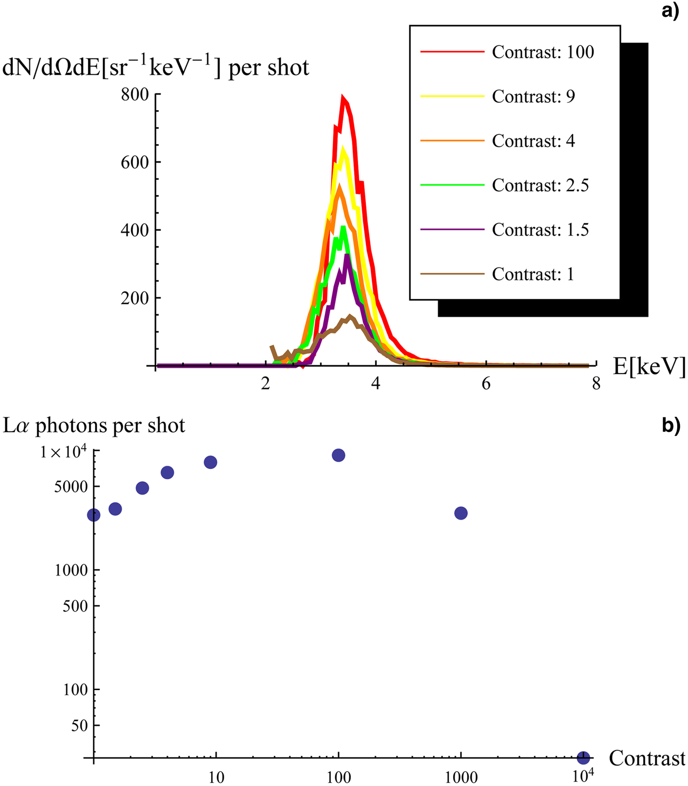
Fig. 4. (a) X-ray spectra for different contrast value. The target is positioned in the optimum point for the yield L α photons, and then with the help of the half-wave plate the energy ratio between the main pulse and the prepulse is adjusted. (b) Number of L α photons versus laser contrast. The target is positioned in the optimum point for the yield L α photons. The spectral and angular integrated number of L α photons is reported with respect to the energy ratio (contrast) between the main pulse and the prepulse.
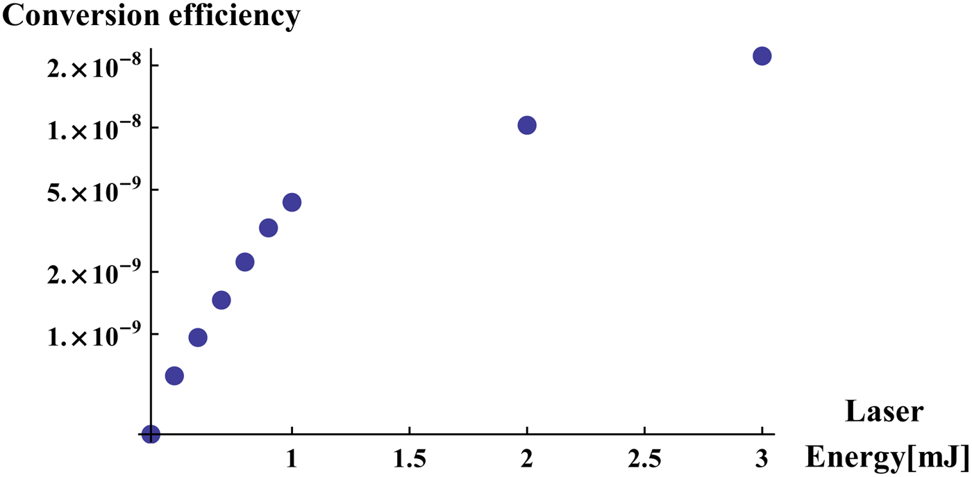
The final part of the X-ray detection study consisted in the characterization of our system as X-ray source. The conversion efficiency from the laser energy to L α radiation was studied for different values of the laser energy. The contrast was fixed at 102 and the target was in the zero (optimum) position. The result is shown in Figure 5, and it is referred like in Figure 3c and 4b to the number of photons emitted from the Sn target. The L α radiation production is very low for energies approximating to some hundreds of μJ probably because of the 102 contrast, which makes the prepulse to be not efficient in modifying the air index refraction. It is worth noting that in previous works K α radiation was found even for laser energy around some tens of μJ, working with a kHz high repetition laser (Faenov et al., Reference Faenov, Pikuz, Zidkov, Skobelev, Komarov, Chefonov, Gasilov and Ovchinnikov2010; Pikuz et al., Reference Pikuz, Chefonova, Gasilova, Komarova, Ovchinnikova, Skobeleva, Ashitkova, Agranata, Zigler and Faenov2010; Zhidkov et al., Reference Zhidkov, Pikuz, Faenov, Chefonov, Ovchinnikov, Agranat and Zigle2012). Even if in these works no punctual mention is given about the laser contrast, the K α production in the same configuration seems to be more efficient.

Fig. 5. Conversion efficiency of the laser energy into L α radiation. The target position and the contrast between the main pulse and the controlled prepulse are chosen in such a way to optimize the yield L α photons. The ratio between the energy irradiated at the L α line and the laser energy is reported for different values of the laser energy.
2.3. Correlation between X-ray and near-infrared spectra
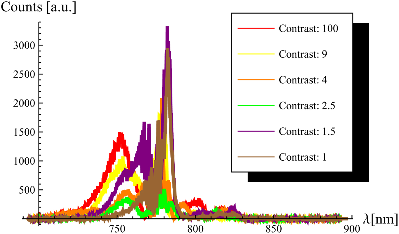
We have demonstrated the correlation between the near-infrared spectra and the X-ray spectra when the laser contrast is adjusted. The spectra were detected at the exit of the plasma channel without the Sn target. We showed that by varying the ratio between the artificial prepulse and the main pulse a significant blue-shift of the self-modulated laser radiation is obtained for ratios approaching to the optimum contrast 102 (Fig. 4b). If varying the contrast value in the region 101 − 102 where the maximum L α production efficiency was found, no significant change in the blue-shifted spectra were registered (Fig. 6). This demonstrates the existence of a parameters region where the laser intensity reaches a maximum value independent from the energy ratio between the prepulse and the main pulse. Previous works demonstrated that blue-shifts of some tens of nanometers can be related to efficient multi-photon ionization, and a constant shift can be related to a fixed laser intensity (Giulietti & Giulietti, Reference Giulietti and Giulietti2015 and references therein; Koga et al., Reference Koga, Naumova, Kando, Tsintsadze, Nakajima, Bulanov, Dewa, Kotaki and Tajima2000). This leads to the conclusion that the role of the prepulse is in modifying the index of refraction of the air plasma, determining a maximum intensity for the main pulse capable to interact with the air–Sn plasma accelerating electrons up to many keV.

Fig. 6. Near-infrared laser spectra at the exit of the plasma channel for different contrast values.
3. CONCLUSIONS
The characterization of L α radiation from air plasma sparks in front of an Sn target was performed by using a Ti:Sa, 3 mJ, 50 fs laser. The focal scan showed the existence of a narrow 27 μm region in which the production of radiation is more efficient. The contrast scan showed that the presence of 1% of the laser energy ultrashort prepulse is required to obtain optimum conversion efficiency to L α radiation. At this level of prepulse, above the damage threshold of the metal, the laser–electron interaction occurs in a region where the plasma composition is determined by the air plasma and the metal surface plasma. The near-infrared spectroscopy points out the influence of the ionization process in air by ultrashort prepulse on the focusing of the main laser pulse on metal target. The correlation found between the laser contrast value and the laser near-infrared spectra at the exit of the plasma spark, without the solid target, could be an indirect diagnostic method for the optimization of the X-ray flux when the target is inserted in the setup.
ACKNOWLEDGEMENTS
This work has been partially supported by Russian Foundation for Basics Research (grant no. 14-22-02089) and by RAS Presidium Program for basic research no. 11.