


Introduction
Toxoplasma gondii is an obligate intracellular protozoan parasite that infects a wide range of host mammals and birds, including humans (Su e Dubey, Reference Su, Dubey and Tonkin2020). Toxoplasmosis affects one-third of the world population; in most cases, the disease is asymptomatic, but immunocompromised individuals may have severe symptoms, such as toxoplasmic encephalitis. Vertical transmission during pregnancy may lead to abortion, or the birth of children with mental retardation, hydrocephalus, psychomotor impairment and chorioretinitis, among others (Robert-Gangneux et al., Reference Robert-Gangneux, Meroni, Dupont, Botterel, Garcia, Brenier-Pinchart, Accoceberry, Akan, Abbate, Boggian, Bruschi, Carratalà, David, Drgona, Djurković-Djaković, Farinas, Genco, Gkrania-Klotsas, Groll, Guy, Hirzel, Khanna, Kurt, Junie, Lazzarotto, Len, Mueller, Munoz, Pana, Roilides, Stajner, van Delden, Villena, Pelloux and Manuel2018).
Pyrimethamine associated with sulphadiazine has been the treatment available and indicated for severe cases for a long period (Silva et al., Reference Silva, Fernandes, Machado, Reis-Cunha, Bartholomeu and Almeida Vitor2019). Both drugs inhibit folate metabolism in the parasite, acting directly on tachyzoites (acute phase of infection); however, this does not eradicate cystic forms (chronic phase of infection) (Konstantinovic et al., Reference Konstantinovic, Guegan, Stäjner, Belaz and Robert-Gangneux2019). The initial treatment of choice must be carried out for several days (up to months) and the medication can cause several side-effects such as haematological abnormalities, increased creatinine and serum liver enzymes and hypersensitivity reactions (Konstantinovic et al., Reference Konstantinovic, Guegan, Stäjner, Belaz and Robert-Gangneux2019).
The investigation of new substances that are capable of eliminating T. gondii tachyzoites and cysts is important for reducing side-effects and serious outcomes, and possibly curing toxoplasmosis (Zwicker et al., Reference Zwicker, Smith, Guerra, Hitchens, Haug, Vander Roest, Lee, Wen, Sun, Wang, Keep, Xiang, Carruthers and Larsen2020). Research with compounds (extracts and molecules) from medicinal plants, which have therapeutic potential, have been used as alternative treatments for some parasitic diseases; this is because, in addition to presenting microbicidal effects, they also confer lower toxicity compared to synthetic drugs (Mirzaalizadeh et al., Reference Mirzaalizadeh, Sharif, Daryani, Ebrahimzadeh, Zargari, Sarvi, Mehrzadi, Rahimi, Mirabediny, Golpour and Montazeri2018).
Moringa oleifera is a plant belonging to the Moringaceae family; studies have shown that parts of the plant contained compounds with bactericidal, fungicidal, anti-tumour, larvicidal and trypanosomicidal properties (Dhakad et al., Reference Dhakad, Ikram, Sharma, Khan, Pandey and Singh2019; Turner et al., Reference Turner, Just, Dasari, Smith, Bissember, Kornienko and Rogelj2019; Arruda et al., Reference Arruda, Freitas, Seabra, Xavier-Júnior, Figueiredo, Napoleão, Paiva, Navarro and Navarro2020; Coriolano et al., Reference Coriolano, Brito, Ferreira, Moura, Melo, Soares, Lorena, Figueiredo, Paiva, Napoleão and Coelho2020; Pagar et al., Reference Pagar, Ghotekar, Pagar, Nikam, Pansambal, Oza, Sanap and Dabhane2020). In addition, M. oleifera has activity against the species of the protozoan Plasmodium (Somsak et al., Reference Somsak, Borkaew, Klubsri, Dondee, Bootprom and Saiphet2016) which, as T. gondii, belongs to the phylum Apicomplexa.
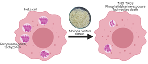
Considering the properties of M. oleifera and the fact that, in relation to T. gondii, there have been no studies on this plant, the aim of the current research was to evaluate the activity of M. oleifera seed extract on HeLa cells infected with T. gondii tachyzoites (RH strain) and directly on tachyzoites forms of T. gondii.
Materials and methods
HeLa cell culture maintenance
The cultivation of HeLa cells was performed in 75 cm2 culture flasks (Ciencor Scientific, Brasil) containing Dulbecco's modified Eagle medium (DMEM) (Gibco, Invitrogen, New York, USA) supplemented with 10% inactivated fetal bovine serum (Sigma-Aldrich, St. Louis, Missouri, EUA), 1% antibiotic (10 000 U mL−1 of penicillin and 10 mg mL−1 streptomycin) (Cultilab, Brazil), l-glutamine, sodium pyruvate and 2β mercaptoethanol. The cells were maintained at 37°C with 5% CO2. For the tests of experimental infection and the maintenance of T. gondii strains in vitro, HeLa cells were used up to the 21st passage.
Toxoplasma gondii RH strain
Tachyzoites from the T. gondii RH strain were kindly provided by Professor João Luís Garcia from the Laboratory of Zoonoses and Public Health at the State University of Londrina, where tachyzoites were obtained from the peritoneal lavage of previously infected Swiss Webster mice. The peritoneal exudates were passed through a 26G needle three times, and washed twice with phosphate-buffered saline (PBS) (pH 7.5) by centrifugation. The sediment was resuspended in PBS and the tachyzoites were counted in a Neubauer chamber.
Part of the tachyzoites was kept in HeLa cells for ‘Anti-T. gondii activity assay in vitro’ (intracellular tachyzoites) and another part was used to evaluate the direct action of M. oleifera extract on T. gondii (extracellular) tachyzoites.
All procedures involving animals in this study were approved by the Animal Experimentation Ethics Committee of the State University of Londrina no. 88/2017/CEUA; 82.862.016.60.
Moringa oleifera seeds extract
Moringa oleifera seeds were collected between December 2018 and March 2019 in the area of Federal University of Sergipe, Aracaju, Sergipe State, located between the geographic coordinates 10°55′56″ south latitude and 37°04′23″ west longitude. Aracaju has a semiarid climate with low pluviometric index (Almeida et al., Reference Almeida, Souza, Loureiro, Pereira, Cruz and Vieira2017). The M. oleifera species was deposited in the herbarium of Federal University of Sergipe, no. ASE8288 (MOTA et al., Reference Mota, Barros, Cunha, Santana, Stevam, Leopoldo and Fernandes2012).
For the preparation of aqueous extracts, the seeds were peeled, weighed (5 g) and processed by turbolysis for 3 min using 100 mL distilled water. Then, the solution was subjected to magnetic stirring (IKA, Germany) for 30 min, and, after this process, subjected to two consecutive filtrations, using qualitative filter paper and a 0.9 μ m glass fibre membrane by vacuum filtration (Madrona et al., Reference Madrona, Serpelloni, Salcedo Vieira, Nishi, Cardoso and Bergamasco2010). The aqueous extract obtained was lyophilized (Freeze Dryer Christ ALPHA 1-2/LD Plus) for 48 h at −45°C (Baptista et al., Reference Baptista, Silva, Gomes, Bergamasco, Vieira and Vieira2017). The material obtained was stored at 4°C until the moment of use. For experiments, the lyophilized material was diluted in DMEM at different concentrations and the solution obtained was called M. oleifera extract.
The extract was characterized by Fourier transform infrared spectroscopy (FTIR) in the form of potassium bromide (KBr) tablets containing approximately 1% of the sample using an FTIR-BOMEN 100 spectrometer with 21 scans per min and a resolution of 4 cm−1. The spectra of FTIR were obtained in the range from 4000 to 400 cm−1 in absorbance mode.
HeLa cell viability by MTT assay
The viability of HeLa cells after treatment with M. oleifera was evaluated based on mitochondrial oxidation, using the tetrazolium salt colorimetric test [3-(4,5-dimethylthiazol-2-yl)-2,5-diphenyltetrazolium bromide or MTT] (Sigma-Aldrich) (Mosmann, Reference Mosmann1983). The cells were grown in 96-well plates (3 × 10⁴ cells per well per 200 μL), for 24 h in DMEM at 37°C and 5% CO2. After this period, M. oleifera extract was added separately at concentrations of 10, 30, 50, 100 and 200 μg mL−1 and maintained for 24 h under the same conditions stated before. As a negative control, cells cultured with DMEM were used, and hydrogen peroxide (H2O2) 0.06% was used as a positive control. Then, the supernatants were removed and the cells received MTT solution (5 mg mL−1) and incubated for 3 h at 37°C and 5% CO2. The formazan crystals were diluted with 100 μL of dimethyl sulphoxide (Sigma-Aldrich), and the absorbance was measured after 30 min at 570 nm in a microplate spectrophotometer (Thermo Plate – TP-Reader). The results were expressed as a relative percentage of MTT reduction in the treated groups compared to the control group, using the following formula:

where OD is the optical density.
Anti-T. gondii activity assay in vitro
To evaluate the effect of the M. oleifera extract on the invasion and proliferation of T. gondii in HeLa cells (1 × 10⁵), 24-well plates were used containing round coverslips of 13 mm (Ciencor Scientific, Brazil), and 5 × 10⁵ T. gondii tachyzoites (1:5 cells/tachyzoites) were added to each one. After 3 h of infection, the supernatant was removed and the cells were washed once with PBS, the plate was manually and slightly shaken and the PBS was discarded. Then, the cells with T. gondii tachyzoites were treated with the M. oleifera extract, at concentrations of 10 and 30 μg mL−1. The negative controls were untreated infected cells (DMEM only), and infected cells treated with a combination of sulphadiazine and pyrimethamine (SDZ + PYR) (50 and 25 μg mL−1) were used as positive controls. They were maintained for 24 h at 37°C in 5% CO2. Afterwards, the supernatant was separated to quantify nitric oxide (NO) and the cells were fixed with 4% paraformaldehyde in PBS for 30 min and stained with 1% toluidine blue (Sigma-Aldrich).
The HeLa cells were analysed by light microscopy (e100, Nikon – led) at 1000× magnification to check the parameters of infection index (number of infected cells per 200 cells examined) and intracellular proliferation of the parasite (total number of parasites per 200 cells examined). Representative images were captured using the conditions described above. The percentages of infection and intracellular proliferation inhibition rates of T. gondii were calculated as: average of the infection or intracellular proliferation rate analysed in untreated cells, corresponding to 100% infection or intracellular proliferation rate. The percentages of inhibition of these parameters submitted to treatments with M. oleifera were calculated by subtracting the percentage values obtained for treated cells from those obtained for untreated cells (Sanfelice et al., Reference Sanfelice, Machado, Bosqui, Miranda-Sapla, Tomiotto-Pellissier, Alcântara Dalevedo, Ioris, Reis, Panagio, Navarro, Bordignon, Conchon-Costa, Pavanelli, Almeida and Costa2017).
Quantification of NO produced by HeLa cells infected by T. gondii tachyzoites
The supernatants in the item above were separated and used to quantify NO, as described by Tomiotto-Pellissier et al. (Reference Tomiotto-Pellissier, Alves, Miranda-Sapla, Morais, Assolini, Silva Bortoleti, Gonçalves, Cataneo, Kian, Madeira, Yamauchi, Nixdorf, Costa, Conchon-Costa and Pavanelli2018). Briefly, the NO was determined through the Griess method in culture supernatant aliquots (60 μL) of T. gondii invasion and proliferation assay. First of all, the aliquots were centrifuged at 2370 × g for 2 min. A 50 μL aliquot of the supernatant was treated with 50 μL of Griess reagent [1% sulphanilamide and 0.1% of N-(1-naphthyl) ethylenediamine in orthophosphoric acid (H3PO4) 5%]. After a 10-min incubation at room temperature, the samples were placed in 96-well microplates. A calibration curve was made using dilutions of NaNO2, and the absorbance was determined at 550 nm on a microplate reader (Thermo Scientific, Multiskan GO).
Quantification of parasitic load by qPCR
A second 24-well plate was prepared in the same way as in the Section ‘Anti-T. gondii activity assay in vitro’ to evaluate the reduction of intracellular tachyzoites observed in light microscopy for the M. oleifera extract of 30 μg mL−1. The prepared plate was used to assess the parasitic load of T. gondii contained in HeLa cells using quantitative real-time polymerase chain reaction (qPCR). DNA extraction was performed using a commercial kit (PureLink™ genomic DNA Mini Kit, Thermo Fisher Scientific®, EUA) following the manufacturer's recommendations. DNA concentration was measured by spectrophotometry (NanoDrop, Thermo Fisher Scientific®, EUA) and qPCR was performed in duplicate with 50 ng of DNA using the TaqMan system, with a standard curve with concentrations from 6.3 × 10⁷ to 6.3 × 10³ parasites per mL. Primers Tox-9F (5′-AGGAGAGATATCAGGACTGTAG-3′) and Tox11-R (5′-GCGTCGTCTCGTCTAGATCG-3′) and probe Tox-TP1 (5′-CCGGCTTGGCTGCTTTTCCT-3′) were used to amplify a 115 bp fragment from the repetitive 529 bp region of T. gondii, as previously described by Opsteegh et al. (Reference Opsteegh, Langelaar, Sprong, den Hartog, De Craeye, Bokken, Ajzenberg, Kijlstra and van der Giessen2010). Each reaction consisted of 1× master mix (Taqman™ Universal PCR Master Mix, Thermo Fisher Scientific®, EUA), 0.7 μ m of each primer (Tox-9F and Tox-11R), 0.1 μ m of probe (Tox-TP-1) and sterile ultrapure water, making the volume up to 20 μL. Amplification was performed on the StepOne™ Plus Real Time PCR System (Thermo Fisher Scientific®, EUA). The thermocycling conditions used were: initial incubation of 95°C for 10 min, denaturation cycles 95°C for 10 s, annealing 58°C for 20 s and extension 72°C for 20 s, followed by a final stage of 40°C for 5 s. The fluorescence signal at 530 nm (Tox-TP1) was measured at the end of each extension step. Sterile ultrapure water without DNA was used as a negative control and was included in all reactions; HeLa cells infected with T. gondii tachyzoites without addition of the M. oleifera extract were used as a negative control of treatment. All samples were analysed in duplicate. The results were visualized and analysed using the program StepOne™ Software v.2.3 (Thermo Fisher Scientific®, EUA).
Scanning and transmission electron microscopy analyses
For scanning electron microscopy (SEM), T. gondii tachyzoites obtained from the peritoneal lavage of previously infected Swiss Webster mice were treated as described in the Section ‘Toxoplasma gondii RH strain’ and counted in the Neubauer chamber. Next, the tachyzoites (1.5 × 10⁶) were treated with the M. oleifera extract (30 μg mL−1), for 1 h at 37°C. Then, they were centrifuged and washed once with PBS by pipetting up and down and centrifuged again. Next, the PBS was discarded and the tachyzoites were fixed with 2.5% glutaraldehyde in 0.1 m sodium cacodylate buffer, containing 1 mm CaCl2. The tachyzoites were fixed on a glass support that was covered with poly-l-lysine, and dehydrated in an ascending series of ethanol (30–100%), before being submitted to critical-point dried with CO2, coated with gold and observed on a high-resolution double beam electron microscope FEI SCIOS.
For transmission electron microscopy (TEM), T. gondii tachyzoites were treated in the same way as described for SEM. After incubation, the parasites were washed with PBS and fixed with 2.5% glutaraldehyde in 0.1 m sodium cacodylate buffer and post-fixed in a solution of 1% OsO4, 0.8% potassium ferrocyanide and 10.0 mm CaCl2 in 0.10 m cacodylate buffer, at room temperature and protected from light. Then, the tachyzoites were washed again with 0.1 m sodium cacodylate buffer, dehydrated in an increasing acetone gradient (30–100%) and embedded in EPON resin and polymerized in an incubator at 60°C for 72 h. Ultrathin sections were obtained, deposited on a copper grid, and stained with uranyl acetate and lead citrate, for 20 and 10 min, respectively. Ultrastructural alterations were observed using a JEOL JEM 1400 transmission electron microscope.
Determination of reactive oxygen species (ROS) generation on T. gondii tachyzoites
ROS generation was evaluated in T. gondii tachyzoites obtained from the peritoneal lavage of previously infected Swiss Webster mice were treated as described above and counted in the Neubauer chamber. The tachyzoites (1.5 × 10⁶) were subjected to treatment with the M. oleifera extract (30 μg mL−1) for 1 h at 37°C in 5% CO2 and analysed by probe H2DCFDA (Bortoleti et al., Reference Bortoleti, Tomiotto-Pellissier, Gonçalves, Miranda-Sapla, Assolini, Carloto, Lima, Silveira, Almeida, Costa, Conchon-Costa and Pavanelli2019). Tachyzoites in DMEM were used as a negative control, and tachyzoites in H2O2 solution (0.4%) were used as a positive control. The micrographs were produced using the EVOS® Microscope FL Auto Cell Imaging System (Thermo Fisher) with a magnification of 200×.
Determination of the mitochondrial membrane potential in T. gondii tachyzoites
For determination of the mitochondrial membrane potential of T. gondii tachyzoites (RH strain), we conducted tetramethylrhodamine ethyl ester (TMRE) staining (Sigma-Aldrich) (Tomiotto-Pellissier et al., Reference Tomiotto-Pellissier, Alves, Miranda-Sapla, Morais, Assolini, Silva Bortoleti, Gonçalves, Cataneo, Kian, Madeira, Yamauchi, Nixdorf, Costa, Conchon-Costa and Pavanelli2018). Briefly, parasites (1.5 × 10⁶), obtained in the same way as described for ROS, were treated with the M. oleifera extract (30 μg mL−1), incubated for 1 h in an incubator at 37°C in 5% CO2, and then washed once with PBS by pipetting up and down and centrifuged. Next, the PBS was discarded and the tachyzoites were incubated with 25 nm TMRE for 30 min at 37°C. Again, they were washed with PBS and analysed immediately in a fluorescence microplate reader (Victor X3, Perkin-Elmer) using the excitation and emission wavelengths of 480 and 580 nm, respectively. Carbonyl cyanide 3-chlorophenylhydrazone (CCCP), 100 μm, was used as a positive control. The micrographs were obtained using the EVOS® Microscope FL Auto Cell Imaging System (Thermo Fisher) with a magnification of 200×.
Lipid staining and fluorescence analysis in T. gondii tachyzoites
In this experiment, the concentration of 1.5 × 10⁶ T. gondii tachyzoites, obtained in the same way as described above, was treated with the M. oleifera extract (30 μg mL−1) and incubated for 1 h at 37°C. Then, the tachyzoites were collected, washed twice with PBS and left to incubate with 10 μg mL−1 Nile Red (Sigma-Aldrich) for 30 min at 37°C in 5% CO2. PBS was used as a positive control. The cytoplasmic lipid bodies of the parasites were detected in a spectrofluorometer (Victor X3, Perkin-Elmer) at the excitation and emission wavelengths of 530 and 635 nm, respectively.
Evaluation of autophagic vacuoles
Toxoplasma gondii tachyzoites (1.5 × 10⁶) obtained in the same way as described above were treated with M. oleifera extract (30 μg mL−1) for 1 h at 37°C in 5% CO2. Then, the autophagic vacuole formation was evaluated using monodansylcadaverine (MDC) 50 μ m labelling, and incubated for 1 h at 25°C in a dark room, according to Bortoleti et al. (Reference Bortoleti, Gonçalves, Tomiotto-Pellissier, Contato, Silva, Matos, Detoni, Rodrigues, Carloto, Lazarin, Arakawa, Costa, Conchon-Costa, Miranda-Sapla, Wowk and Pavanelli2021). After this period, the tachyzoites were washed with PBS and analysed on the spectrofluorimeter (Victor X3, Perkin-Elmer), using excitation and emission wavelengths of 380 and 525 nm, respectively. Tachyzoites in PBS solution were used as a positive control (Bortoleti et al., Reference Bortoleti, Gonçalves, Tomiotto-Pellissier, Contato, Silva, Matos, Detoni, Rodrigues, Carloto, Lazarin, Arakawa, Costa, Conchon-Costa, Miranda-Sapla, Wowk and Pavanelli2021).
Co-determination of annexin-V–fluorescein isothiocyanate (FITC) and propidium iodide (PI) in tachyzoites of T. gondii
Toxoplasma gondii tachyzoites (1.5 × 10⁶) obtained in the same way as described above were treated with the M. oleifera extract (30 μg mL−1), for 1 h at 37°C in 5% CO2. Then, they were washed with PBS, and resuspended in 100 μL of binding buffer (Santa Cruz Biotechnology) with the addition of a mixture of 1 μL of annexin-V–FITC and 1 μL of PI (Santa Cruz Biotechnology). The analysis was performed by flow cytometry using the BD Accuri C6 (BD Biosciences, San Jose, CA) (Bortoleti et al., Reference Bortoleti, Tomiotto-Pellissier, Gonçalves, Miranda-Sapla, Assolini, Carloto, Lima, Silveira, Almeida, Costa, Conchon-Costa and Pavanelli2019). The negative controls were untreated tachyzoites.
Statistical analysis
All data represent the average and standard error of the mean of three independent experiments performed in triplicate. The differences between treatments and controls were assessed using analysis of variance (one-way ANOVA), followed by Tukey's test, and using GraphPad Prism 5.0 software (GraphPad Software, Inc., San Diego, CA, USA). Statistical significance was considered when P < 0.05.
Results
Characterization of M. oleifera extract by FTIR
From the FTIR spectrum of the aqueous extract of M. oleifera (Fig. 1), a band is observed from 3200 to 3500 cm−1 which can be attributed to the presence of hydroxyl groups (OH) in phenolic compounds (Sharaf et al., Reference Sharaf, Higazy and Hebeish2013) and also in proteins, fatty acids, carbohydrates and lignin (Adebisi et al., Reference Adebisi, Adedayo, Oluwaseye, Adekunle, Michael, Olutayo, Clement, Ikokoh, Rasheed and Ademola2014). The peak at 1645 cm−1 could be related to the C=C stretching vibration of aromatic rings and to the vibration of N–H of amines, C=O of amides and carboxylic groups; also, this band could be related to flavonoids and amino acids (Oliveira et al., Reference Oliveira, Mancini, Oliveira, Passos, Quilty, Thiré and McGuinness2016).

Fig. 1. FTIR spectra of Moringa oleifera extract.
Moringa oleifera extract in low concentrations does not alter the viability of HeLa cells
When analysing the viability of HeLa cells treated with the M. oleifera extract using the MTT cytotoxicity assay, we observed that the lowest concentrations studied (10, 30 and 50 μg mL−1) did not significantly reduce the viability of the investigated cells (Fig. 2). On the contrary, concentrations of 100 and 200 μg mL−1 significantly altered the viability of HeLa cells (P < 0.001). The concentrations of 10 and 30 μg mL−1 of the M. oleifera extract were chosen for subsequent experiments.

Fig. 2. MTT cell viability assay of the M. oleifera seed aqueous extract in HeLa cells. HeLa cells were treated with M. oleifera extract (10, 30, 50, 100 and 200 μg mL−1) for 24 h. Control: cells treated with DMEM. H2O2 (positive control). ***Significant difference in relation to the negative control (P < 0.001). Three independent experiments were carried out in triplicate (one-way ANOVA followed by Tukey's test).
Moringa oleifera extract reduces infection and proliferation in the face of increased NO and reduces the parasitic burden and replication of T. gondii in HeLa cells
To assess the effect of M. oleifera treatment during in vitro infection with T. gondii tachyzoites, we analysed the number of infected cells and the number of tachyzoites within these cells (Fig. 3A and B).

Fig. 3. HeLa cells experimentally infected with Toxoplasma gondii tachyzoites RH strain, treated with the M. oleifera extract (30 and 10 μg mL−1. (A) Percentage of cells infected with T. gondii tachyzoites. (B) Amount of tachyzoites inside the infected cells. (C) NO production by HeLa cells infected with T. gondii tachyzoites and treated with the aqueous extract of M. oleifera 30 and 10 μg mL−1, during 24 h of treatment. Griess method for nitrite levels in supernatant of culture cells. (D) Parasitic load by qPCR of HeLa cells infected with T. gondii tachyzoites and treated with M. oleifera (30 μg mL−1). The values represent the mean ± s.e.m. of three independent experiments performed in duplicate. The values represent the mean ± s.e.m. of three independent experiments. *Significant difference compared to negative control (P < 0.05), **(P < 0.01), ***(P < 0.001).
A significant reduction was observed in the infection rate of HeLa cells by tachyzoites treated with the M. oleifera extract at a concentration of 30 μg mL−1 (Fig. 3A) (P < 0.01). This was similar to that found in infected cells treated with SDZ + PYR, with reductions of 20.50 and 20.25%, respectively. Both reductions were significant when compared to the control group, infected with untreated HeLa cells (Fig. 3A). The concentration of 10 μg mL−1 of the M. oleifera extract was not able to significantly reduce the number of infected HeLa cells (9.25%), with results similar to that observed in the control without treatment (Fig. 3A) (P > 0.05). However, for both concentrations of M. oleifera, 30 and 10 μg mL−1 (P < 0.01; P < 0.05, respectively), there was a significant reduction in the amount of tachyzoites observed inside HeLa cells, in the same way as in the treated control group (SDZ + PYR) (P < 0.05) (Fig. 3B).
In view of the promising results of the concentration of 30 μg mL−1 of M. oleifera, both in reducing the rate of infection of cells and in the intracellular proliferation of tachyzoites, this concentration was chosen for the next experiments.
Knowing that NO is the main microbicidal mediator responsible for the elimination of intracellular pathogens, we verified whether treatment with the extract could induce the production of this agent. It was observed that the concentration of 30 μg mL−1 of the M. oleifera extract promoted a significant increase of NO in HeLa cells infected with T. gondii compared to untreated infected cells (P < 0.001) (Fig. 3C). When we evaluated the parasitic load by qPCR, we observed that treatment with M. oleifera (30 μg mL−1) was able to reduce the parasitic load of HeLa cells infected with T. gondii (strain RH) by 65.8% compared to untreated cells (P < 0.001) (Fig. 3D).
The optical microscopy images demonstrate that HeLa cells treated with 30 μg mL−1 of the M. oleifera extract had a smaller number of tachyzoites inside their parasitophorous vacuoles when compared to the untreated control (Fig. 4).

Fig. 4. HeLa cells experimentally infected with tachyzoites from the T. gondii RH strain. (A) Negative control – cells without treatment. (B) Cells treated with aqueous M. oleifera seed extract of 30 μg mL−1. The arrows indicate the parasitophorous vacuoles with tachyzoites inside.
Moringa oleifera extract changes the morphology and ultrastructure of T. gondii tachyzoites
In Fig. 5A–F, SEM images are presented, whereas Fig. 5G–L show TEM photomicrographs. In Figure 5A and B, we can see the untreated tachyzoites. They were half-moon-shaped, with an enlarged centre and pointed ends. In Fig. 5C–F, we can see the changes in morphology of membrane surface after treatment with the M. oleifera extract (30 μg mL−1), with the deformation and rupture of the outer membrane with the leakage of the cytoplasmic contents.

Fig. 5. Morphological and ultrastructural alterations on tachyzoites of T. gondii treated with 30 μg mL−1 of M. oleifera extract for 1 h. SEM images: (A, B) untreated tachyzoites; (C–F) M. oleifera extract-treated parasites. TEM images: (G, H) untreated tachyzoites; (I–L) M. oleifera extract-treated parasites. a, apicoplast; am, amylopectin granule; c, conoid; dg, dense granule; Gc, Golgi complex; m, mitochondria; mi, microneme; n, nucleus; r, rhoptry; *, autophagic vacuoles; ♦, swelling mitochondrial; ▴, lipid-storage bodies; black arrow and white arrow head, plasma membrane damage; white arrow, leakage of cytoplasmic contents. Scale bars = 2 μ m (A–E), 1 μ m (F), 0.5 μ m (G, I–L), 0.2 μ m (H).
In Fig. 5G and H, we can see that the internal structure of the untreated tachyzoites is preserved. After 1 h of treatment, we can see that the tachyzoites lose their normal morphology and become rounded. It is possible to verify swelling mitochondria, lipid bodies and plasma membrane damage.
Moringa oleifera increases levels of ROS, autophagic and lipid vacuoles and interferes with the mitochondrial integrity of T. gondii tachyzoites
With the knowledge that M. oleifera has an action on HeLa cells infected by T. gondii, causing increasing of NO synthesis, and also induces changes in tachyzoites morphology and ultrastructure, we aimed to understand the possible mechanism implicated in tachyzoites death. For that, tachyzoites were treated with the M. oleifera extract at the most effective concentration, 30 μg mL−1, and possible changes in organelles involved with cell death were investigated.
First, we investigated the synthesis of ROS, important microbicidal molecules. It was found that the treatment increased ROS levels by 23.5-fold when compared to the untreated control (P < 0.001), with values similar to those produced by the positive control (H2O2) (Fig. 6A). In addition, we found that M. oleifera induced mitochondrial depolarization, reducing the total fluorescence intensity of the TMRE in 10% in relation to the untreated control group (P < 0.01), without differing from the positive control (CCCP) (Fig. 6B and C).

Fig. 6. Toxoplasma gondii tachyzoites subjected to treatment with 30 μg mL−1 aqueous M. oleifera seed extract. The following methods were used for the respective assessments: (A) H2DCFDA probe for reactive species of oxygen measurement and (B) TMRE assay for fluorometric analysis of the mitochondrial membrane potential. (C) Fluorescence microscopy images of tachyzoites after H2DCFDA and TMRE labelling. Data represents the mean ± s.e.m. of three independent experiments performed in duplicate. **Significant difference compared to control (P < 0.01), ****(P < 0.0001).
Furthermore, we found that T. gondii tachyzoites treated with M. oleifera showed an increase of up to 19.2-fold in the formation of lipid bodies when compared to the untreated control (P < 0.0001) (Fig. 7A), as well as increase of 185% in the intensity of the MDC fluorescence (P < 0.0001), indicating the formation of autophagic vacuoles (Fig. 7B). In both assays, treatment with M. oleifera induced similar increases to the positive control (PBS).

Fig. 7. Toxoplasma gondii tachyzoites subjected to treatment with aqueous M. oleifera seed extract of 30 μg mL−1. The following methods were used for the respective assessments: (A) Nile Red and (B) MDC. Data represent the mean ± s.e.m. of three independent experiments performed in duplicate. ****Significant difference compared to control (P < 0.0001).
Moringa oleifera induces death by apoptosis in T. gondii tachyzoites
To determine whether the mechanism of action induced by M. oleifera in tachyzoites leads to cell death by apoptosis, we evaluated the externalization of phosphatidylserine (PS), a phospholipid confined to the inner face of the plasma membrane and translocated to the cell surface in apoptotic cells, by staining with annexin-V and PI. Figure 8 shows that treatment with 30 μg mL−1 of M. oleifera for 1 h induced a significant increase in annexin-V fluorescence intensity of 119% compared to the negative control, indicating apoptosis. In addition, 34% of parasites exhibited double labelling for annexin-V+/PI+, when compared to untreated parasites, demonstrating that M. oleifera also induced characteristic signs of the process of late apoptosis.

Fig. 8. Moringa oleifera extract induces PS exposure in T. gondii tachyzoites subjected to 1 h of treatment with 30 μg mL−1 of the extract using annexin V/FITC and PI. (A) Fluorescence intensity of parasites stained with annexin V/FITC and PI. (B) Typical dot plots of at least three independent experiments are shown. Data represent the mean ± s.e.m. of three independent experiments performed in duplicate. *Significant difference in relation to control (P < 0.05).
Discussion
The current study was undertaken to investigate, for the first time, the in vitro effect of the M. oleifera extract on T. gondii tachyzoites and the results indicate that it was able to interfere with both the invasion and intracellular replication of tachyzoites.
It was found that the most striking substances present in the extract were phenolic compounds and flavonoids, since the presence of aromatic rings, structures which are characteristic of these compounds, was observed in the tests performed (Tanase et al., Reference Tanase, Coșarcă and Muntean2019).
Moringa oleifera extract at concentrations of 10, 30 and 50 μg mL−1 presented low toxicity in HeLa cells. In general, other studies that evaluated the in vitro and in vivo effects of different parts of M. oleifera plants concluded that this is a safe plant for medicinal and nutritional purposes (Jafarain et al., Reference Jafarain, Asghari and Ghassami2014; Padayachee and Baijnath, Reference Padayachee and Baijnath2020).
When the microbicidal activity was analysed, it was quite evident that the M. oleifera extract is capable of reducing intracellular forms of T. gondii tachyzoites, as well as interfering with both invasion and intracellular replication. Possibly this anti-T. gondii activity may be due to flavonoids and phenolic compounds. Plants rich in flavonoids have shown an effect against protozoa, including anti-T. gondii action (Si et al., Reference Si, Xu, Zhang, Zhang, Li and Zhou2018). For example, the flavonoid Lyco A, isolated from the root of Glycyrrhiza species, belonging to the Fabaceae family, showed activity on T. gondii tachyzoites (in vitro) and cysts (in vivo) (Zhang et al., Reference Zhang, Jin, Cui, Zhang, Wu, Park, Quan and Jin2016; Si et al., Reference Si, Xu, Zhang, Zhang, Li and Zhou2018), in Plasmodium falciparum trophozoites (Chen et al., Reference Chen, Theander, Christensen, Hviid, Zhai and Kharazmi1994), and in Leishmania major and Leishmania donovani promastigote and amastigote forms (Chen et al., Reference Chen, Christensen, Blom, Lemmich, Nadelmann, Fich, Theander and Kharazmi1993).
Moringa oleifera extract had a dose-dependent effect in relation to the cellular invasion of T. gondii tachyzoites, being more efficient at a dose of 30 μg mL−1 when compared to the group without treatment. This concentration had results similar to those of infected HeLa cells and cells treated with SDZ + PYR. The reduction in parasitic load on HeLa cells was confirmed by qPCR. The decrease in intracellular invasion and proliferation of T. gondii tachyzoites has been reported with the use of other plant derivatives (Si et al., Reference Si, Xu, Zhang, Zhang, Li and Zhou2018; Chen et al., Reference Chen, Dong, Qin, Yang, He, Li, Zheng, Chen and Chen2019). For example, resveratrol reduced the tachyzoites’ population, probably due to a disturbance in the redox homoeostasis of the protozoan, cellular stress (apoptosis) of infected cells and the elimination of intracellular tachyzoites (Chen et al., Reference Chen, Dong, Qin, Yang, He, Li, Zheng, Chen and Chen2019).
Moringa oleifera extract also interfered with the redox homoeostasis of T. gondii tachyzoites and the consequent release of cellular stress substance NO, that is a highly reactive free radical and is synthesized by several cells of the immune system, involved in the intracellular inactivation of pathogens by cytotoxic mechanisms (Coleman, Reference Coleman2001); at high concentrations, it can directly destroy T. gondii tachyzoites (Pimenta et al., Reference Pimenta, Chaves, Rodrigues, Diniz, DaMatta and Diniz2018).
SEM and TEM analyses showed that the tachyzoites treated with the M. oleifera extract present rupture and membrane deformation, lost internal organization, becoming rounded and developing lipid bodies. Similar results were obtained by Si et al. (Reference Si, Xu, Zhang, Zhang, Li and Zhou2018), who used another plant derived from T. gondii tachyzoites and observed alterations in morphology and ultrastructure of the parasite. The changes observed in SEM and TEM were also confirmed by the tests that were carried out to verify the mechanisms of tachyzoites’ death.
The treatment with the M. oleifera extract increased ROS production by T. gondii tachyzoites. ROS act as microbicides and can lead to cell damage and parasite destruction (Bortoleti et al., Reference Bortoleti, Tomiotto-Pellissier, Gonçalves, Miranda-Sapla, Assolini, Carloto, Lima, Silveira, Almeida, Costa, Conchon-Costa and Pavanelli2019), organelle rupture and cell death by necrosis (Zong and Thompson, Reference Zong and Thompson2006). It is worth remembering that the increase in ROS production causes, directly or indirectly, severe damage to biological macromolecules such as lipids, proteins and DNA, and a loss of membrane integrity (Desoti et al., Reference Desoti, Lazarin-Bidóia, Sudatti, Pereira, Alonso, Ueda-Nakamura, Dias Filho, Nakamura and Silva2012).
Another interesting observation was that M. oleifera extract directly affected the mitochondrial function of T. gondii tachyzoites detected by a reduction in the total fluorescence intensity (TMRE). In other studies, it has been reported that flavonoids inhibit tyrosine, protein kinases, topoisomerase activity, mitochondrion function and fatty acid type II synthesis; they also disrupt cytoplasmic and plasma membrane integrity (Abugri and Witola, Reference Abugri and Witola2020). By compromising the integrity of the only mitochondria existing in this protozoan, we suggest that the M. oleifera extract has important pharmacological properties in terms of inhibiting the signalling of cell survival or death factors (Szewczyk and Wojtczak, Reference Szewczyk and Wojtczak2002). The observed changes can lead to apoptosis, ROS production and increased mitochondrial permeability (Belyaeva et al., Reference Belyaeva, Dymkowska, Wieckowski and Wojtczak2006). The loss of mitochondrial integrity of T. gondii tachyzoites with the use of other plant-derived compounds such as artemisine and other active compounds have also been reported by other authors (Giovati et al., Reference Giovati, Santinoli, Mangia, Vismarra, Belletti, D'Adda, Fumarola, Ciociola, Bacci, Magliani, Polonelli, Conti and Kramer2018; Rosenberg et al., Reference Rosenberg, Luth, Winzeler, Behnke and Sibley2019).
Treatment with the M. oleifera extract caused vacuolization of the cytoplasm of T. gondii tachyzoites, these results may suggest that autophagy mechanisms are involved in the elimination of parasites. Many studies have evaluated flavonoids as potential autophagy-inducing agents in toxoplasmosis infection and other diseases (Prieto-Domínguez et al., Reference Prieto-Domínguez, Garcia-Mediavilla, Sanchez-Campos, Mauriz and Gonzalez-Gallego2018; Lee et al., Reference Lee, Choi, Han, Kim, Song, Byun, Byun, Lee and Yuk2020). Studies indicate that autophagy mechanisms are involved in the elimination of intracellular and extracellular T. gondii tachyzoites (Lavine and Arrizabalaga, Reference Lavine and Arrizabalaga2012; Lee et al., Reference Lee, Choi, Han, Kim, Song, Byun, Byun, Lee and Yuk2020).
Treatment with the M. oleifera extract may have influenced the metabolism of intracellular lipids and the formation of neutral lipid aggregates inside the tachyzoites, as evidenced by the increase in fluorescence after staining with Nile Red. The influence of plant-derived compounds on the T. gondii lipid metabolism was also observed by Si et al. (Reference Si, Xu, Zhang, Zhang, Li and Zhou2018), who evaluated the flavonoid LicoA (4 μg mL−1) in T. gondii tachyzoites inside human foreskin fibroblast (HFF) cells, found an increase in intracellular lipids.
By staining with annexin-V/PI, it was possible to verify that the main action of the M. oleifera extract in T. gondii tachyzoites was compatible with late apoptosis, as indicated by the translocation of PS from the inner surface of the tachyzoites membrane to the outer surface (Shamseddin et al., Reference Shamseddin, Akhlaghi, Razmjou, Shojaee, Monavari, Tajik, Ebrahimi and Meamar2015). The apoptosis-like death mechanism was identified in other studies against T. gondii. Giovati et al. (Reference Giovati, Santinoli, Mangia, Vismarra, Belletti, D'Adda, Fumarola, Ciociola, Bacci, Magliani, Polonelli, Conti and Kramer2018), studying anti-T. gondii activity in vitro (tachyzoites RH strain) for a synthetic decapeptide (killer peptide KP), identified an apoptosis-like mechanism verified by annexin-V labelling, the modification of mitochondrial membrane potential, DNA fragmentation and morphological changes observed by TEM, similar to the results presented in this study.
Conclusions
In view of the results obtained, it is possible to infer that M. oleifera extract, more precisely flavonoids and phenolic compounds, have important pharmacological properties. An increase in the production of NO by HeLa cells, leading to the reduction of intracellular forms of T. gondii tachyzoites and also acting directly on the parasite, inducing cell death with expressive alterations in T. gondii machinery components and their function, probably in order to induce mechanisms of late apoptosis, stimulate the production of ROS and interfere with lipid metabolism, promoting alterations to the parasite mitochondrial membrane.
The results demonstrate that the M. oleifera extract exhibits potential in vitro activity against T. gondii, and that it is extremely important to conduct studies to continue these findings, in an attempt to seek treatment options for acute phase of toxoplasmosis and mainly in the chronic phase of infection, since the inefficacy of drugs in this phase is the main motivators for the studies with alternatives treatments for toxoplasmosis.
Acknowledgements
The authors thank Prof. Dr João Luís Garcia of the State University of Londrina for ceding the RH strain of T. gondii. The authors also acknowledge Complexo de Centrais de Apoio a Pesquisa of the State University of Maringá (COMCAP-UEM) for the support in electron microscopy analysis.
Author contributions
Conceived and designed the experiments: LN, RASS, INC, WRP and ALFG. Performed the experiments: LN, RASS, BTSB, FTP, TFS, FFE, DLB and ATAB. Analysed the data: LN, RASS, FTP, BTSB, TFS, DLB, ATAB, RB, INC, WRP and ALFG. Contributed reagents/materials/analysis tools: LN, RB, INC, WRP, ICC and ALFG. Wrote the paper: LN, RASS, DLB, INC and ALFG.
Financial support
This study was supported by Coordination for the Improvement of Higher Education Personnel Brazil (CAPES) – Financing Code 001.
Conflict of interest
The authors declare there are no conflicts of interest.


