


Introduction
Non-invasive positive pressure ventilation (NIPPV), or non-invasive ventilation, is used to treat acute respiratory distress and can potentially avert endotracheal intubation, reduce hospital length-of-stay (LOS), and reduce mortality. Non-invasive positive pressure ventilation has been used in various clinical scenarios; however, it is used most commonly in the settings of acute cardiogenic pulmonary edema/heart failure and acute exacerbations of chronic obstructive pulmonary disease (AECOPD). High-quality systematic review evidence demonstrated that NIPPV use in AECOPD with respiratory failure resulted in decreased mortality, decreased need for endotracheal intubation, reduction in treatment failure, and rapid improvements within the first hour in pH, PaCO2, and respiratory rate (RR).Reference Ram, Picot, Lightowler and Wedzicha 1 In acute cardiogenic pulmonary edema, NIPPV prevents alveolar collapse and redistributes intra-alveolar fluid which improves pulmonary compliance and reduces the pressure of breathing. High-quality systematic review evidence demonstrated that NIPPV significantly reduced hospital mortality, endotracheal intubation, and intensive care unit (ICU) LOS in patients with pulmonary edema.Reference Vital, Saconato and Ladeira 2
Although NIPPV is used commonly in the emergency department (ED) and in-patient settings, it also has become a valuable treatment option for prehospital care providers for patients in acute respiratory distress. There is growing evidence of the efficacy of early use of NIPPV in an out-of-hospital environment, where it has been shown to have similar benefits to in-hospital initiated NIPPV, such as a decreased frequency of intubations and reduced ICU LOS, in addition to reducing the need for prehospital intubation.Reference Roesslaer, Schmid and Michels 3 - Reference Thompson, Petrie, Ackroyd-Stolarz and Bardua 6 Nevertheless, NIPPV is still a relatively new modality in the prehospital setting, and NIPPV is associated with some complications. Therefore, appropriate patient selection is key to NIPPV success.Reference Gay 7
The National Association of Emergency Medical Services (EMS) Physicians (Olathe, Kansas USA) believes that, “EMS agencies must conduct quality assurance and inspection efforts to verify the safety and effectiveness of NIPPV.” 8 The authors of this study sought to identify factors associated with NIPPV failure and to evaluate the impact of NIPPV on scene times.
Methods
Study Setting
Shock Trauma Air Rescue Society (STARS; Calgary, Alberta, Canada) is a critical care helicopter Emergency Medical Service (HEMS) operating in Western Canada that flies approximately 1,500 high-acuity scene and interfacility missions per year. The STARS Air Medical Crew (AMC) consists of a nurse and a paramedic. A transport physician provides medical control and accompanies the crew on select missions. In 2007, STARS implemented the use of NIPPV for patients in acute cardiogenic pulmonary edema and/or AECOPD.
Study Design
This was a retrospective cohort study. Medical chart reviews were completed on a consecutive sample of patients who received NIPPV by STARS from January 1, 2010 through December 31, 2012. The STARS AMC are required to record all patient-related data on an electronic Patient Care Record (ePCR), which is stored in an ePCR database. The data for this study were collected in a standardized data collection form using a secure web platform for managing online surveys and databases (Research Electronic Data Capture Institute for Clinical and Translational Research, Vanderbilt University; Nashville, Tennessee USA).Reference Harris, Taylor, Thielke, Payne, Gonzalez and Conde 9 Data abstraction was performed by one researcher (DO). Abstraction performance was monitored, and the first 12 charts were reviewed independently by a second, senior researcher (MM) to assure accuracy.
Exposure of Interest—NIPPV Application
The standard operating procedure at STARS is to initiate NIPPV in consultation with the transport physician. Initial settings are titrated in response to patients’ tolerance and clinical condition. The program’s ventilator is a Newport HT50 (Newport Medical Instruments, Inc.; Costa Mesa, California USA). Point-of-care blood gas testing is used routinely to supplement clinical assessment in this setting. A determination of NIPPV success or failure optimally is made prior to initiating patient transport in an effort to avoid a difficult in-transit intubation.
Study Outcomes
The primary outcome was to determine risk factors of NIPPV failure, defined as the need for airway intervention or other means of ventilatory support (eg, endotracheal intubation, supraglottic airway device, and bag-valve mask ventilation). Secondary outcomes included determining the incidence of NIPPV failure and evaluating scene times when NIPPV has failed compared to scene times with NIPPV success. Scene time is defined as the elapsed time from the arrival of STARS (either in a prehospital scene location or at a sending hospital) to when patient transport is initiated.
Data Analysis
Continuous data were summarized as means and standard deviations or median and interquartile ranges, as appropriate. Comparisons of continuous data were performed using t-tests or Mann-Whitney tests, as appropriate. Proportions were calculated for categorical variables and compared using chi-square tests. Adjusted analyses (multivariable logistic regression) were performed to examine the association between clinically relevant (based on clinical experience and expert opinion) and statistically significant factors for NIPPV failure identified in the univariate analyses (P value <.1). Adjusted odds ratios with a 95% confidence internal were used to present the results of the final model. P values ≤.05 were considered statistically significant. Analyses were performed using STATA Statistical Software: Release 11.0 (Stata Corporation; College Station, Texas USA).
Ethics
The University of Alberta Research Ethics Office (Edmonton, Alberta, Canada) approved this study and waived informed consent.
Results
Study Sample
From January 2010 through December 2012, 76 charts were identified and 45 patients were treated with NIPPV (Figure 1). One patient failed NIPPV prior to STARS arrival and was in need of emergent intubation for imminent respiratory arrest. Non-invasive positive pressure ventilation was applied prior to STARS arrival and continued as a method of pre-oxygenation prior to intubation by the STARS AMC. There was no intent to use NIPPV in this patient as a definitive treatment, and thus, this patient was excluded, leaving 44 patients available for analysis (Table 1). There were five prehospital (scene) and 39 inter-hospital (interfacility) missions. Overall, a total of 14 (32%) patients failed NIPPV. Thirteen patients required endotracheal intubation and one patient required bag-mask ventilation as a rescue therapy (Table 2). All patients who failed NIPPV were from inter-hospital missions.

Figure 1 Study Flow Diagram.
Abbreviations: BIPAP, bi-level positive airway pressure; CPAP, continuous positive airway pressure; HEMS, helicopter Emergency Medical Service; NIPPV, non-invasive positive pressure ventilation.
Table 1 Patient Characteristics

Abbreviations: IQR, interquartile range; NIPPV, non-invasive positive pressure ventilation.
Table 2 NIPPV Failure Criteria

Abbreviation: NIPPV, non-invasive positive pressure ventilation.
Factors Associated with NIPPV Failure
Vital signs on STARS arrival (RR, heart rate, systolic blood pressure, diastolic blood pressure, temperature, and oxygen saturation) and initial blood gases (pH, arterial PCO2, arterial PO2, and bicarbonate) were not associated significantly with NIPPV failure in the univariate analyses (Table 3). After controlling for age, sex, and RR, a Glasgow Coma Scale (GCS) <15 on STARS arrival was associated independently with NIPPV failure (aOR 13.9; 95% CI, 2.4-80.3; P=.003).
Table 3 Risk Factors Associated with NIPPV Failure
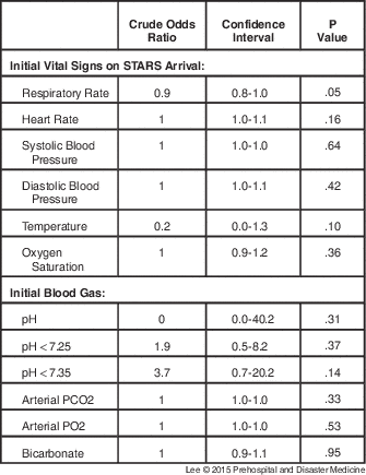
Abbreviations: NIPPV, non-invasive positive pressure ventilation; STARS, Shock Trauma Air Rescue Society.
Factors Associated with Prolonged Scene Times
Shock Trauma Air Rescue Society spent significantly more time at the sending hospital with patients who failed NIPPV (Table 4). Furthermore, after controlling for age, sex, RR, and GCS, scene time for those failing NIPPV was 39.4 minutes longer than those who did not fail (95% CI, 16.2-52.5; P=.001).
Table 4 Scene, Transport,Footnote a and UsageFootnote b Times

Abbreviations: IQR, interquartile range; NIPPV, non-invasive positive pressure ventilation; STARS, Shock Trauma Air Rescue Society.
a Transport time defined as elapsed time from when patient transport initiated to handover of care.
b Usage time defined as time elapsed from dispatch to handover of care.
Discussion
There is growing evidence of the efficacy of NIPPV in prehospital care where it has been shown to have similar benefits to in-hospital initiated NIPPV, such as a decreased frequency of intubations, reduced ICU LOS, as well as the added benefit of a reduction of prehospital intubations.Reference Roesslaer, Schmid and Michels 3 - Reference Thompson, Petrie, Ackroyd-Stolarz and Bardua 6 This is believed to be the first study examining the factors associated with NIPPV failure in a critical care HEMS.
Although this study had a relatively high incidence of NIPPV failure (32%), prehospital NIPPV has been shown to reduce the need for invasive ventilation with a number needed to treat (NNT) of eight and decreased in-hospital mortality with a NNT of 18.Reference Mal, McLeod, Iansavichene, Dukelow and Lewell 10 Of the 14 patients who failed NIPPV, 12 patients were intubated in the sending hospital prior to departure, one patient was intubated in-flight without complication, and one patient required rescue bag-mask ventilation due to a depleted oxygen supply in the helicopter. An internal investigation of the case with a depleted oxygen supply from NIPPV use revealed that the oxygen supply was depleted from a poor mask seal and a high minute ventilation, causing excessive high-flow oxygen usage and waste. The patient was successfully bag-mask ventilated at 10-15 litres/minute until another oxygen source became available upon landing where the patient was placed back on NIPPV. Non-invasive positive pressure ventilation procedure changes were implemented to highlight the risk and mitigate the occurrence of oxygen source depletion when using this modality. This case demonstrates the unique patient safety factors that need to be considered in the setting of potentially long transport times. It should be noted that all cases of failure occurred during inter-hospital transfers. It is likely that different clinical decisions are made in a supportive and controlled hospital environment than in an uncontrolled environment of a prehospital scene. All five cases of prehospital scene response in the study cohort were managed successfully with NIPPV. Although these numbers are small, NIPPV use in this context was reassuring; in three of the cases, the need to manage very high-risk airways with limited resources was avoided successfully through the use of NIPPV.
Appropriate prehospital NIPPV use is advocated by the authors of this study; however, careful patient selection is vital to the successful use of NIPPV in the transport environment.Reference Le Cong and Robertson 11 There are situations where a secured airway is preferred for a patient eligible for NIPPV treatment, after taking into account the unique environment of HEMS, patient safety, and resource allocation. Patients with a decreased level of consciousness (GCS<15) on AMC arrival were 14 times more likely to fail NIPPV. This finding is in keeping with previously published risk factors associated with NIPPV failure in the EDReference Merlani, Pasquin, MGranier, Treggiari, Rutschmann and Ricou 12 and in-hospital settings.Reference Confalonieri, Garuti and Cattaruzza 13 , Reference Schettino, Altobelli and Kacmarek 14 Furthermore, the ED and hospital-based studies determined that a low GCS, tachypnea, and acidosis were risk factors of failure one or two hours after the initiation of NIPPV.Reference Merlani, Pasquin, MGranier, Treggiari, Rutschmann and Ricou 12 - Reference Schettino, Altobelli and Kacmarek 14 Other than initial GCS on arrival, no other risk factors of NIPPV failure were identified based on initial arrival vital signs and blood gases. Determining a patient’s response to NIPPV requires time, which has different implications in the transport environment than in the definitive in-hospital environment. Patients who failed NIPPV were found to have significantly longer scene times (up to 39.4 minutes longer) compared to those in whom NIPPV was deemed successful. These prolonged scene times likely reflect a combination of increased severity, need for monitoring prior to transport, failed intervention, and the need for an alternative airway strategy; however, with this retrospective study, the authors were unable to determine the primary cause with certainty.
Providers need to consider the time sensitivity of the condition when applying NIPPV; for example, the patient in pulmonary edema may benefit greatly from NIPPV, but the patient in pulmonary edema with ischemic changes on electrocardiogram may benefit more from minimizing transport time. Helicopter Emergency Medical Service providers need to balance the benefits of NIPPV against the impacts of long scene times, potential oxygen supply depletion, and inability to respond to other mission requests.
Limitations
This study has several limitations that require discussion. First, there are inherent limitations to medical record reviews.Reference Worster and Haines 15 Missing data, poor documentation, and variability in clinical care limit the validity of these results; however, this study employed valid methods in an effort to reduce these biases. For example, all medications were recorded accurately by the HEMS crew, which enhanced the validity of the data. In addition, strategies to avoid selection bias (methods employed to choose the study sample), strategies to minimize error during measurements (standardized forms and assessment of data abstraction), objective definition of the primary outcome (NIPPV failure/success), and controlling for possible confounders (adjusted analyses) were all employed. Second, the study was conducted at one Canadian HEMS organization, which may not allow the study results to be extrapolated to other organizations. The participating site is a leading HEMS research site in Canada, affiliated with a strong network of ED researchers, and likely represents the best-case scenario for the estimation of variability. Third, original medical charts were the primary source used at all sites. Variability across providers was reduced with an ePCR, however, suggesting eligible medical records were less likely to have been missed. Once again, these cases likely represent the most severe cases of persistent respiratory distress for NIPPV use. Finally, these results are not population-based, and this likely does not represent all of the cases in a confined area. Moreover, this level of individual granular detail would not be possible in a population-based study.
Conclusion
Using accepted high-quality chart review methods,Reference Worster and Haines 15 the authors of this study found that a decreased level of consciousness at HEMS crew arrival was associated with NIPPV failure. Non-invasive positive pressure ventilation failure is associated with long scene times, and in some cases, high oxygen consumption. These factors should be taken into consideration when NIPPV is applied to critical care HEMS patients.
Acknowledgements/Support
Author Dr. Villa-Roel is supported by the Canadian Institutes of Health Research (CIHR; Ottawa, Ontario, Canada) in partnership with the Knowledge Translation branch. Author Ms. Couperthwaite is supported by the Emergency Medicine Research Group in the Department of Emergency Medicine at the University of Alberta (Edmonton, Alberta, Canada). Author Dr. Rowe is supported by the CIHR as a Tier I Canada Research Chair in Evidence-based Emergency Medicine through the Government of Canada (Ottawa, Ontario, Canada). All authors would like to acknowledge Peter Hepburn and Angela Kelter (STARS IT) who assisted with data collection and database access.


