


Introduction
The order Suberitida was recognized in 2015 by Morrow & Cárdenas after a major overturning of the Demospongiae classification. Some years before, Chombard & Boury-Esnault (Reference Chombard and Boury-Esnault1999) already created the suborder Suberitina, grouping the families Suberitidae and Halichondriidae, according to morphological and genetic traits. However, this taxonomic group did not appear in the Systema Porifera (Hooper & van Soest, Reference Hooper and van Soest2002) due to several inconsistencies in systematics. In summary, the order is presently defined as a group of species without an obvious cortex and a skeleton devoid of microscleres. Megascleres are mostly styles, oxeas and/or tylostyles. The ectosome arrangement is tangential or paratangential. The skeleton of the choanosome is frequently confused, but a radial arrangement and/or ascending multispicular tracts can be found in several representatives (van Soest, Reference van Soest, Hooper and van Soest2002a, Reference van Soest, Hooper and van Soest2002b; Erpenbeck & van Soest, Reference Erpenbeck, van Soest, Hooper and van Soest2002). The molecular synapomorphy that characterizes Suberitida is a deletion of a small loop of 15 base pairs in the secondary structure (D2) of the 28S rDNA when compared with other Heteroscleromorpha (Morrow & Cárdenas, Reference Morrow and Cárdenas2015).
The order encompasses three families, 26 genera and 491 species distributed across the oceans. Along the Brazilian coast, only two families, nine genera, and, so far, 20 valid species were inventoried (Muricy et al., Reference Muricy, Lopes, Hajdu, Carvalho, Moraes, Klautau, Menegola and Pinheiro2011; van Soest et al., Reference van Soest, Boury-Esnault, Hooper, Rützler, de Voogd, Alvarez, Hajdu, Pisera, Vacelet, Manconi, Schoenberg, Janussen, Tabachnick and Klautau2019, Table 1). The north-east region of Brazil seems to harbour the richest sponge fauna of Brazil; however, most of the studies were conducted along the eastern littoral (Boury-Esnault, Reference Boury-Esnault1973; Muricy & Moraes, Reference Muricy and Moraes1998; Hajdu et al., Reference Hajdu, Peixinho and Fernandez2011; Leonel et al., Reference Leonel, Mothes, Lerner, Gama and Campos2011; Cedro et al., Reference Cedro, Hajdu and Correia2013; Sandes & Pinheiro, Reference Sandes and Pinheiro2013; Santos et al., Reference Santos, Nascimento and Pinheiro2018) and around seamounts or oceanic islands (Salani et al., Reference Salani, Lotufo and Hajdu2006; Muricy et al., Reference Muricy, Esteves, Santos, Silva, Klautau and Lanna2008; Moraes, Reference Moraes2011; Carvalho et al., Reference Carvalho, Da Silva and Pinheiro2013), whereas the northern coast has received much less attention (Mothes et al., Reference Mothes, Campos, Lerner and Ferreira-Correia2004; Campos et al., Reference Campos, Mothes, Eckert and van Soest2005).
Table 1. State of the art on the occurrence of Order Suberitida in Brazil
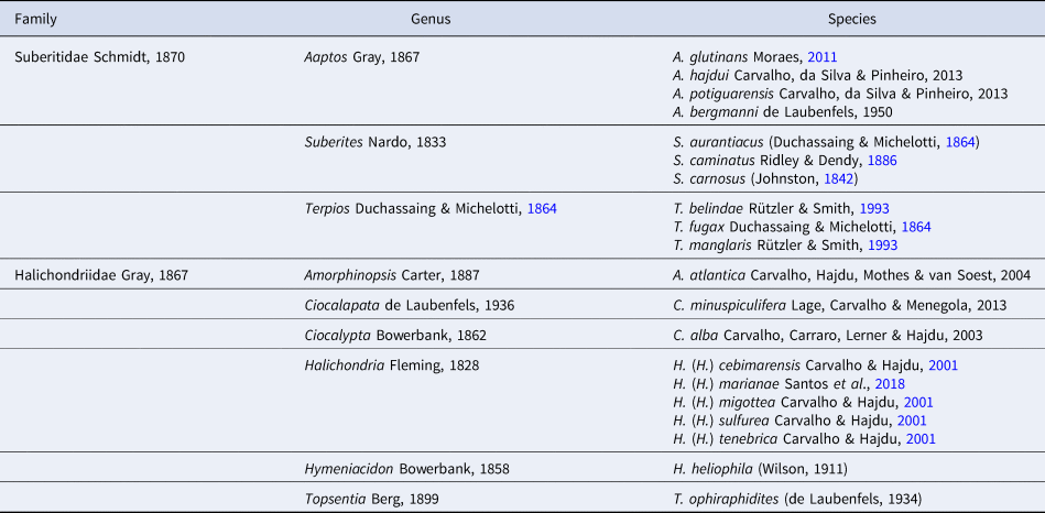
All Brazilian records can be seen in Muricy et al. (Reference Muricy, Lopes, Hajdu, Carvalho, Moraes, Klautau, Menegola and Pinheiro2011).
In a recent study, van Soest (Reference van Soest2017) has shown that the Caribbean Sea and the Brazilian coast share several representatives of the sponge fauna, the northern coast of Brazil thus appearing as a transitional area in the Tropical Western Atlantic (TWA) (Spalding et al., Reference Spalding, Fox, Allen, Davidson, Ferdaña, Finlayson, Halpern, Jorge, Lombana, Lourie, Martin, McManus, Molnar, Recchia and Robertson2007). Aiming at a better knowledge of the sponge fauna of this transitional region, surveys were realized at shallow waters lengthways at the northern coast of Brazil. This work presents a focus on the order Suberitida with the description of six species from the intertidal zone, two considered new for science.
Materials and methods
Study area
The sampling was realized in the framework of the expeditions TAXPOMOL/2016 and Maranhão/2017 in the unexplored part of the northern coast of the north-east region of Brazil, which is named the Brazilian Semi-Arid Coast (Diniz & Oliveira, Reference Diniz and Oliveira2016). All samples were collected in the intertidal zone, on sandy beaches, with rocky tide pools, from the states of Maranhão and Ceará (Figure 1).

Fig. 1. Part of the Brazilian Semi-Arid Coast at the northern region of the Brazilian coast, with the location of sampling sites. The highlight in the right corner shows the position of the states on the map of Brazil. The small letters indicate the sites where sponge species were collected: (A) Araçagi beach, (B) Meio beach, (C) Panaquatira beach, depicted enlarged at the highlight in the left corner; and (D) Dois Coqueiros beach. MA, Maranhão State; PI, Piauí State; CE, Ceará State.
The Brazilian Semi-Arid Coast includes Maranhão, Piauí, Ceará and part of Rio Grande do Norte states, the shoreline being about 1065 km long. In this region, the average temperature is 25.8°C, the pluviometry is 262.3 ppm (Climate-data, 2019, accessed online, 3 April 2019), and it experiences the strongest winds of the Brazilian coast (Muehe, Reference Muehe, Cunha and Guerra1998). According to the oceanographic conditions, the region possesses the largest tidal range (8–12 m) along the Brazilian coast (Muehe, Reference Muehe, Cunha and Guerra1998). The seawater temperature average is always higher than 27°C, the chlorophyll a concentration varies from 0.2 to 4.3 mg m–3, and the particulate organic carbon concentration (POC) is about 60 mg m–3 at the sandy beaches of Ceará and Rio Grande do Norte, and an average of 397 mg m–3 at the estuary of São Luís (Maranhão) (compiled data from Giovanni platform of NASA).
Sample collection and identification
Sponge samples were preserved in 92% ethanol and deposited in the Porifera Collection of the Museu Nacional (MN/UFRJ). Spicule and skeletal preparations followed classic protocols for Demospongiae species (Hajdu et al., Reference Hajdu, Peixinho and Fernandez2011). For each sponge individual, 50 spicules were measured in order to provide basic descriptors (minimal, mean, standard deviation and maximal values) in comparative tables.
Systematics
Phylum PORIFERA Grant, 1836
Class DEMOSPONGIAE Sollas, 1885
Subclass HETEROSCLEROMORPHA Cárdenas, Pérez and Boury-Esnault, 2012
Order SUBERITIDA Chombard & Boury-Esnault, 1999
Family SUBERITIDAE Schmidt, 1870
Genus Suberites Nardo, 1833
Synonymy
For synonymy see van Soest (Reference van Soest, Hooper and van Soest2002b).
Definition
Suberitidae harbouring tylostyles in two size categories, large at the choanosome, and small forming bouquets at the ectosome. The choanosomal skeleton is confused or alveolar with sub-radiate bundles of megascleres. Microscleres are not common, but spined centrotylote microstrongyles can be observed (van Soest, Reference van Soest, Hooper and van Soest2002b).
Type species
Alcyonium domuncula Olivi, 1792: 241 (by original designation).
Suberites purpura sp. nov.
(Figures 2–4, Table 2)
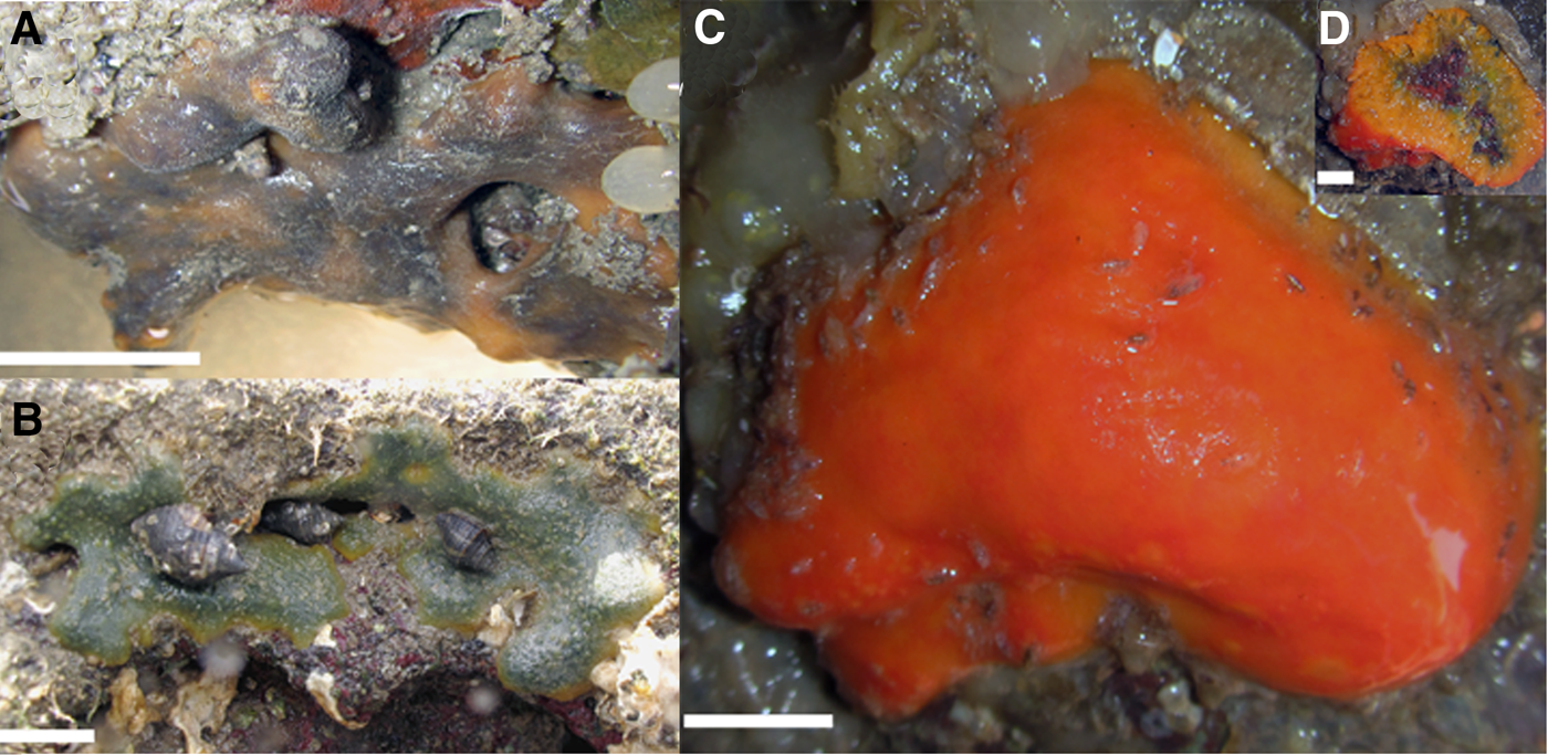
Fig. 2. In situ images presenting the external morphology of Suberites specimens: (A) Suberites purpura sp. nov. (MNRJ 21343); (B–D) Suberites aurantiacus (Duchassaing & Michelotti, Reference Duchassaing and Michelotti1864) (B – MNRJ 21430, C, D – MNRJ 21500). Scale: A, D = 1 cm; B, C = 2 cm.

Fig. 3. Optical microscopy images presenting the skeleton architecture of Suberites specimens. Suberites purpura sp. nov. (MNRJ 21343), (A) ectosomal skeleton with short bouquets of small tylostyles and confused choanosomal skeleton with longer tylostyles forming rare branches, (B) detail of short bouquets; Suberites aurantiacus (Duchassaing & Michelotti, Reference Duchassaing and Michelotti1864), (C) ‘Suberites-like skeleton’ – small tylostyles forming wide bouquets in the ectosome (MNRJ 21405), (D) Orange morphotype of Suberites aurantiacus, confused choanosomal skeleton, with bundles in direction to the surface (MNRJ 21500). Scale: A, C, D = 200 μm; B = 100 μm.

Fig. 4. Tylostyle size and fusiform form of Suberites species. (A) Suberites purpura sp. nov.; (B, C) Suberites aurantiacus (Duchassaing & Michelotti, Reference Duchassaing and Michelotti1864). Scale: tylostyles = 100 μm; tyles = 10 μm. The orange morphotype of Suberites aurantiacus is represented in C.
Table 2. Comparison of the tylostyles and tyles, growth forms and colour of all Suberites species from the Tropical Western Atlantic and the type species Suberites domuncula Olivi, 1792

Spicules values are in micrometres (μm) and presented as minimal–mean (standard deviation)–maximal length/width. N = 50.
a van Soest (Reference van Soest, Hooper and van Soest2002b);
b Ridley & Dendy (Reference Ridley and Dendy1886);
c Boury-Esnault (Reference Boury-Esnault1973);
d van Soest (Reference van Soest2017;
e Schmidt (Reference Schmidt1870);
f Wilson (Reference Wilson1902);
g Wilson (Reference Wilson1931);
h Rützler & Smith (Reference Rützler and Smith1993);
i Duchassaing & Michelotti (Reference Duchassaing and Michelotti1864);
j Moraes (Reference Moraes2011);
k Muricy & Hajdu (Reference Muricy and Hajdu2006);
l Present study.
Type Material
Holotype. MNRJ 21343, Praia de Araçagi, São José de Ribamar, tidal pool (2°27′46.84″S 44°12′14.94″W), Maranhão State, Brazil, 0.5 m depth, coll. H. Fortunato, 5/XI/2017.
Diagnosis
Suberites purpura sp. nov. is the only Suberites species from the Western Atlantic Ocean, purple in colour, with a combination of tylostyles up to 1500 μm long and short and open bouquets in the ectosomal skeleton.
Description
External morphology (Figure 2A). Thickly encrusting to massive sponge, 10 × 5 cm size, and small oscules (<1 mm diameter). The surface is smooth but also microhispid and the consistency is firm, little compressible, and somehow cartilaginous. The colour is purple outside, and orange at the basis and inside.
Ecology
Only one specimen was found in a crevice of a tide pool, in a dark habitat.
Distribution
Brazil: north-east region: Maranhão State.
Etymology
In Latin, the feminine noun, ‘purpura’ = purple coloured, refers to the external colour of the species (Brown, Reference Brown1956).
Remarks
The combination of tylostyles size (>1500 μm), the purple colour, the short and open bouquets, and the geographic distribution make this species different from all other species from the TWA. The most similar species from the TWA is S. aurantiacus (Duchassaing & Michelotti, Reference Duchassaing and Michelotti1864); however, the spicule length of this species is never greater than 1000 μm. Suberites caminatus Ridely & Dendy, Reference Ridley and Dendy1886, was described from the Prince Edward Islands in the Antarctic Indian Ocean, but the original description also included slightly variant material from off Rio de la Plata, in the Uruguay-Buenos Aires shelf; the name was also used by Boury-Esnault (Reference Boury-Esnault1973) for material from south-east Brazil (see Ridley & Dendy, Reference Ridley and Dendy1886 in van Soest et al., Reference van Soest, Boury-Esnault, Hooper, Rützler, de Voogd, Alvarez, Hajdu, Pisera, Vacelet, Manconi, Schoenberg, Janussen, Tabachnick and Klautau2019). It has tylostyle sizes up to 600 μm, half of that of the new species. The disjunct distribution of this species calls for a comparative study to decide if the SW Atlantic material belongs to a different, yet unnamed species. The name Suberites carnosus (Johnston, Reference Johnston1842) was used by Ridley & Dendy (Reference Ridley and Dendy1886) and by Boury-Esnault (Reference Boury-Esnault1973) for material respectively from Fernando de Noronha and south-east Brazil (see Johnston, Reference Johnston1842 in van Soest et al., Reference van Soest, Boury-Esnault, Hooper, Rützler, de Voogd, Alvarez, Hajdu, Pisera, Vacelet, Manconi, Schoenberg, Janussen, Tabachnick and Klautau2019). The former has spicules up to 560 μm, and the latter up to 430 μm and in a single size (see details in Table 2), unlike S. purpura sp. nov. Moreover, as S. carnosus from the Eastern Atlantic and the Mediterranean has a stalked plum-like shape and a large central osculum (Ackers et al., Reference Ackers, Moss, Picton, Stone and Morrow2007), and Boury-Esnault (Reference Boury-Esnault1973) material is described as thinly encrusting, this record seems inaccurate. We suggest calling it Suberites aff. carnosus until a new taxonomic study is carried out. Suberites purpura sp. nov. also differs from the other species distributed in the Caribbean by its internal and external characteristics. Suberites lobatus (Wilson, Reference Wilson1902) has an encrusting growth form; S. crispolobatus van Soest, Reference van Soest2017 has a greyish brown colour, with erect rounded branches, and dwells in rather deep-water (50–85 m) habitats; and S. distortus Schmidt, Reference Schmidt1870 presents a club-shaped yellow form. Unfortunately, the comparison with Suberites heros Schmidt, Reference Schmidt1870, from the Eastern Caribbean, is difficult due to the lack of anatomical data for the latter species. In fact, there are reasons to doubt if it is a valid species, as the author had mentioned that S. heros presents quite similar habit to the type species, Suberites domuncula, also in association with molluscs and possession of tylostyles with short and not pronounced tyles. Moreover, van Soest (Reference van Soest, Hooper and van Soest2002b) informed that the lectotype conforms to S. domuncula due to the presence of a hermit crab hole in the sponge and tylostyle in two categories. Thus, this species still needs revision. Lastly, S. purpura sp. nov. differs from the Chesapeake Bay (USA) representative, Suberites paradoxus Wilson, Reference Wilson1931, because the last has a lamellar and buried habit with abundant sand grains incorporating the sponge tissue, and the tylostyles are slightly curved, with overlapping categories varying from 220 to 350 μm in length.
Suberites aurantiacus (Duchassaing & Michelotti, Reference Duchassaing and Michelotti1864)
(Figures 2–4, Table 2)
Synonymy
For synonymy see Rützler & Smith (Reference Rützler and Smith1993).
Type locality
Caribbean Sea, Virgin Islands, St. Thomas.
Material Examined
MNRJ 21405, MNRJ 21430 Araçagi beach, São José de Ribamar, tidal pool (2°27′46.84″S 44°12′14.94″W), Maranhão State, Brazil, coll. H. Fortunato, 5/XI/2017, MNRJ 21500, Meio beach, São José de Ribamar, tidal pool (2°28′19.11″S 44°13′8.73″W), Maranhão State, Brazil, coll. H. Fortunato, 5/XI/2017.
Redescription
External morphology (Figure 2B–D)
Sponges vary from encrusting to massive or subspherical habitat. The size of the examined individuals was ~12 × 8 cm (length × width). Their surface was velvety and rugose, with vesicles. Their consistency was firm and little compressible. Small oscula can be seen dispersed at the surface (1–5 mm in diameter). The colour in vivo is mostly green externally (Figure 2B), but also bright orange and always yellow internally (Figure 2C, D).
Ecology
The species has been observed on semi-exposed surfaces, mostly in shallow waters.
Distribution
Tropical Western Atlantic: Gulf of Mexico, Caribbean Sea, and Brazilian Tropical and Temperate coasts.
Remarks
Suberites aurantiacus is a widely distributed species in the TWA, showing a great polymorphism of growth form and colour, especially when rocky shore and mangrove individuals are compared. The specimens found along the northern coast of the north-east region of Brazil are similar to others from the TWA by the presence of non-curved tylostyles in two size categories, varying from 140 to 800 μm, globose tyles, mostly green colour externally and yellow internally, and large ectosomal bouquets. An orange, subspherical morphotype, with ectosomal bouquets being narrower and with longer spicules, found living upside down in crevices of a tidal pool at Maranhão State (MNRJ 21500, Figures 2C and D, 3D and 4C), was assigned to S. aurantiacus owing to the known variability and wide tolerance of this species. Molecular investigations may highlight how and why these polymorphisms occur and would also inform if this morphotype is really S. aurantiacus.
Key to the Genus Suberites from the Tropical Western Atlantic Comparing with the Type Species
1a Two categories of tylostyles………2
1b One category of tylostyles………S. aff. carnosus
2a Colonization of gastropod shells………3
2b No colonization of gastropod shells………4
3a Slightly curved tylostyles producing subterminal (drop-shaped), annular swellings or well-formed tyles………S. domuncula
3b Slightly curved tylostyles possessing short and not pronounced tyles………S. heros
4a Shallow water habitat………5
4b Mesophotic sponge (50–85 m) with erect rounded branches habit………S. crispolobatus
5a (Sub)spherical, massive or encrusting growth form sponges………6
5b Encrusting growth form………S. lobatus
5c Club-shaped sponge with 2 mm oscula diameter………S. distortus
6a Tylostyles length do not reach 1000 μm………7
6b Tylostyles length exceed 1000 μm………8
7a (Sub)spherical sponge with tylostyle length up to 600 μm………S. caminatus
7b Thick encrusting to massive polychromatic sponge with tylostyle reaching 900 μm………S. aurantiacus
8 Thick encrusting to massive purple sponge with larger tylostyles exceeding 1500 μm length………S. purpura sp. nov.
Genus Terpios Duchassaing & Michelotti, Reference Duchassaing and Michelotti1864
Synonymy
For synonymy see van Soest (Reference van Soest, Hooper and van Soest2002b).
Definition
Thin encrusting Suberitidae. Spicules are exclusively tylostyles, with a lobate tyle. The colours of the species of this genus are commonly associated with those present in their symbiotic bacteria or cyanobacteria (Rützler & Smith, Reference Rützler and Smith1993).
Type species
Terpios fugax Duchassaing & Michelotti, Reference Duchassaing and Michelotti1864 (by subsequent designation; Topsent, 1900).
Terpios fugax Duchassaing & Michelotti, Reference Duchassaing and Michelotti1864
(Figure 5, Table 3)
Synonymy

Fig. 5. Terpios fugax Duchassaing & Michelotti, 1864. (A) Specimen in a tide pool, at Ceará state. (B) The tylostyle of T. fugax with detail for flattened and round multilobate tylostyle. Scale: tylostyle 50 μm, tyle 10 μm.
Table 3. Comparative tylostyles dimensions and head shape, locality and colour of Terpios spp. from the Tropical Western Atlantic

Values are expressed in micrometres (μm) as minimum–mean (standard deviation)–maximum length/width. N = 100.
Comparative data in aRützler & Smith (Reference Rützler and Smith1993). bPresent study.
see van Soest (Reference van Soest, Hooper and van Soest2002b).
Type locality
U.S. Virgin Islands, St. Thomas.
Type material
Lectotype BMNH 1928.11.12.11, designated by van Soest et al. (1983).
Material examined
MNRJ 18940, MNRJ 21696, Dois Coqueiros beach, Caucaia, tidal pool (3°41′17.19″S 38°36′35.25″W), Ceará State, Brazil, 0.5 m depth, coll. H. Fortunato, 11/IV/2016.
Redescription
External morphology (Figure 5A)
The sponge is thinly encrusting with small oscula (<1 mm) randomly distributed. The surface is uneven, microhispid, not detachable and without sub channels. The consistency is soft and fragile. The colour is vivid dark blue in vivo and maintained after fixation in alcohol.
Skeleton
The ectosomal skeleton is not specialized, not detachable, but spicules can protrude the surface. The choanosome presents a low density of spicules, most directionless, but some arranged with the tyles facing the substratum and the acerate ends protruding the sponge surface.
Ecology
The species has been observed on exposed and semi-exposed hard substrates in the subtidal zone.
Distribution
Tropical Western Atlantic: Caribbean and Brazil (north-east and south-east regions).
Remarks
The records from Ceará described here are similar to the type species. All three species of the genus distributed at the TWA are shared between the Caribbean and Brazil. Although T. fugax and T. manglaris are both encrusting and blue, the latter has exclusively flattened and quadrilobate tyles, while in the former there are both flattened and round tyles, and these are multilobate. Terpios belindae distinguishes itself by the red to orange colour and a marked flattened tyle.
Family Halichondriidae Gray, 1867
Genus Hymeniacidon Bowerbank, 1858
Synonymy
For synonymy see Erpenbeck & van Soest (Reference Erpenbeck, van Soest, Hooper and van Soest2002).
Definition
Halichondriidae that have an encrusting or massive and lobate growth form, and fistules covering the apical portion. Megascleres are exclusive small styles or stylotes (>500 μm). The ectosomal skeleton is tangential with intercrossing bundles made of a single type of megasclere. The choanosomal skeleton is mostly loose and confused, but it harbours some ascending vague bundles of megascleres (Erpenbeck & van Soest, Reference Erpenbeck, van Soest, Hooper and van Soest2002).
Type species
Hymeniacidon perlevis (Montagu, 1814).
Hymeniacidon upaonassu sp. nov.
(Figure 6, Table 4)

Fig. 6. External and internal anatomy of Hymeniacidon upaonassu sp. nov. (A) External traits of the new species, scale: 5 cm, (B) Architecture of the skeleton, scale: 500 μm, (C) styles, scale: 50 μm.
Table 4. Comparison of the main traits of all Hymeniacidon species from the Tropical Western Atlantic, with addition of Rio de Janeiro State, south-east (SE) region of Brazil and the type species Hymeniacidon perlevis (Montagu, 1814) data

Values are in micrometres (μm), as minimal–mean (standard deviation)–maximal length/width. N = 50.
a Erpenbeck & van Soest (Reference Erpenbeck, van Soest, Hooper and van Soest2002);
b Burton (Reference Burton1954);
c Díaz et al. (Reference Díaz, Pomponi and van Soest1993);
d Muricy & Hajdu (Reference Muricy and Hajdu2006);
e Present study.
Type Specimens
Holotype
MNRJ 21390, Panaquatira beach, São José de Ribamar, tidal pool (2°31′4.65″S 44°1′38.16″W), Maranhão State, Brazil.
Paratypes
MNRJ 21360, Panaquatira beach, São José de Ribamar, tidal pool (2°31′4.65″S 44°1′38.16″W), Maranhão State, Brazil, 0.5 m depth, coll. H. Fortunato, 5/XI/2017, MNRJ 21346, Araçagi beach, São José de Ribamar, tidal pool (2°27′46.84″S 44°12′14.94″W), Maranhão State, Brazil.
Diagnosis
Hymeniacidon upaonassu sp. nov. has a yellow colour and harbours conical fistules uniformly arranged covering the entire body without oscula at their apex.
Description
External morphology (Figure 6A)
The sponge has a massive base and conical fistules covering the entire upper portion of the body. The holotype consists of a fragment of 13 × 9 × 3 cm (length × width × thickness). Its surface is rugose and rough, its consistency is friable and fleshy. No oscula can be seen. The colour in vivo and after fixation is pale yellow.
Skeleton (Figure 6B)
The ectosomal skeleton is not easily detachable from the choanosomal one, nor much defined. It tends to form a paratangential layer with intercrossing styles and subectosomal cavities. The choanosomal skeleton is confused, but multispicular tracts (30 μm thick) are present, and axial condensation of longitudinal tracts are observed in the fistules.
Ecology
The species is found in tide pools, covered by sand.
Distribution
Brazil: north-east region: Maranhão State.
Etymology
‘Upaon–Açu’ is the historical indigenous name (Tupi–Guarani) of São Luís Island in Maranhão State, where all specimens here described were recorded.
Remarks
Hymeniacidon upaonassu sp. nov. is the only species of the genus recorded in the north-east region of Brazil. Indeed, the Brazilian record of H. heliophila (Wilson, 1911) is exclusive for the south-east region, and H. perlevis (Montagu, 1814) is considered misidentified (Muricy et al., Reference Muricy, Lopes, Hajdu, Carvalho, Moraes, Klautau, Menegola and Pinheiro2011). The new species has the classical internal anatomy of the genus Hymeniacidon sharing many similarities with the above-mentioned species. However, the whole body covered by fistules represents the main diagnostic trait of the new species. The absence of oscula in the apex of the fistules, and its yellow colour also differ from the other species. The new Brazilian species also differs from the two Caribbean species, H. caerulea Pulitzer-Finali, Reference Pulitzer-Finali1986, which is blue in colour, and Hymeniacidon glabrata Burton, Reference Burton1954, which was found only by the species' author, and possesses styles exceeding 500 μm, that shapes more likely to strongyloxeas. Moreover, both Caribbean species dwell in subtidal habitats.
Genus Halichondria Fleming, 1828
Subgenus Halichondria Fleming, 1828
Synonymy
For synonymy see Erpenbeck & van Soest (Reference Erpenbeck, van Soest, Hooper and van Soest2002).
Definition
Halichondria with smooth or digitate surface (Erpenbeck & van Soest, Reference Erpenbeck, van Soest, Hooper and van Soest2002).
Type species
Spongia panicea Pallas, 1766: 388 (by original designation).
Halichondria (Halichondria) melanadocia de Laubenfels, 1936
(Figures 7–9, Table 5)
Synonymy

Fig. 7. External anatomy variations of Halichondria (Halichondria). (A–D) Halichondria (H.) marianae Santos et al. (Reference Santos, Nascimento and Pinheiro2018); (E) Halichondria (H.) melanadocia de Laubenfels, 1936. Scale: A–C = 1 cm, D–E = 2 cm. Morphotype of H. (H.) marianae with oxeas exceeding 1500 μm is represented in A.

Fig. 8. Skeleton architecture of Halichondria (Halichondria). (A) Halichondria (H.) melanadocia de Laubenfels, 1936; (B) Halichondria (H.) marianae Santos et al. (Reference Santos, Nascimento and Pinheiro2018). Scale: 100 μm.

Fig. 9. Smooth oxeas Halichondria (Halichondria). A: Halichondria (H.) melanadocia de Laubenfels, 1936; B–C: Halichondria (H.) marianae Santos et al. (Reference Santos, Nascimento and Pinheiro2018). Scale = 100 μm. The morphotype of H. (H.) marianae with oxeas exceeding 1500 μm is represented in B.
Table 5. Comparison of the main traits of some Halichondria spp. from the Tropical Western Atlantic, with addition of the type species Halichondria (Halichondria) panicea (Pallas, 1766) from Europe.

Values are in micrometres (μm), as minimal–mean (standard deviation)–maximal length/width. N = 50. aErpenbeck & van Soest (Reference Erpenbeck, van Soest, Hooper and van Soest2002); bPulitzer–Finali (Reference Pulitzer-Finali1986); cDíaz et al. (Reference Díaz, Pomponi and van Soest1993); dSantos et al. (Reference Santos, Nascimento and Pinheiro2018); eCarvalho & Hajdu (Reference Carvalho and Hajdu2001); fPresent study.
For synonymy see Hajdu et al. (Reference Hajdu, Peixinho and Fernandez2011), Leonel et al. (Reference Leonel, Mothes, Lerner, Gama and Campos2011) and Santos et al. (Reference Santos, Nascimento and Pinheiro2018).
Material examined
MNRJ 21396, Panaquatira beach, São José de Ribamar, tidal pool (2°31′4.65″S 44°1′38.16″W), Maranhão State, Brazil, 0.5 m depth, coll. H. Fortunato, 5/XI/2017.
Redescription with Modifications
External morphology (Figure 7A)
The observed specimen was moss green in vivo, and black in spirit. It had a thick encrusting to massive habit, forming a mat size of 12.0 × 10.0 cm (length × width). The surface was irregular but mostly rough, reticulated. The consistency was soft and compressible, easily torn, with several oscula on small projections (1.1–2.7 mm).
Skeleton (Figure 8A)
The ectosomal skeleton is anisotropic, tangentially arranged with parallel spicules arrangement. The choanosomal skeleton is disorganized with randomly arranged spicules, although some multispicular tracings towards the ectosome are visible.
Spicules (Figure 9A)
Oxeas are smooth, slightly curved, and in two distinct sizes: smaller 129.1–156.2–176.0/3.6–8.4–13.4 μm, larger 373.2–513.8–654.3/4.5–9.7–19.5 μm.
Ecology
This is a shallow water species, found in rocky shores and sea grass bed from the tidal zone to ~20 m depth.
Distribution
Tropical Western Atlantic: Caribbean and Brazil (north-east region).
Remarks
The Halichondria (H.) melanadocia specimen described here is distinguished from the original description (Díaz et al., Reference Díaz, Pomponi and van Soest1993) by the presence of two categories of oxeas, reaching 600 μm and no odour after collection. On the other hand, the skeleton architecture, external habit and dark colour in spirit are shared traits in comparison to the Caribbean specimens (Díaz et al., Reference Díaz, Pomponi and van Soest1993). Halichondria (H.) melanadocia was registered as H. (H.) aff. melanadocia for Brazil in two field guides, by Hajdu et al. (Reference Hajdu, Peixinho and Fernandez2011), for the state of Bahia, and by Leonel et al. (Reference Leonel, Mothes, Lerner, Gama and Campos2011), for the state of Paraíba; while Santos et al. (Reference Santos, Nascimento and Pinheiro2018) defined that specimens of H. (H.) aff. melanadocia should be synonymized with Halichondria (H.) marianae Santos, Nascimento & Pinheiro, Reference Santos, Nascimento and Pinheiro2018. The specimen from this work differs from other congeners, including H. (H.) marianae, chiefly by the black colour after fixation, and vulcaniform oscula. Moreover, H. (H.) marianae has oxeas exceeding 800 μm and more variation in colour (see details below) than H. (H.) melanadocia. According to our data, specimens with spicules up to 600 μm must be named H. (H.) melanadocia, while those with oxeas exceeding 600 μm are H. (H.) marianae. Here the Halichondria (H.) melanadocia species is validated to the entire littoral of the north-east region of Brazil.
Halichondria (Halichondria) marianae Santos, Nascimento & Pinheiro, Reference Santos, Nascimento and Pinheiro2018
(Figures 7–9, Table 5)
Synonymy
For synonymy see Santos et al. (Reference Santos, Nascimento and Pinheiro2018).
Type locality
Brazil, Paraíba State, Carapibus beach, Conde City.
Type specimens
UFPEPOR 1861, Carapibus beach (7°17′57.66″S 34°47′52.93″W), Conde City, Paraíba State, Brazil, 0.5–1 m depth, coll. G. G. Santos, 21/II/2015.
Material examined
MNRJ 21339, 21345, MNRJ 21363, Araçagi beach, São José de Ribamar, tidal pool (2°27′46.84″S 44°12′14.94″W), Maranhão State, Brazil, 0.5 m depth, coll. H. Fortunato, 5/XI/2017; MNRJ 21706, Dois Coqueiros beach, Caucaia, tidal pool (3°41′17.19″S 38°36′35.25″W), Ceará State, Brazil, 1 m depth, coll. H. Fortunato, 11/IV/2016.
Redescription with Modifications
External morphology (Figure 7B, C)
Halichondriidae with an encrusting to massive growth form, with some erect lobes where the oscula are located, ranging from 1.1 to 2.7 mm in diameter. Surface is rough and rugose, detachable. Consistency is firm, fleshy, and compressible. Colour in vivo is usually dark green but greyish and yellow are possible too and brown to greyish after fixation.
Skeleton (Figure 8B)
The ectosomal skeleton is detachable with a regular reticulated tangential layer of intercrossing oxeas. The choanosomal skeleton is typically halichondroid with loose oxeas in confusion, but some ascending tracts can be seen. Few sponging fibres could be observed.
Spicules (Figure 9B, C)
Oxeas are smooth, slightly curved, with two possible size categories: smaller 103.2–228.8–489.1 μm/2.4–7.9–17.5 μm, larger 302.7–609.1–837.1 μm/4.5–13.6–25.0 μm. An exception was found in MNRJ 21339, which possesses twofold higher oxeas: smaller 244.5–411.3–925.7 μm/4.8–12.4–26.8 μm, larger 1114.2–1286.6–1560.1 μm/21.7–32.3–45.9 μm (length/width).
Ecology
The species was found in tide pools, exposed to air at low tide. Association with algae was observed.
Distribution
Tropical Western Atlantic: Brazil (north-east region).
Remarks
According to the new traits observed here, Halichondria (H.) marianae needs an improvement in its diagnosis. We suggest the combination of colours varying from moss green to yellow and smooth oxeas in two categories exceeding 800 μm as diagnostic traits. In this sense, the species significantly differs from all other species recorded in the Tropical Western Atlantic by the presence of oxeas larger than 1200 μm, especially H. (H.) melanadocia, the most similar congener. The other four Brazilian species are exclusive to the Western Temperate South-eastern Atlantic (see Carvalho & Hajdu, Reference Carvalho and Hajdu2001) and do not share external or internal morphological traits.
Discussion
The description of two new species of Suberitida from intertidal marine habitats of Maranhão State emphasizes that the sponge biodiversity of the Brazilian northern coast is underestimated. This research also shows that many new species of sponges are waiting to be discovered in easily accessible habitats, and with low financial and logistical cost.
Although the order Suberitida is accepted, it remains a matter of debate because of the non-monophyly in some families and genera, and because the main diagnostic traits are overlapping or absent in some groups (Morrow & Cárdenas, Reference Morrow and Cárdenas2015). However, some traits could be highlighted in the two new species described here. For the genus Suberites, the tylostyles distinction in two size categories (Rützler & Smith, Reference Rützler and Smith1993), and the type of ectosomal bouquet are important diagnostic traits, which must be added in the diagnosis of the genus, instead of colours, which may vary hugely (e.g. Suberites aurantiacus, see Collin et al., Reference Collin, Díaz, Norenburg, Rocha, Sánchez, Schulze, Schwartz and Valdés2005; Muricy & Hajdu, Reference Muricy and Hajdu2006; Hajdu et al., Reference Hajdu, Peixinho and Fernandez2011). The genus Hymeniacidon does not vary much concerning internal characteristics (Díaz et al., Reference Díaz, Pomponi and van Soest1993; Erpenbeck & van Soest, Reference Erpenbeck, van Soest, Hooper and van Soest2002); however, the external features, such as colour and type of the fistules, seem to be diagnostic traits at the species level. In this sense, morphological traits must be applied carefully as one characteristic may be useful for one group but not for another.
Suberites purpura sp. nov. and S. aurantiacus from Maranhão State were found at the intertidal zone, sometimes totally outside the water. These records contrast with the literature that indicates Suberites as a genus commonly found in subtidal cold waters (van Soest, Reference van Soest, Hooper and van Soest2002b). The colour variability is not a peculiarity of S. aurantiacus, since such a polymorphism is also known in Suberites diversicolor Becking & Lim, 2009 from the Indian Ocean. Four species of Suberites are now recorded along the Brazilian coast, one restricted to the north-east coast (S. purpura), two widely distributed (S. aurantiacus and S. carnosus), and S. caminatus being the only one exclusive of temperate waters (Boury-Esnault, Reference Boury-Esnault1973). Suberites aurantiacus from Brazil, Caribbean and Gulf of Mexico coast and S. caminatus from South and North Atlantic, may include more than one genetic lineage, but phylogeographic studies are needed to solve the question (see example in Becking et al., Reference Becking, Erpenbeck, Peijnenburg and de Voogd2013). For now, we recommend using Suberites aff. carnosus until the completion of taxonomic studies of this species comparing all populations distributed in the Atlantic Ocean (Brazil, Açores, North-west Africa and North-east Atlantic Ocean) and the Mediterranean Sea with the type material of the Irish coast.
Terpios fugax is common along the Caribbean and Brazilian coasts, and very easily recognizable thanks to its bright blue colour and thinly encrusting growth form. Rützler & Smith (Reference Rützler and Smith1993) indicated that the tyle form was the major trait for the genus, even to distinguish the two blue sympatric species of the Caribbean Sea (T. fugax and T. manglaris). Another blue Terpios is T. gelatinosus (Bowerbank, 1866), which was recognized as a junior synonym of T. fugax for the Mediterranean Sea(Voultsiadou-Koukoura & van Soest, Reference Voultsiadou-Koukoura and van Soest1993). However, the low spicular density (van Soest, Reference van Soest, Hooper and van Soest2002b), the elongated lobate tylostyle heads, and the loss of colour after fixation distinguish both species (HFMF, personal observations). Therefore, sponges recorded outside TWA (see Topsent, Reference Topsent1934; Thomas, Reference Thomas and James1985) call for an urgent revision.
Hymeniacidon upaonassu sp. nov. is the first species of this genus described for the North-east region of Brazil. It has the same pale yellow colour of the type species Hymeniacidon perlevis from Europe. Other specimens recorded from the cold waters of China and Argentina coasts possess the same colour and morphology (Gastaldi et al., Reference Gastaldi, De Paula, Narvarte, Lôbo-Hajdu and Hajdu2018). In turn, the orange H. heliophila originally from Florida, is widespread in the Caribbean Sea, and is also recorded for the South-east region of Brazil (Muricy et al., Reference Muricy, Lopes, Hajdu, Carvalho, Moraes, Klautau, Menegola and Pinheiro2011). These three species have similar morphologies, except for the type of the fistules. The new species differs from the other two distributed in the Caribbean, H. glabrata and H. caerulea. Both have spicules larger than 500 μm, not common to Hymeniacidon species. Moreover, the former is subtidal and H. caerulea is blue, differing from H. upaonassu, which seems to be exclusively intertidal and pale yellow.
Halichondria (Halichondria) melanadocia and H. (H.) marianae are the only species of the genus recorded in the north-east of Brazil. Both share many morphological traits but differ in the size of spicules and main colour. Although these characteristics can be considered plastic, the genus has few diagnostic traits (Alvarez & Hooper, Reference Alvarez and Hooper2011), therefore, the taxonomy of the group needs an urgent revision, including additional tools, such as ecology, genetics, reproduction and metabolomics. This need is corroborated by phylogenetic reconstructions with both 18S and 28S nuclear markers, which indicate a paraphyly for the genus (Redmond et al., Reference Redmond, Morrow, Thacker, Diaz, Boury-Esnault, Cárdenas, Hajdu, Lôbo-Hajdu, Picton, Pomponi, Kayal and Collins2013; Thacker et al., Reference Thacker, Hill, Hill, Redmond, Collins, Morrow, Spicer, Carmack, Zappe, Pohlmann, Hall, Diaz and Bangalore2013; Gastaldi et al., Reference Gastaldi, De Paula, Narvarte, Lôbo-Hajdu and Hajdu2018).
In this survey, we were able to identify new species of Suberitida according to morphological traits. We observed that not all traits are diagnostic for all genera, for example, colour in Suberites and spicules size in Hymeniacidon are unsatisfactory to describe species. However, these features work quite well the other way around. Considering this, we suggest spongiologists to meticulously examine the ectosomal bouquets, and whether the tylostyles are divided into different size categories for all Suberites representatives. Hymeniacidon external features, mainly fistule form, must be carefully described in order to differentiate species. Finally, Terpios should be further evaluated according to the tyle form and the colour after fixation in ethanol, which are crucial features to decipher differences between species.
Acknowledgements
We thank Dr Eduardo Hajdu (Museu Nacional, Universidade Federal do Rio Janeiro) for the invitation to participate on both expeditions for the northern coast of Brazil (TAXPOMOL/2016 and Maranhão/2017 Expeditions, permit No. 013/2016 NUC–IDEMA, permits SISBIO Nrs. 52968–1 and 10357–1). This research on the sponge diversity of the TWA takes place in the framework of the Associated International Laboratory MARRIO (LIA CNRS/UERJ/UFRJ).
Financial support
This study was funded by Coordenação de Aperfeiçoamento de Pessoal de Nível Superior (CAPES), Brasil (AUXPE–CIMAR–1986/2014 nr. 23038.004313/2014–19 and nr. 23038.001427/2014–15), and also partly funded by Conselho Nacional de Desenvolvimento Científico e Tecnológico (CNPq) (grant number 476953/2013–8 to G.L.H.). H.F.M.F. gratefully acknowledges CAPES for granting the Doctorate (CAPES Finance Code 001) scholarships from 2015 to 2019. G.L.H. thanks Programa de Incentivo à Produção Científica, Técnica e Artística, Prociência – UERJ, for providing a fellowship of productivity in research.


