


Introduction
Leishmaniasis is endemic in most countries across Africa, the Middle East and South Asia, as well as some European countries.Reference Herwaldt 1 In any one year, an average 1.5–2 million children and adults develop symptomatic disease;Reference Desjeux 2 subclinical infection adds to this significant disease burden. In endemic areas, up to 9 per cent of the population may have a positive leishmanianin skin test, indicating previous contact but not necessarily infection. Self-healing anthroponotic leishmaniasis is endemic across south-eastern Anatolia and the Mediterranean regions of Turkey.Reference Uzun, Uslular, Yücel, Acar, Ozpoyraz and Memisoglu 3
Leishmaniasis comprises a group of diseases caused by a protozoan parasite belonging to the genus leishmania. Leishmania species have a complex life cycle and exist in two main morphologies. Within the sand fly, the parasites have flagella and are termed promastigotes. Within infected cells, leishmania species take on an ovoid morphology and are known as amastigotes (without flagella); this latter is the only clinically relevant form. The presence of amastigotes on dermal scrapings of lesions, or in histopathological specimens, is diagnostic.Reference David and Craft 4
Leishmaniasis is transmitted by the bite of infected female sand flies, which typically seek blood meals at dusk.Reference Murray, Berman, Davies and Saravia 5 After a sand fly feeds on the blood of an infected human or animal, the parasite replicates within the gut of the sand fly; it is then injected into the skin during the sand fly's next blood meal.Reference Davies, Kaye, Croft and Sundar 6 Leishmaniasis is an intracellular parasite that targets and multiplies within phagocytic cells of the innate immune system, such as macrophages, dendritic cells and neutrophils.
The geographical distribution of cutaneous leishmaniasis is classified into two groups: New World cutaneous leishmaniasis (in South America, transmitted by lutzomyia sand fly species) and Old World cutaneous leishmaniasis (in the Middle East, Asia and North Africa, transmitted by phlebotomus species). In the south of Turkey (i.e. mainly the Cukurova region) the sand fly vector is Phlebotomus sergenti.Reference Alptekin, Kasap and Lüleyap 7 Old World cutaneous leishmaniasis is caused by Leishmania infantum, L aethiopica, L tropica and L major. The latter two are well-known causes of cutaneous leishmaniasis involving the uncovered parts of the body (including the head and neck).Reference Martinelli, Giorgini, Minu, Orsi and Leoncini 8 In the south of Turkey, cutaneous leishmaniasis is caused by L tropica.Reference Uzun, Uslular, Yücel, Acar, Ozpoyraz and Memisoglu 3 Cutaneous leishmaniasis is endemic in the Cukurova region of Turkey (especially in the city of Adana).Reference Akman, Homan and Eryilmaz 9
Case report
A 35-year-old man had attended various healthcare centres because of a growing rash and swelling of his right auricle. He had been treated with systemic antibiotics but his symptoms had persisted despite this therapy. One of the medical centres had performed an incisional biopsy. This biopsy had been reported as showing angiolymphoid hyperplasia, and the patient was subsequently referred to our clinic.
On clinical history-taking, the patient reported having no previous, similar lesions, nor being aware of any in other people with whom he had been in contact.
The initial physical examination revealed oedema and diffuse hyperaemia of the right auricle. There were erythematous, ulcerated, crusted areas on the helix, the antihelix and the external auditory canal entrance (Figure 1). Otoscopic examination revealed an intact tympanic membrane. Other than the ear lesion, the ear, nose and throat examination identified no pathological findings.

Fig. 1 The erythematous, crusted, nodulo-ulcerative lesion located on the patient's right helix, antihelix and external auditory canal entrance.
The patient was evaluated in the dermatology clinic. Histological examination of a direct smear obtained from the auricular lesion showed the amastigote form of the leishmania parasite (Figure 2). Thus, the patient was diagnosed with auricular cutaneous leishmaniasis, and was commenced on meglumine antimoniate therapy (15 mg/kg/day intramuscularly) for 15 days.

Fig. 2 Photomicrograph of a scraping from the lesion, showing a leishmania body (amastigote; arrow) in the extracellular space. (Giemsa; ×100)
Following this treatment, the patient's complaints resolved and the auricular lesion was replaced by scar tissue (Figure 3).
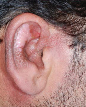
Fig. 3 The patient's right auricle after treatment.
Discussion
There are almost 20 infectious agents that may cause leishmaniasis in humans.Reference Herwaldt 1 The clinical manifestations of the disease are influenced by the characteristics of both the human host and the parasite species, and range from asymptomatic exposure, through self-healing skin ulcers, to widespread, destructive, life-threatening ulceration.Reference David and Craft 4
Certain leishmania strains have a predilection for specific organs and tissues; therefore, the clinical manifestations of the disease show a wide spectrum including cutaneous, mucocutaneous and visceral forms.
Visceral leishmaniasis is a systemic infection characterised by fever, weight loss and hepatosplenomegaly; if untreated, it is usually fatal.
In contrast, the majority of cutaneous leishmaniasis cases do not exhibit systemic symptoms, although, rarely, disseminated disease can occur.Reference David and Craft 4 Systemic symptoms such as lymphadenopathy, fever and hepatomegaly have been reported to precede ulcerative lesions caused by L braziliensis.Reference al-Gindan, Kubba, el-Hassan, Omer, Kutty and Saeed 10
The lesions themselves are often chronic and unresponsive to antibiotics or steroids. They consist of simple cutaneous pathology such as papules, ulcerations or infiltrations, of varying degrees of severity, and may show clinical polymorphism.
The clinical history is very important for successful diagnosis of cutaneous leishmaniasis. In endemic regions, cutaneous leishmaniasis should be borne in mind, and actively suspected in the presence of typical history features and/or clinical signs.Reference Sabri, Khatib, Kanj-Sharara, Husseini, Nuwayri-Salti and Semaan 11 Patients who live in nonendemic regions should be asked about recent travel to endemic areas.
The diagnosis of cutaneous leishmaniasis is established by identification of the organism in skin scrapings acquired from ulcerated, crusted lesions in the acute phase. Other tests which confirm the diagnosis include microbial culture (on Novy-MacNeal-Nicolle (NNN) media), polymerase chain reaction analysis and the leishmanianin skin test.Reference Sabri, Khatib, Kanj-Sharara, Husseini, Nuwayri-Salti and Semaan 11 However, examination of skin scrapings is one of the most common and effective methods, with a sensitivity of 75 per cent.Reference Herwaldt 1 Cutaneous leishmaniasis is diagnosed upon observation of amastigote forms within monocytes or in the extracellular space. If skin scrapings do not reveal amastigotes but clinical suspicion remains high, or if the lesion is nodular, a 4 mm punch biopsy should be performed at the edge of the lesion or ulcer in question.Reference David and Craft 4 In our case, the diagnosis of cutaneous leishmaniasis was reached by examination of skin scrapings, using Giemsa staining.
Upon histopathological examination, cutaneous leishmaniasis shows diffuse, nodular, epithelioid cell granulomas within the deep and superficial dermis. However, these findings are nonspecific if the pathogen cannot be identified histopathologically. Typical infected specimens show a mixed cell granulomatous inflammatory response in the dermis containing numerous parasitised macrophages, mononuclear cells, eosinophils and lymphocytes.Reference Maurer, Dondji and von Stebut 12
Cutaneous leishmaniasis can be confused with other diseases, both on physical examination and histological evaluation (Table I). The differential diagnosis of ulcerative auricular lesions includes other infections, which can be bacterial, fungal, atypical mycobacterial or tubercular.Reference Magill, Hunter and Strickland 13 Moreover, nodular, papillomatous and ulcerative lesions can be observed on the auricle secondary to immunological diseases or neoplasms. Neoplasms may cause misdiagnosis due to their similar physical and histopathological appearance. While samples obtained from the lesion facilitate diagnosis, some cases with granulomatous infiltration may result in diagnostic delay. However, by bearing in mind the specific symptoms observed in granulomatous diseases, the correct differential diagnosis can be established.
Table I Differential diagnosis of cutaneous leishmaniasisReference Magill, Hunter and Strickland 13 , Reference Uzun, Acar, Uslular, Kavukcu, Aksungur and Culha 14

Another disease which may be confused with cutaneous leishmaniasis is angiolymphoid hyperplasia. It may be mistaken for leishmaniasis due to auricular localisation and ulcerated, nodular, papular, crusted lesions. Histopathologically, angiolymphoid hyperplasia exhibits perivascular lymphocytic infiltration and vascular proliferation,Reference Abrahamson and Davis 15 whereas cutaneous leishmaniasis demonstrates amastigote forms in monocytes and/or the extracellular space when correctly stained. Our case had previously been diagnosed with angiolymphoid hyperplasia as a result of clinical examination and histopathological analysis in another centre. The clinical history indicated that the patient lived in a region of Turkey in which cutaneous leishmaniasis was endemic. Physical examination showed diffuse oedema along with a nodular, ulcerated auricular lesion, which suggested several other diseases including neoplasia. However, since the patient lived in a region in which cutaneous leishmaniasis was known to be endemic, smear samples were acquired from the lesion, which subsequently confirmed the diagnosis of cutaneous leishmaniasis.
-
• Rarely, cutaneous leishmaniasis presents as ear auricle lesions
-
• These may mimic neoplasia
-
• Consider leishmaniasis in cases of stubborn, pruritic, crusted or ulcerated auricular lesions
When treating cutaneous leishmaniasis, the parasite type, disease dissemination and lesion localisation should be considered. Old World cutaneous leishmaniasis usually heals spontaneously within three to 18 months, leaving a depressive cicatrice. Local treatment may be sufficient for early, localised, cutaneous lesions and for Old World cutaneous leishmaniasis.Reference Sabri, Khatib, Kanj-Sharara, Husseini, Nuwayri-Salti and Semaan 11 Treatment is also encouraged in cases in which there are multiple (i.e. more than five to 10) or large (i.e. more than 4–5 cm) lesions, or lesions present for more than six months, or lesions located in a cosmetically sensitive area (i.e. the face) or over joints.Reference Weina, Neafie, Wortmann, Polhemus and Aronson 16 Treatment in such cases may include cryotherapy, local use of infrared heat lamps, paromomycin ointment, and infiltration of the base of the lesion with pentavalent antimony salts. One study found that intralesional meglumine antimoniate solution had an efficacy of 97 per cent with no adverse side effects.Reference Uzun, Durdu, Culha, Allahverdiyev and Memisoglu 17 Antimonial drugs induce faster healing of lesions while preventing relapse and local dissemination. Other treatment agents include amphotericin B, pentamidine isethionate and paromomycin. Our patient received 15 mg/kg/day meglumine antimoniate. He responded to the treatment within 15 days, and the lesion was eventually replaced by scar tissue.
Conclusion
We present a case of Old World cutaneous leishmaniasis with atypical localisation. It should be borne in mind that cutaneous leishmaniasis presenting as an isolated auricular lesion may mimic neoplasia. Auricular cutaneous leishmaniasis is one of the infectious diseases that should be considered in the differential diagnosis of cases with pruritic, crusted, ulcerated lesions which do not heal upon medical therapy, in patients who live in or travel to endemic regions.


