


Introduction
The poultry red mite (PRM), Dermanyssus gallinae, is the most detrimental ectoparasite on poultry farms, especially for layer and breeding birds (Flochlay et al., Reference Flochlay, Thomas and Sparagano2017). Extensive infestations of D. gallinae have been reported in Europe, China, Japan, Korean, Australia, USA and Brazil (Chu et al., Reference Chu, Murano, Uno, Usui and Yamaguchi2015; Oh et al., Reference Oh, Do, Kim, Yi and Yoo2020). Severe infestation of PRMs could occur in a short period because of their short life cycle (within 1 week under optimal conditions) and high fecundity, resulting in rapid increase of the mite population (Pritchard et al., Reference Pritchard, Kuster, Sparagano and Tomley2015). Infestation of PRMs can cause bird anaemia and malnutrition, resulting in decreased egg production, poor quality and increased morbidity as well as mortality of hosts (Sleeckx et al., Reference Sleeckx, Van Gorp, Koopman, Kempen, Van Hoye, De Baere, Zoons and De Herdt2019). Besides these directly negative effects, it also indirectly affects the host health by acting as potential vectors for bacterial and viral pathogens (Moro et al., Reference Moro, De Luna, Tod, Guy, Sparagano and Zenner2009a, Reference Moro, Thioulouse, Chauve, Normand and Zenner2009b), including Spirochetes anoseiferi, Salmonella enteritidis, Pasteurella multocida, Coxiella burnetii, Erysipelothrix rhusiopathiae, Chlamydia psittaci, Listeria monocytogenes, porcine lupus erythematosus, fowlpox virus, equine encephalitis viruses, Newcastle disease virus and avian influenza virus (Hubert et al., Reference Hubert, Erban, Kopecky, Sopko, Nesvorna, Lichovnikova, Schicht, Strube and Sparagano2017; Cocciolo et al., Reference Cocciolo, Circella, Pugliese, Lupini, Mescolini, Catelli, Borchert-Stuhltrager, Zoller, Thomas and Camarda2020; Schiavone et al., Reference Schiavone, Pugliese, Circella and Camarda2020). Although PRM is one of the small pests, e.g. the engorged adult mite is ~1.5 mm in length and 300 μg in body weight, it causes huge economic loss, for example estimated to 231 million € per year in Europe. In addition, D. gallinae can bite human beings, causing erythematous, papulae and dermatitis (George et al., Reference George, Finn, Graham, Mul, Maurer, Moro and Sparagano2015; Cafiero et al., Reference Cafiero, Barlaam, Camarda, Radeski, Mul, Sparagano and Giangaspero2019).
Adults and nymphs of D. gallinae feed on blood from birds in a short period at night while spend most of their life hiding in cracks in walls and troughs. PRMs are highly resistant to starvation and desiccation, allowing them to survive up to 8 months without a blood meal (Chauve, Reference Chauve1998; Sparagano et al., Reference Sparagano, George, Harrington and Giangaspero2014). These biological characteristics make it very difficult to eliminate D. gallinae from poultry farms.
The blood from the host is essential for the survival, development and reproduction of haematophagous arthropods (e.g. ticks, mosquitos, PRMs, etc.). After engorgement the blood is digested by a series of proteolytic enzymes in the intestine of haematophagous arthropods, producing amino acids and other components to provide energy and nutrition for various life activities, ovary development and egg production for mosquitos (Brackney et al., Reference Brackney, Isoe, Black, Zamora, Foy, Miesfeld and Olson2010; Sojka et al., Reference Sojka, Franta, Horn, Caffrey, Mares and Kopacek2013), ticks, PRMs (Bartley et al., Reference Bartley, Huntley, Wright, Nath and Nisbet2012) and bugs (Leyria et al., Reference Leyria, Orchard and Lange2020). It has been demonstrated that the engorgement level has a positive relationship with the fecundity of PRM (Wang et al., Reference Wang, Ma, Huang, Xu, Cai and Pan2018). Dozens of drugs and vaccines have been found to inhibit the blood digestion, interfering in the growth, development and reproduction of haematophagous arthropods. For example, 2 antigens to digestive protease of D. gallinae, Dg-CatD and Dg-CatL could prevent and control D. gallinae by reducing the survival (Bartley et al., Reference Bartley, Huntley, Wright, Nath and Nisbet2012), reproductive capacity (Price et al., Reference Price, Kuster, Oines, Oliver, Bartley, Nunn, Barbero, Pritchard, Karp-Tatham, Hauge, Blake, Tomley and Nisbet2019) and blood digestion (Xu et al., Reference Xu, Wang, Zhang, Huang, Pan, Wang and Pan2019) of mite. Two ferritins are crucial to the survival, reproduction and blood digestion of D. gallinae, showing good potential as an antigen of vaccine against D. gallinae (Xu et al., Reference Xu, Wang, Liu, Huang, Sun and Pan2022). Orally administrated macrocyclic lactones (eprinomectin, ivermectin) to chicks could significantly delay the blood-meal digestion of PRMs, reducing egg production (Xu et al., Reference Xu, Wang, Zhang, Huang, Pan, Wang and Pan2019). Therefore, developing reliable methods to evaluate the blood digestion of PRMs is a significant step in the development of novel drugs and vaccines.
Currently, several methods have been used in the assessment of the blood digestion of haematophagous arthropods. The first one is to calculate blood digestion rate based on the change in pest body weight during the digestion (Xu et al., Reference Xu, Wang, Zhang, Huang, Pan, Wang and Pan2019). Although this method has been widely used, it shows some limitations for small-sized pests (e.g. PRMs). The application of this method in PRMs needs not only a large number of mites, but also an analytical balance with high precision. The second method is to determine the blood digestion status by observing the appearance of the insect. The inhibition effects of moxidectin on the blood digestion of bed bugs were evaluated by observing the congestion in the intestines of the bugs (Zha et al., Reference Zha, Wang and Sheele2017). This method could intuitively but not quantitatively reflect the status of blood digestion, since no standards or criteria were provided. Third, a fluorescence-based assay was used to track the feeding and digestion status of Aedes (mosquitoes). However, in this method an individual homogenate was required, making continuous observation of an individual impossible (Jove et al., Reference Jove, Venkataraman, Gabel and Duvall2020). Finally, the blood digestion status can be evaluated by quantifying the amount of haematin defecated by blood-sucking pests (Briegel, Reference Briegel1980). However, this method requires special equipment and kits, such as high-performance liquid chromatography (HPLC), spectrophotometry or expensive haematin detection kits, and large amounts of excrement, bringing challenges to the detection of haem in miniature haematophagous arthropods, such as PRMs.
The aim of the present study was to establish a visually and convenient scoring method to evaluate the blood digestion status of D. gallinae, and to provide a new approach for evaluating the efficacy of drugs or vaccines against PRMs.
Materials and methods
Animals and D. gallinae colonies
All chicks (females, Jingbai 939 strain) used in the study were obtained from a commercial hatchery on day 1, which were kept in metal cages with cardboard over the bottom grids and placed in plastic storage boxes, and allowed to access feed and water ad libitum until usage. The storage boxes with chicks were placed in an artificial climate incubator (RXZ-500B-LED, Ningbo Jiangnan Instrument Factory, Ningbo, China), at 30°C and 75% relative humidity (RH), with a 12/12 h light/dark photoperiod. All procedures and chick maintenance were conducted according to the recommendations of the Guide for the Care and Use of Laboratory Animals, Ministry of Science and Technology, China, which was ratified by the Institutional Animal Care and Use Committee of China Agricultural University (approval no. CAU20190909-1).
The D. gallinae colony used in the present study was originally obtained from a commercial poultry farm in China, which was reared in the in vivo system with chicks at 30°C, 75% RH under laboratory conditions (Wang et al., Reference Wang, Ma, Huang, Xu, Cai and Pan2018). The number of mites in the rearing system were monitored weekly by counting mites in 2 randomly chosen trap tubes (as sample survey) and the total number of mites in the system with 8 trap tubes were calculated based on the number in 2 trap tubes. When the number of mites was more than 50 000 per bird, some tubes were taken out of the system to reduce the mite population. The D. gallinae densities of 50 000 mites per bird are considered ‘normal’, causing no significant harm to poultry health, while serious health problems appeared when the infection level attained more than 150 000 mites per bird (Kilpinen et al., Reference Kilpinen, Roepstorff, Permin, Norgaard-Nielsen, Lawson and Simonsen2005). In addition, 1 month after infestation, the old chicks were replaced with new ones to avoid severe welfare problem. The animal experiments were approved by the Institutional Animal Care and Use Committee of China Agricultural University (approval no. CAU20200310-1).
Mite infestation and recovery
Infestation and recovery of mites were carried out according to the previously described method (Wang et al., Reference Wang, Ma, Huang, Xu, Cai and Pan2018; Xu et al., Reference Xu, Wang, Zhang, Huang, Pan, Wang and Pan2019). Briefly, when chicks were 6 weeks old, they were challenged with PRMs. Female mites used for infestation were starved for 3 days prior to infestation. For infestation, tubes containing 600 starved adult mites were placed at the bottom of the metal cage and were opened to let the mites move out and feed on 3 chicks. Then, the cages were placed in plastic storage boxes and the rearing systems were kept in an artificial climate incubator in the dark for 12 h to enable the mites to feed. The engorged mites hiding in trap tubes, which were installed at the bottom of cage before infestation, were collected to observe blood digestion.
Scoring of blood digestion
Three hundred engorged mites were collected from cage and this time point at the digestive state was defined as day 0 (d0). The engorged mites were transferred into 100 mL centrifuge tubes and placed in an incubator at 30°C and 75% RH for blood digestion. Then, the mites were observed continuously for 6 days. During this period, some of them were randomly picked out and fixed on a slide by double-sided tape; the blood digestion status of mites was closely observed and an image was taken under a stereomicroscope (SteREO-Discovery.V12; Carl Zeiss, Jena, Germany). A blood digestion scoring criterion was set-up based on the intestinal change of PRMs during the digestion.
Afterwards, 300 engorged mites were re-collected on d0–d6; then 20 mites were randomly selected and scored by scoring criterion established above. The remaining mites were weighed daily by an electronic balance (Sartorius PB310s, Göttingen, Germany) during the 6-day digestion period. For daily weighing, the mites were transferred to a new centrifuge tube to eliminate feces, eggs and exuviae. After weighing, the mites were placed back in the incubator at 30°C and 75% RH for blood digestion. The average weight of each mite at each time point was calculated, and the blood digestion rate was calculated as below. This experiment included 3 replicates.

To compare the results obtained by these 2 methods, the correlation between blood digestion rate and blood digestion score was calculated by Pearson correlation coefficient.
Blood digestion scoring by volunteers
To confirm the practicability of the blood digestion scoring for colleagues besides the researchers in the present study, 20 adult mites at various digestion statuses were randomly chosen from the rearing system and scored by 10 volunteers who had not received professional training according to the established digestion scoring criterion.
Effect of doramectin on blood digestion of D. gallinae
To assess the applicability of blood digestion scoring method in the efficacy study, the effects of doramectin on the blood digestion of PRMs was evaluated. Doramectin was chosen based on these considerations, as an avermectin, doramectin shows good efficacy against a variety of parasites, such as mites, ticks and nematodes (Lohmeyer et al., Reference Lohmeyer, Miller, Pound and Oehler2009; Edmonds et al., Reference Edmonds, Vatta, Marchiondo, Vanimisetti and Edmonds2018; Larroza et al., Reference Larroza, Soler, Robles, Cabrera, Ballent, Lanusse and Lifschitz2020; Mauger et al., Reference Mauger, Kelly, Annandale, Robertson, Waichigo and Aleri2022), and its effects on the blood digestion of PRMs was not reported. Doramectin (with a purity of 95%, purchased from Nanning Guangtai Agricultural Co. Ltd, China) was dissolved in an ethanol/PBS/Tween 80 mixture at a 33:66:1 ratio (v/v) to formulate a 2 mg mL−1 solution. Afterwards doramectin was orally administrated to 3 chicks of 6 weeks old at a dose of 1, 2 and 5 mg kg−1 body weight by gavage on d0. One control group was included, where birds were given placebo solvent without drug. After treatment, the chicks were placed back into the metal cage and were immediately challenged with 300 starved female mites as described above. After 12 h in the dark, all engorged female mites (~150 mites per replicate) found in the rearing system were collected, including mites from all trap tubes, the metal cage and plastic storage box. Fifty live engorged mites were chosen to observe the change in appearance in the digestive tract and body of the mites, and the digestive status was scored daily according to the established blood digestion scoring criterion. The remaining live engorged mites (more than 100 mites in the control and 1 mg kg−1 groups, more than 50 mites in the 2 and 5 mg kg−1 groups because some mites died) were counted and placed into 100 mL centrifuge tubes for the evaluation of blood digestion rate by weighing. This experiment included 3 replicates.
Statistical analysis
All statistical analyses were performed using Prism 8.0.1 (GraphPad Software, Inc.). The differences in the digestion rate (Fig. 5B) and the digestion score (Fig. 5C) between 3 treatment groups and control group was analysed by the analysis of variance (ANOVA) (parametric) method and Kruskal–Wallis (non-parametric), respectively. Statistically significant differences are shown with asterisks as follows: *P < 0.05, **P < 0.01, ***P < 0.001; whereas ns indicates no significant difference. Pearson correlation coefficient was calculated to examine the relationship between the digestion rate and digestion score.
Results
Changes in appearance of adult mites during blood digestion
The appearance of digestive tracts of PRMs is similar to that of ticks, e.g. the digestive tract extends from the gnathosoma posteriorly through the oesophagus, midgut and caeca and ends in the hindgut (Fig. 1). In starved mites, the intestine shrinks (Fig. 2, d6), but in engorged ones, the intestine expands to fill most of the body cavity (Fig. 2, d0). The blood digestion mainly occurs in the 3 expanded caecal pairings (Ca I–III) and central midgut (Mg) (Fig. 1).

Fig. 1. Digestive tract system of PRM. Mites were observed from the dorsal side under a stereomicroscope. Ca I–III: caeca I–III, Mg: midgut. Scale bar = 500 μm.

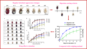
Fig. 2. Appearance of adult mites at different digestion periods. During the blood digestion period (0–6 days), the mites were randomly chosen and carefully observed by a stereomicroscope. Scale bars = 500 μm.
As shown in Fig. 2, on d0 after blood fed (ABF), the body of engorged female mites was full of blood, appearing bright red, round and no transparent part in the body. On d1 ABF, a white or transparent area appeared in the centre part (midgut) of the hinder body, while the other parts of the body still appeared red, especially for the areas locating the 3 pairs of caeca. Interestingly, an egg appeared in most of the females, indicating the blood digestion was related to the formation of eggs. On d2 ABF, the blood was further digested, the white and transparent areas in centre of the hinder enlarged, some parts of caeca became transparent and the blood no longer appeared bright red, but dark brown or reddish black. On d3 ABF, as most blood was digested, most part of the hinder of PRMs became white or transparent, and only some part remained dark red blood in caeca. The whole profile of caeca could be clearly recognized. On d4 ABF, the blood in the caeca was nearly completely digested, only a few of intermittent dark red lines were observed, and the whole profile of caeca became indistinct and hard to be clearly recognized. On d5 ABF, most of the adult mite's body appeared transparent or grey-white, with only sporadic black blood spots remaining in every caecum. On d6 ABF, the body of adult mites appeared greyish-white or transparent; there was almost no residual blood in any part of the body, showing the blood digestion completed. The obvious changes in appearance in the blood digestion suggest that it is possible to evaluate the blood digestion status through appearance observation.
Establishment of blood digestion scoring method for D. gallinae
Based on the intestinal tract changes of mites during blood digestion, 0–4 point criterion for the blood digestion scoring was established, which is described in Table 1, and the corresponding typical images of mite appearance are shown in Fig. 3. Briefly, when the digestive tract of mite was full of red blood, it was scored as 0. When there was almost no blood in the midgut and caeca, and the mite became grey and white, it was scored as 4. When the trunk of mites was mostly bright red or dark red, with only a small part of areas of the body was transparent, the score was 1. When 3 pairs of caeca and hindgut have blood, most centre parts of the hinder body become transparent or white, and the intestinal profile can be clearly recognized, it was scored as 2. When most of the body becomes transparent or white, only a small amount of blood remains in the caeca and the intestinal profile cannot be clearly recognized, the score was 3.

Fig. 3. The corresponding appearance of different digestion score of adult mites. Scale bars = 500 µm.
Table 1. Digestion score and appearance description of adult mites

The digestion status of PRMs during the blood digestion process (0–6 days) was scored according to the scoring criterion, and the results are shown in Fig. 4A. On d0, the mites had just finished feeding and were scored as 0; most mites were scored as 1 on d1 and 2 on d2 after collection. On d3–d5, the blood digestion rate of mites slowed down and the scores were mostly concentrated at 3. On d6, the blood meal was basically digested, and the score at this point was 4.

Fig. 4. Correlation analysis of weighing method and scoring method. (A) Blood digestion score and blood digestion rate in the blood digestion process of PRMs. Error bars represent the standard error of the calculated mean based on biological replicates. (B) Correlation between blood digestion score and blood digestion rate. A strong positive correlation was observed (r = 0.9302, P < 0.0001).
The blood digestion of PRMs was evaluated by the weighing method to compare with the results obtained by the digestion scoring method. For 3 days, the digestion rate was high, which was 46.5 ± 1.89% on d1 and attained 82.03 ± 3.31% on d3. Afterwards, the blood digestion rate of mites slowed down, attaining 93.70 ± 0.68% on d4, and 95.10 ± 2.69% on d5. On d6, the blood in mites was almost completely digested and the digestion rate attained 99 ± 0.6% (Fig. 4A). As shown in Fig. 4B, the blood digestion scores and blood digestion rate showed a generally consistent trend, and a strongly positive correlation was observed between the digestion score and digestion rate (Fig. 4B, r = 0.9302, P < 0.0001). This result indicated that the blood digestion scoring method could accurately evaluate the blood digestion under natural blood digestion, e.g. without effects of drugs or vaccines.
Blood digestion scoring of D. gallinae by volunteers
Twenty adult mites at various digestion states were provided to volunteers for digestion scoring, and the scoring result is shown in Table 2. The coefficient of variation of scores for these 20 mites ranged from 0 to 19%, showing low variability for scores. Interestingly the values obtained by volunteers were close to the scores from the researcher who established the criterion, indicating that the scoring method has good practicability for others who may concern about and want to use this method.
Table 2. Blood digestion scoring results of adult mites by volunteers

‘–’ represents the coefficient of variation cannot be calculated (average score is 0).
Evaluating the effect of drugs on blood digestion of D. gallinae by blood digestion scoring
The blood-meal digestions of mites from the 2 and 5 mg kg−1 doramectin treatment groups were significantly inhibited than those in the control group (Fig. 5A), where the blood digestion score was finally concentrated in 1–2 vs 3–4 in the control group at the end of the observation (d6). There was a good consistency between the profiles of blood digestion rate and blood digestion score, showing a strongly positive correlation between the digestion score and digestion rate in each group (Con: r = 0.8097, P < 0.0001; 1 mg kg−1: r = 0.7008, P < 0.01; 2 mg kg−1: r = 0.7896, P < 0.0001; 5 mg kg−1: r = 0.8648, P < 0.0001) (Fig. 5B and C). These results indicated that the blood digestion score could accurately reflect the blood digestion state of mites, and be used to evaluate the effect of drugs on the blood digestion of mites.

Fig. 5. Effects of doramectin on blood digestion of adult mites. (A) The appearance of mites during digestion periods. Scale bars = 500 μm. (B) Effects of doramectin on digestion rate of mites [one-way ANOVA (parametric)]. (C) The digestion scores of mites after administration of doramectin (Kruskal–Wallis test). Values are shown as average and error bars represent s.d. *P < 0.05, **P < 0.01, ***P < 0.001.
Discussion
Scoring is widely used to quantitatively describe the severity of tissue lesion or symptoms caused by parasites. It is also used to evaluate the efficacy of drugs or vaccines against parasites. For example, Martineau et al. evaluated the efficacy of ivermectin against scabies in pigs using the lesion scoring method (Martineau et al., Reference Martineau, Vaillancourt and Frechette1984). Johnson et al. established a lesion scoring method (0–4) for chicken intestinal lesion caused by Eimeria (Johnson and Reid, Reference Johnson and Reid1970). Studies have shown that there is a good correlation between the dairy calf feces scoring and the number of Cryptosporidium oocysts shedding (Bellosa et al., Reference Bellosa, Nydam, Liotta, Zambriski, Linden and Bowman2011). As a key physiological process of haematophagous arthropods, the effect on blood digestion is a commonly used parameter for evaluating the efficacy of drugs and vaccines against these pests. However, there are only a few methods for intuitively evaluating the blood digestion of haematophagous arthropods, especially for those with small size.
Dermanyssus gallinae, a miniature blood-sucking ectoparasite in poultry, has a very small size and is very light weight. An engorged female PRM weighs ~300 μg and its body length is no more than 1.5 mm, while a starved one weighs <100 μg. Thus, a large number of mites are needed to evaluate the blood digestion status by weighing or other detection methods, such as quantifying the amount of haematin defecated by PRMs with HPLC. In addition, for the vaccine or drug showing high efficacy, the small number of surviving mites is generally insufficient to weigh or extract metabolites (e.g. haematin) for detection to evaluate the blood digestion. Therefore, an intuitive blood scoring method would be helpful to evaluate the blood digestive state of PRMs.
In mosquitoes, a blood digestion score method called Sella scoring has been established, where the digestion status of mosquito blood meals was scored visually from 0 (unfed mosquitoes) to 7 (female without visible blood and eggs fully developed in their abdomen) (Detinova, Reference Detinova1962; Martinez-de la Puente et al., Reference Martinez-de la Puente, Ruiz, Soriguer and Figuerola2013). Later, this method was modified to evaluate the extent of blood digestion of mosquitoes, where the blood meals were scored 1–3 based on the colour and size of the blood meal and the state of developing ovaries (Reeves et al., Reference Reeves, Gillett-Kaufman, Kawahara and Kaufman2018). Since the morphological structure of mosquitoes is quite different from that of PRMs, the Sella scoring method is not suitable for PRMs.
In the present study, the changes in the intestinal tract of mites during the blood digestion process were firstly observed. Afterwards, the blood digestion state of mites can be evaluated based on changes in appearance. A blood digestion scoring criterion of 0–4 was established, which corresponds to typical and easily distinguished changes in appearance. based on the present scoring criterion, the digestive status of 0, 1 and 4 were easily determined, but the digestive statuses of 2 and 3 were occasionally confused because the blood digestive rate is slow during this process and the changes in appearance in the digestive tract or body size of the PRMs is little bit difficult to recognize, which needs close observation and careful discrimination. We also tried a wider scale of scoring points (higher than 4), which often leads to overlapping when moving from one score to the next one.
The results obtained by the blood digestion scoring method were compared with those obtained by the weighing method under natural conditions (Fig. 4) and drug treatment (Fig. 5), and found that the profiles from the 2 methods have good consistency, indicating that the blood digestion of PRMs can be accurately evaluated by the scoring method. Since the blood digestion scoring method is based on individual, this method not only can reduce the tedious work involved in collecting a large number of PRMs for weighing, but also can accurately reflect the individual differences in the efficacy of drugs or vaccines. The practicability of a novel method is crucial to its wide application. The scoring values obtained by volunteers were close to those obtained by the researcher who established the criterion, and low variability was observed (Table 2), indicating that the scoring method has good practicability.
Although the robustness of this method has been demonstrated under laboratory conditions, its potential limits should be not be neglected. For those drugs or vaccines with the possibility of disruption of blood sucking, which has been demonstrated in ticks (Wilson et al., Reference Wilson, Hair, Sauer and Weeks1991), partially fed mites will appear when they fed on birds treated with such drugs or vaccines. In these circumstances, how can the scoring be adjusted, given that partially fed mites might visually resemble a mite partially digested? This issue would be prominent, especially for haematophagous arthropods, which need long period for engorgement (such as ticks). However for PRMs, this problem may occur less. It takes only 0.5–1.5 h for PRMs to fully feed (Chauve, Reference Chauve1998), which is probably not long enough for most of the drugs or vaccines to interfere in blood taking, resulting in partially fed mites. For example, the present results (Fig. 5) along with a previous one (Xu et al., Reference Xu, Wang, Zhang, Huang, Pan, Wang and Pan2019) showed that orally administered avermectin did not interfere in blood taking of PRMs, since the engorgement level of mites was similar between the drug treatment groups and control groups. In addition, based on our observation, it was found that most of the PRMs (more than 90%) were engorged when they had a blood meal from birds under natural conditions.
In summary, in the present study a blood digestion scoring method for PRM was established under laboratory conditions. This method has good accuracy and applicability, which can quickly assess the blood digestion status of PRMs, and is useful to evaluate the effect of drugs on blood digestion of PRMs. However, several issues need to be studied further or confirmed, such as the feasibility of this method under field conditions, where the situation is more complex than laboratory conditions, and the suitability of this method to nymph because the blood digestion of nymph is rapid, which is generally completed within 1–2 days, and the adaptability of this method to other blood-sucking arthropods, where changes in appearance of digestive system during the blood digestion in different species may be quite different.
Conclusion
In the present study, a blood digestion scoring method with 0–4 point scoring criterion was first described based on the intestinal change in appearance of PRMs during the blood digestion process, which can intuitively and accurately assess the blood digestion status of mites, and is useful to evaluate the effects of drugs on blood digestion of D. gallinae.
Author contributions
Q. L. and Y. Y. M. conceived the study; B. L. P. and W. W. S. supervised the project; Y. Y. M. performed the experiment; Q. L. and X. W. analysed the data; Q. L. led the writing of the manuscript; B. X. L. and P. L. W. supervised the analyses and writing. All authors contributed critically to the drafts and approved the final manuscript.
Financial support
This work was supported by the ‘National Natural Science Foundation of China’ (Grant Nos. 32072882 and 31873008). The funding bodies had no role in the design of the study, in the collection, analysis and interpretation of data or in writing the manuscript.
Conflict of interest
The authors declare there are no conflicts of interest.
Ethical standards
The chicks in the study were housed under an approved protocol, and research was performed in strict accordance with the recommendations of the Guide for the Care and Use of Laboratory Animals, Ministry of Science and Technology, China, which was approved by the Institutional Animal Care and Use Committee of China Agricultural University (approval no.: CAU20190909-1 and CAU20200310-1).


