


Introduction
Post-auricular cutaneous mastoid fistula refers to the rare occurrence where the mastoid cavity is connected with the post-auricular skin surface, with limited cases reported in the literature.Reference Luetje1–Reference Saki, Araghi, Abshirini and Nikakhlagh16 These fistulas are formed following repeated soft tissue injury, and are commonly caused by chronic suppurative otitis media, repeated surgical treatments such as radical mastoidectomy, or, more rarely, spontaneous exteriorisation of a cholesteatoma.Reference Luetje1–Reference Wadhera, Gulati, Kalra, Ghai and Garg3,Reference Orji, Ukaegbe, Alex-Okoro, Ofoegbu and Okorafor5–Reference Migirov, Carmel, Dagan, Duvdevani and Wolf7 As a result of the fistula, patients may present with symptoms of otorrhoea or soft tissue infection, or, in some cases, for cosmetic reasons whilst being asymptomatic.Reference Luetje1,Reference Choo, Shaw and Chong2,Reference Chew, Cheong, Khir, Brito-Mutunayagam and Prepageran4,Reference Bhat, Udayashankar, Venkatesha and Kumar8,Reference Gök, Kanlikama and Özsaraç9
Management is challenging, with conservative treatment often being unsuccessful. The difficulty arises as cutaneous mastoid fistulas tend to heal very slowly, because of the necrosis and epithelisation of the inverted skin edges and the presence of the empty mastoid cavity.Reference Luetje1,Reference Pendolino, Pavone and Zanoletti10,Reference Müderris, Berçin, Sevil and Kırış11 As such, surgery remains the cornerstone of definitive management. Options include primarily simple closure and muscle flap coverage. Primary closure is the simplest technique that may be used, but this has a high failure rate because of the increased tissue tension, created as the open fistula runs through the deeper anatomical structures. Local advancement temporalis muscle flaps, with or without fascial and periosteal advancement, have therefore been a preferred option, with reported good outcomes, at the cost of cosmesis.Reference Pendolino, Pavone and Zanoletti10,Reference Olusesi and Opaluwah15 Collectively, the rarity of post-auricular mastoid fistulas means there is only a small number of reported successful surgical techniques, often with limited applicability.
We hereby present a case of post-auricular cutaneous mastoid fistula, successfully treated with a new technique utilising a sternocleidomastoid rotational and cervical-fascial advancement flap in the ENT department of the Lewisham and Greenwich NHS Trust.
Case report
A 58-year-old male patient underwent two mastoidectomies with obliteration for chronic otitis media, which resulted in a persistent right-sided post-auricular fistula. This was closed on two previous occasions via simple primary closure, but recurred.
We subsequently proceeded to excise the fistula, and repair it with a sternocleidomastoid rotation flap and lateral cervical-fascial advancement reconstruction.
Surgical technique
The patient is placed supine, with the affected ear positioned uppermost and the face facing away from the surgeon. The post-auricular is cleaned, and a head and neck drape is applied. The fistula and fistula tract are exposed and marked. Lidocaine–adrenaline 1:10 000 solution is injected in the post-auricular region in the place of the incision (Figure 1).
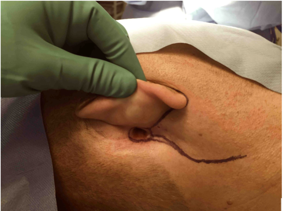
Fig. 1. Exposure of the post-auricular cutaneous mastoid fistula.
A modified facelift (cervical-fascial) incision is made, starting from the superior tragus, advancing to the ear lobule, and then continuing to the mastoid region to the borders of the fistula tract. The flap is then raised (Figure 2). The greater auricular nerve is identified and preserved. After, the sternocleidomastoid muscle is dissected longitudinally and inferiorly towards its posterior aspect. The resultant postero-superior flap of the muscle is then rotated superiorly to obliterate the mastoid cavity. Here, it is attached to the soft tissue superiorly. The cervical-fascial flap is then advanced superiorly and posteriorly, and sutured with freshened edges of the fistula margins (Figure 3).

Fig. 2. The facial flap following the cervical-fascial incision.

Fig. 3. Direct closure of the fistula with flap advancement (a), and the immediate post-operative result (b).
Results
The patient was reviewed at two weeks, one month, three months and one year post-operatively. The flap had healed well by the time of the first follow up, with no concerns. At the one-year follow up, the flap had completely healed without recurrence of the fistula, and the patient was discharged from our clinic (Figure 4).
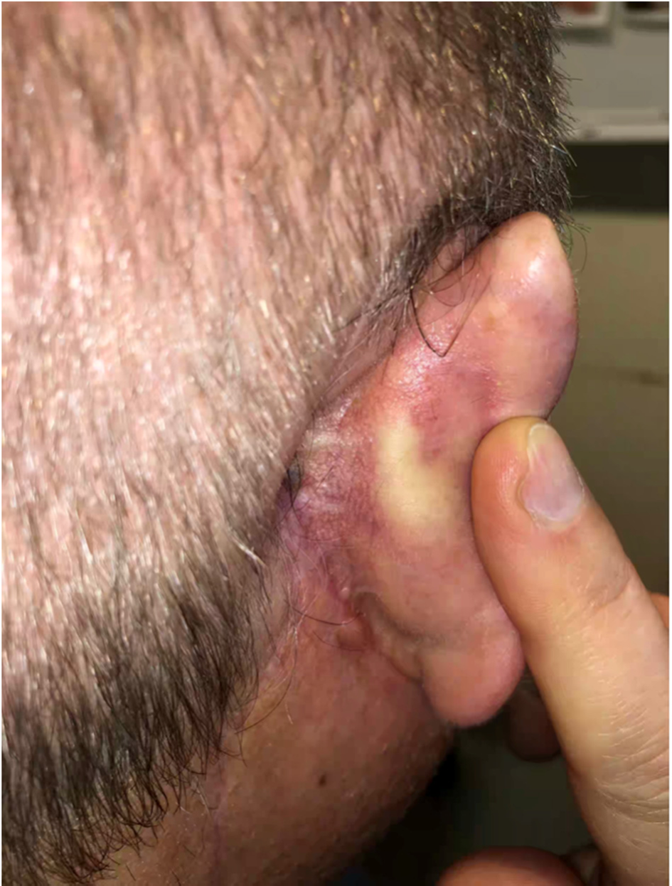
Fig. 4. The healed cutaneous mastoid fistula.
Discussion
Post-auricular cutaneous mastoid fistula is a rarely reported complication of chronic suppurative otitis media, cholesteatoma and radical mastoidectomy. Treatment is challenging: a conservative approach will result in post-auricular discharge, and simple closure is often unsuccessful because of the necrotic skin edges. This report demonstrates a novel technique of closure, which provides complete fascio-musculo-cutaneous reconstruction with excellent cosmesis.
Literature on the optimal management of cutaneous mastoid fistulas is scarce. Attempts at conservative management, comprising the removal of keratin debris with regular suctioning, have been unsuccessful at achieving spontaneous closure or hearing improvement.Reference Chew, Cheong, Khir, Brito-Mutunayagam and Prepageran4 Thus, surgical management is often advocated.
• Post-auricular mastoid fistula is a rare occurrence
• Management is challenging, with few reports of successful surgical techniques, which often have limited applicability
• Post-auricular cutaneous mastoid fistulas may recur despite multiple simple primary closures, requiring more complex reconstruction for complete closure
• A new technique utilising a sternocleidomastoid rotational and cervical-fascial advancement flap is described
• A double-layered flap and facelift incision results in excellent functional and cosmetic outcomes
• There was definitive obliteration of the persistent cutaneous mastoid fistula at 12 months’ follow up
Asherson was the first surgeon to report a closure technique, in 1933. Specifically, closure was achieved by elevating the edges of the fistula and transplanting a portion of the temporalis muscle into the mastoid cavity.Reference Asherson14 Subsequently, a number of case reports have described similar surgical techniques using temporalis muscle flaps. These can be either single rotation or transposition flaps, or part of a double rotation flap consisting of temporalis muscle with fascia and medially based conchal cartilage and skin.Reference Choo, Shaw and Chong2,Reference Lee, Lee, Rhie and Ahn12,Reference Vira and Andrews13 The limitations of temporalis muscle flaps, however, include the risk of denervation atrophy; there is also an aesthetic disadvantage of using a temporal muscle flap, as it depresses the temporal area of the scalp.Reference Pendolino, Pavone and Zanoletti10,Reference Olusesi and Opaluwah15
The use of fibro-muscular-periosteal flaps for the closure of persistent cutaneous mastoid fistulas has also had favourable results. These have been utilised as simple advancement flaps, or in combination with bone pâté and free abdominal fat transfer.Reference Luetje1,Reference Olusesi and Opaluwah15,Reference Saki, Araghi, Abshirini and Nikakhlagh16 More recently, Pendolino and colleagues proposed a surgical refinement involving a fibro-muscular-periosteal flap to fill the mastoid cavity, with the addition of a bilobed flap from the lateral neck and mastoid region to complete skin closure. This technique aimed to reduce suture tension, and had successfully obliterated a mastoid fistula by the six-month follow up.Reference Pendolino, Pavone and Zanoletti10
This is the first report of a sternocleidomastoid rotational flap. Its benefits are functional and cosmetic. The flap has a reliable blood supply from the sternocleidomastoid artery, thus reducing the risk of failure. Further, it is a double-layered flap. The deeper flap provides the muscle bulk for obliteration of the fistula, whereas the superficial flap provides adequate and tensionless surface closure, with each layer having its own blood supply. In addition, with the scars hidden by the facial lift incision, this technique results in an optimal cosmetic outcome.
This technique provides a novel way of reconstructing these rare defects, adding to the techniques described in the literature for closure of a persistent cutaneous mastoid fistula.
Competing interests
None declared


